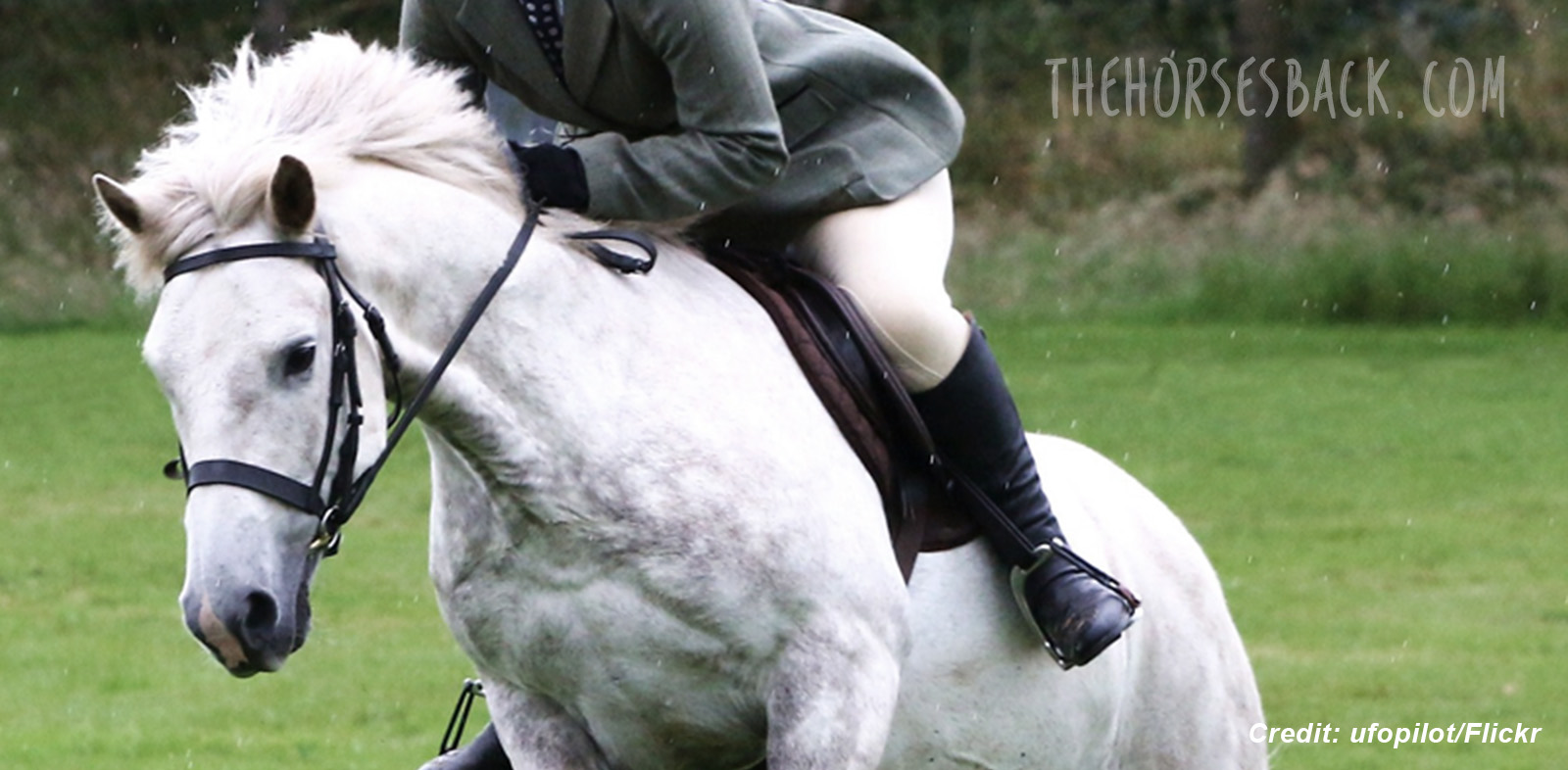
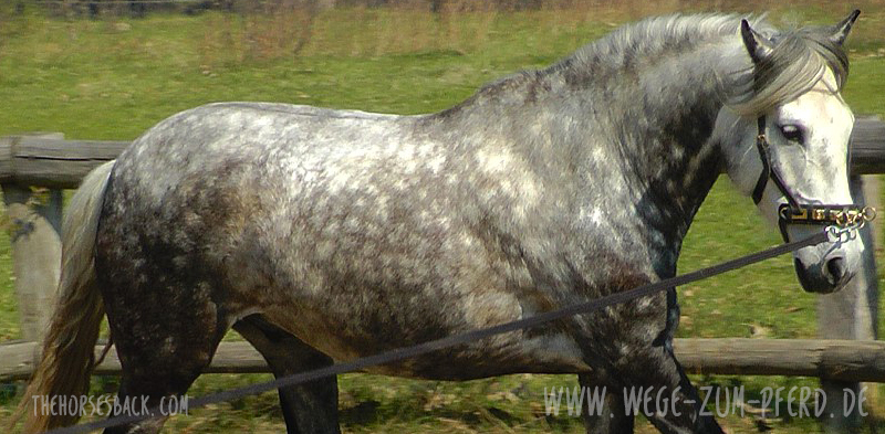
 There’s a basic premise of saddle fitting that hasn’t occurred to a lot of people. It can’t have done, not if the tack they’re riding in is anything to go by.
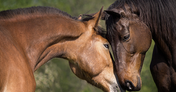
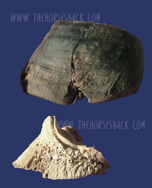
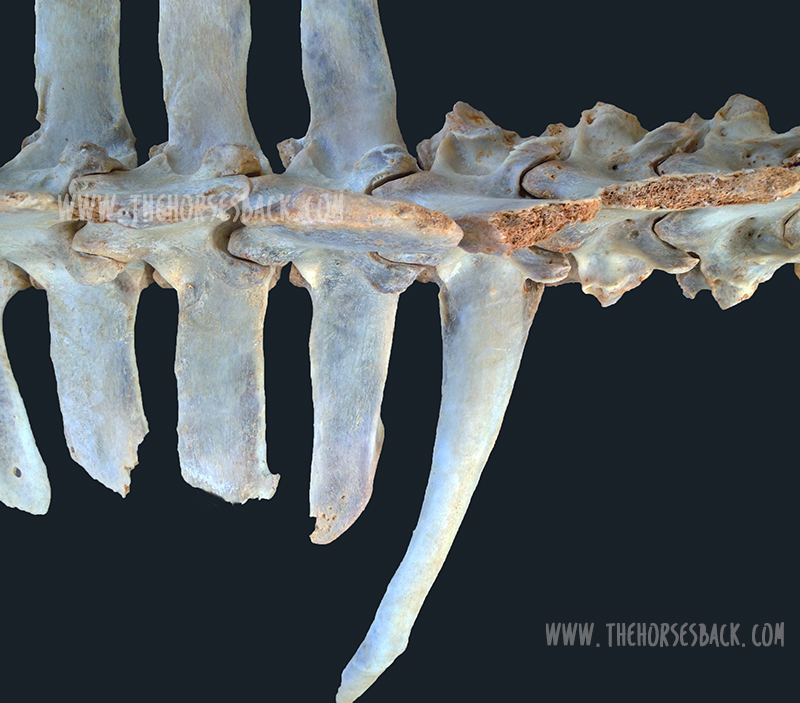
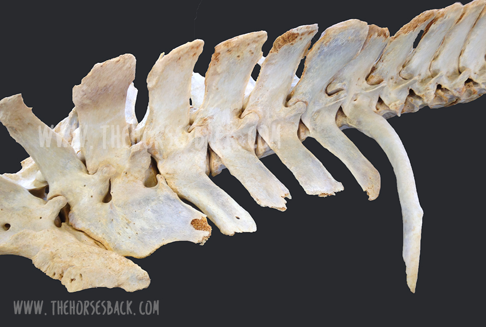
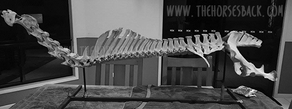
To start, let’s remind ourselves that the horse’s back wasn’t designed to be sat on and it certainly wasn’t designed for saddles. This isn’t to pursue an argument about what is natural for horses or not. Yes we ride them and it’s not natural, ta-de-da.
 The reason for this statement is simply to highlight the fact that inserting a relatively static piece of equipment between two living organisms in motion is, at best, always going to be fraught with complexity.
We have to acknowledge that when it doesn’t work, it’s usually to the horse’s detriment. Yes, sometimes also the rider, but if the rider’s copping it, the horse is usually getting it too.
One of my favourite statements about saddle fitting
It’s possibly a bit odd to have a fave sentence on this subject, but here’s one that leapt out at me a few years back and is still hanging around.
Ken Lyndon-Dykes, Master Saddler expressed this particular concept so succinctly in his book, Practical Saddle Fitting. Enjoy this – it’s a biggie:
“The best designed, most beautifully crafted saddle made specifically to fit an individual horse will not improve the horse’s ability to perform, benefit his welfare or increase his comfort.”
Say what? Do you feel the impact of that? Maybe you knew it already, but it never hurts to have certain truths reflected back at us as a reminder.
Certainly, having used this quote on a presentation slide in my Saddle Fit Essentials for Horse Owners clinics, I’ve found that these words often pull people up short. There’s usually a moment or two when you can almost hear the implications sinking in.
Let’s take that sentence apart a bit.
“The best saddle will not improve the horse’s ability to perform …”
Let’s be clear, this is when compared to how the horse performs when free of its saddle and rider. The horse is always going to move better with no restriction or load on its back.
“…benefit his welfare… “
Again, nope, we’re not improving the horse’s welfare, but minimizing and ideally eliminating the negative impact on the horse’s welfare that comes with restriction or pain due to poor saddle fit.
“… or increase his comfort.”
Same. When did you ever see a horse look more comfortable after you put the saddle on? What we are aiming for is simply neutral, in that the horse is as comfortable with the saddle (and rider) as it is without.
The fact is that saddles are there to assist the rider and beyond that are an exercise in damage limitation to minimize the effect of their own presence.
And that’s it. There’s the crazy logic, right there.
It’s a paradox, the definition of a parodox being “a seemingly absurd or contradictory statement or proposition which when investigated may prove to be well founded or true.”
Saddles may not make a horse go better, but the well-fitted saddle definitely minimizes the negative effect that a saddle and rider may have in preventing normal action and, at worse, causing discomfort and pain.
Interesting, isn’t it? Yet once we recognize this fundamental truth, the more likely it is that we’re on the road to making wise and sensible choices that increase comfort for our horses and improve performance as a result.
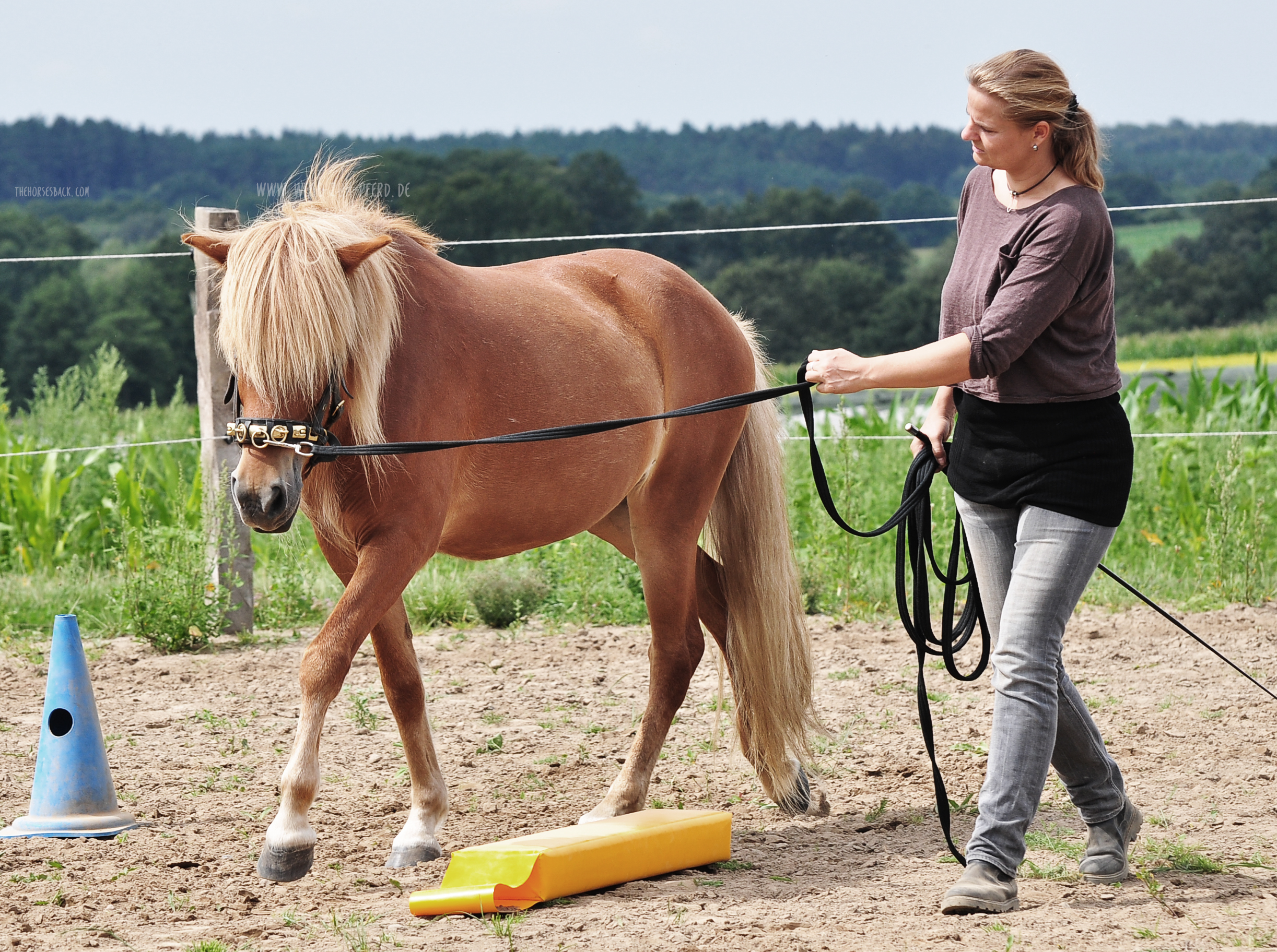
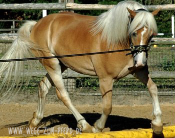
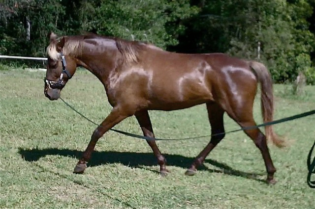
 Thinking as a bodyworker, if there were one thing I could change in the training of ridden horses, it would be the way that many people lunge their animals.
 Here is a simple and affordable resource that will help you to do it better:
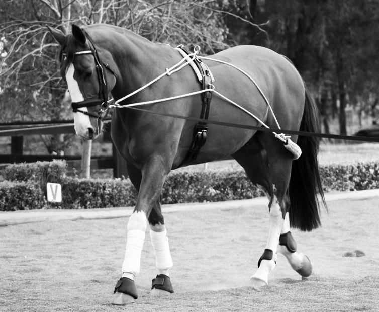
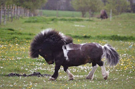
 In the last few years, a renewed interest in a more classical, biomechanically correct approach to training has brought simple, in-hand training techniques to wider audiences. Older methods that aimed to prepare horses for ridden careers, which have been overlooked in the great rush to do everything faster, have come back into focus.
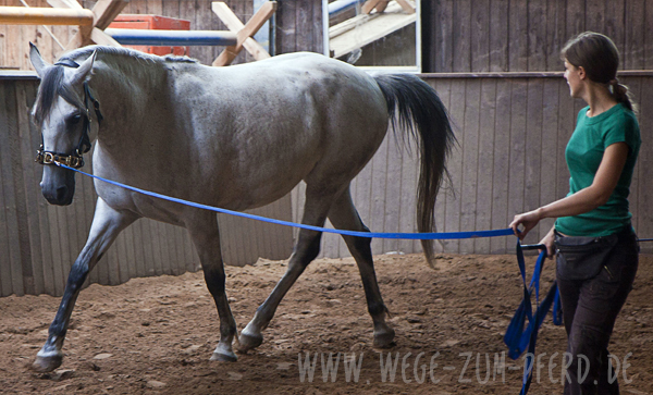
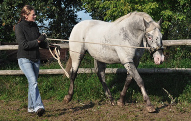 Biomechanically correct lungeing helps to develop the back and teach self-carriage, which makes it humane as well as effective.
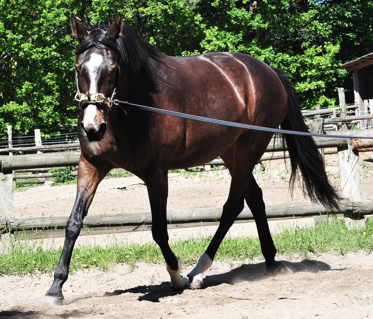
 In this guest post,

 Australia has declared a new war on worms – and this time it’s biological.