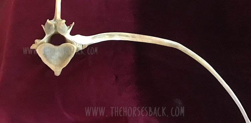
It’s been a question of mine for a while. Can diagnostic imaging show the presence of transitional vertebrae?
We’re seeing many bone samples from dissections, as shown in my previous article on transitional vertebrae.
But if we’re to help our horses that live with this issue, we need to identify it before they’re dead. (Yes, right?!)
Allow me to introduce a practicing vet and educator who is doing just that.
Imaging for Transitional Vertebrae
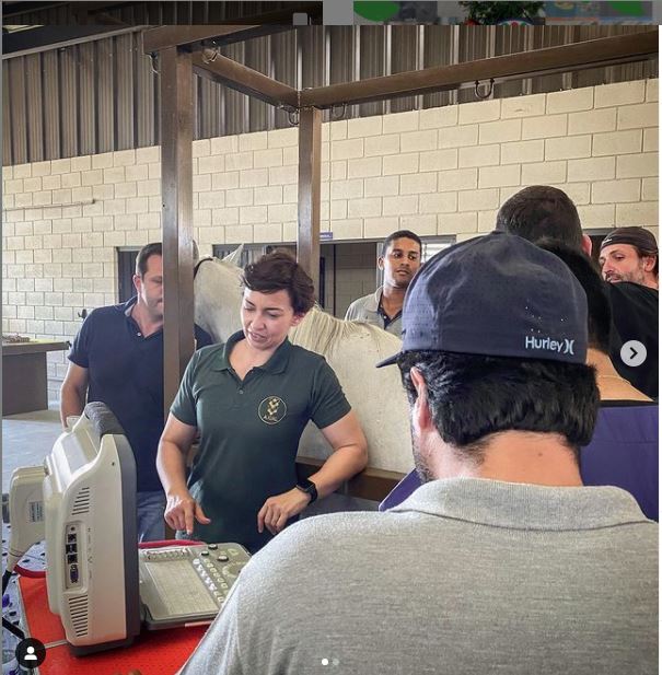 Meet Dr Brunna Fonseca, Associate Professor, educator and specialist in equine orthopedics, focusing on the spine and nervous system. She’s based in São Paulo, Brazil.
I’ve been following her Instagram for a while, because she posts brilliant videos and photos explaining what she does, and how, and why.
I was delighted to see a recent post on imaging for a transitional vertebra, which included fantastic visuals. Such a great communicator!
Dr Brunna has kindly given me permission to repost her images and descriptions here. So without further ado…
-
All images copyright of Axial Vet
Ultrasonograms
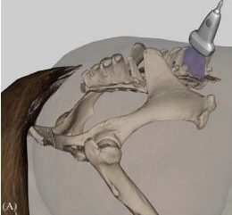
The following ultronographic images are each a composite of two images, one showing the left side and the other the right.
This textbook illustration helps to show the angle the image is taken at. This angle is usually used for imaging the articular facets of the vertebrae.
Additionally, the image at the top of this article shows a transitional vertebra at T18, like the mare being diagnosed by Dr Brunna.
1. Can we recognise transitional vertebrae?
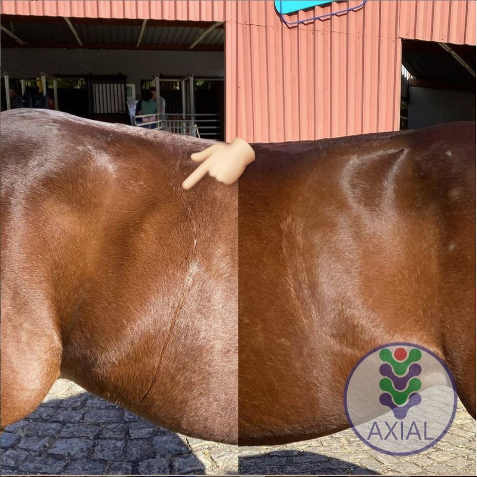
The first image shows two sides of a mare’s body. The hand icon gives us a strong hint of where to look… This appearance is very similar to that of the TB mare in my previous post.
Dr Brunna writes, “This mare has the T18 transitional vertebra, presenting a transverse process similar to the lumbar vertebrae on the right side, which causes the appearance of the horse to have the most visible rib on that side.
The occurrence of transactional vertebrae in the horse is not uncommon, especially in the thoracolumbar transition, which can occur in T18 or L1.”
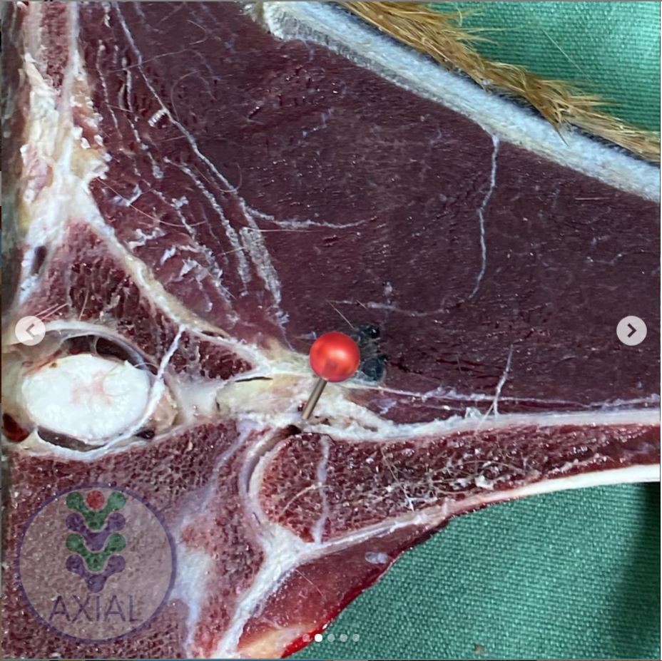
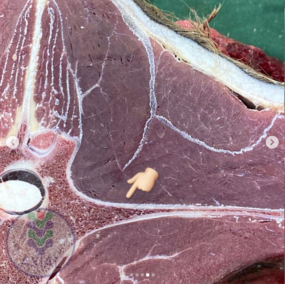
2. Section of a thoracic vertrebra
This image is from a different horse showing a normal rib head and its joint with the vertebra.
Dr Brunna writes, “This is the image of a thoracic vertebra, showing the costotransverse joint.”
3. Image of a normal vertebra
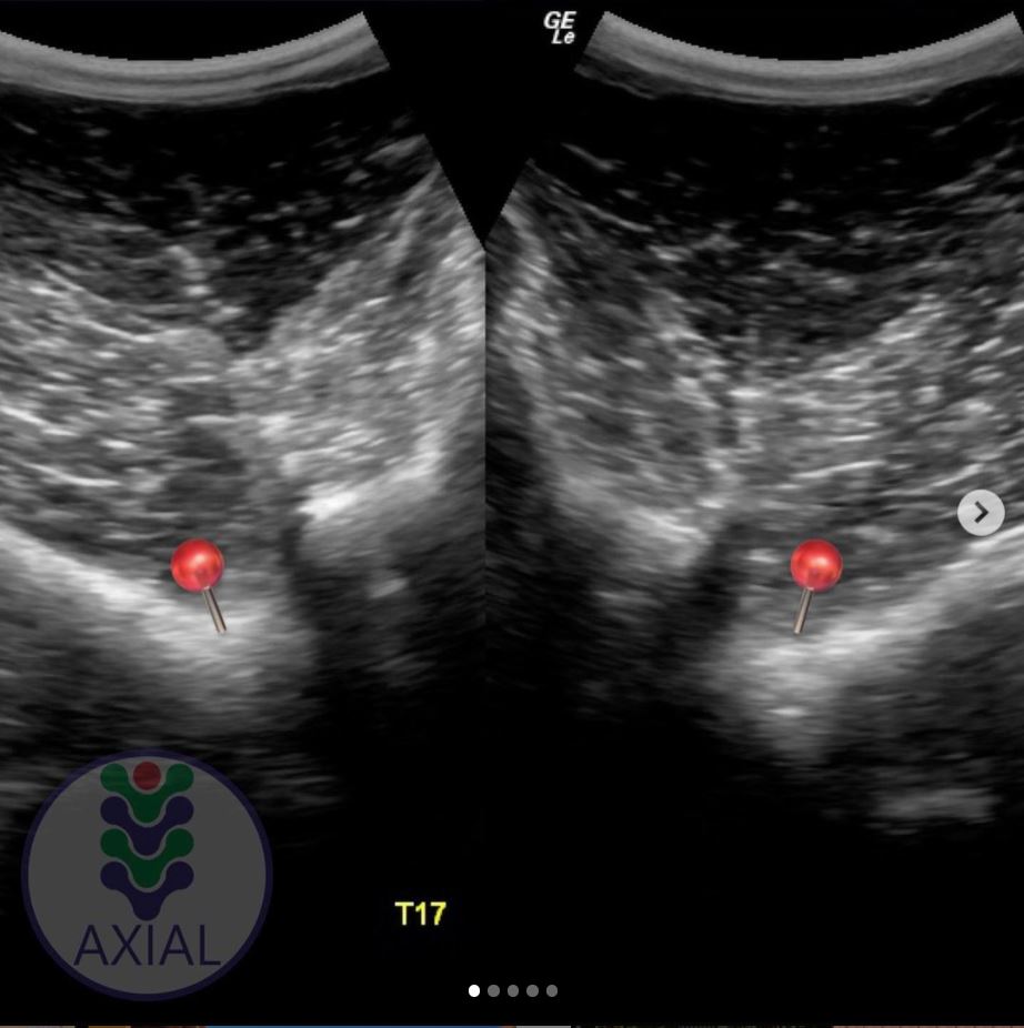
Dr Brunna writes, “This is a T17 ultrasound image, where we can see the image of the normal costotransverse joints.”
This is the bay mare again.
As with the previous cross section, the red pins which show the facet joint between rib head and vertebra.
4. Section of a lumbar vertebra
This is cross section is of a normal lumbar vertebra from a different horse.
As you can see, there is no joint between the transverse process and the vertebral body.
The process is wide and flat, and integral to the vertebra.
 5. Image of a lumbar vertebra
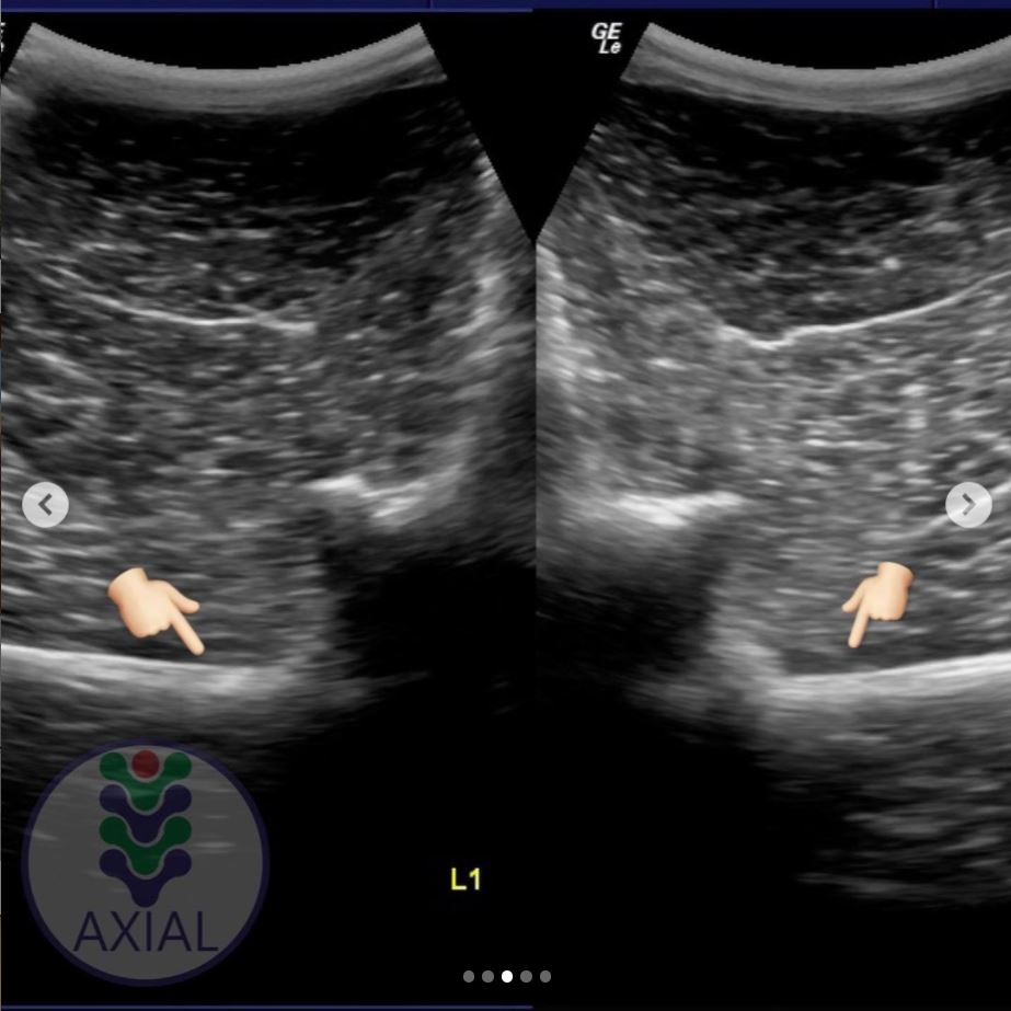
Here’s an ultrasound of the first lumbar vertebra (L1) in the bay mare.
As in the above cross section (picture 4), there is no joint between the transverse processes and the vertebral body.
We now have ultrasound images of the normal T17 and normal L1. As we will see, the transitional vertebra mixes elements from both.
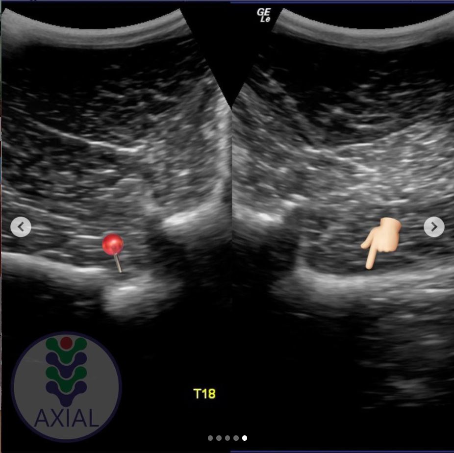 6. Imaging transitional vertebrae
“This is an ultrasound image of T18, where we can see the image of the costotransverse joint on the left side (red pin) and image of the transverse process on the right side.”
So here’s the underlying skeletal issue in the bay mare.
The left side is a normal joint, being the same as the T17 thoracic vertebra (picture 3).
The right side is similar to the previous image of the lumbar vertebra (picture 5).
It is not identical, for while the process-like rib is joined to the vertebra, it is not the same shape and does not lie as flat as the lumbar process.
Want to Hear More From Dr Brunna Fonesca?
 You can follow her Axial Vet Instagram page to see examples of her equine cases and their assessment, in images and videos.
An increasing number of captions are now translated into English.
Jane, I’ll admit to not being an Instagram user so was not able to figure out how to access the IGTV videos. Would you kindly provide some clues, thanks!!
I believe you need to be an IG account holder to view them.
Thankyou Jane for keeping us informed!
Thank you for sharing this she’s great.
Not easy with subtitles but already learned something about nerve roots to pelvis I didn’t know.
Hi Jane
Just saw Dr Brianna has book out about Equine Spine.
Fascinating insights on transitional vertebrae! It’s great to see advancements in imaging that can help diagnose these issues early. Thanks for sharing Dr. Brunna’s work!
I’m so pleased that this blog is helping to link professionals and their work. Thank you for your feedback!