• Skip to main content
  • Skip to footer

The Horse's Back

Body Talk for Thinking Owners

  • Home
  • Articles
  • BEMER
  • Store
    • Store
    • Shipping & Returns
    • Support
  • Equine Healthworks
    • Bodywork for Horses
    • Reviews
    • Leave a Review
  • About & Contact
    • About
    • Contact

Thoroughbred

An Unwelcome Side Effect: Transitional Vertebrae in Horses

May 1, 2018 by Jane @ THB 24 Comments


They can lead to scoliosis, spinal arthritis, flexion and straightness problems, saddle fit issues, secondary lameness, hoof problems and soft tissue trauma. So, what on earth are transitional vertebrae, and why haven’t we heard more about them?

To answer the first part of that question, transitional vertebrae are hybrids that appear where one group of vertebrae changes to another. They show mixed features of each group.

They can be found along the spine, where:

  • the cervical (neck) meet the thoracic vertebrae,
  • the thoracic meet the lumbar vertebrae,
  • the lumbar meet the sacral vertebrae (sacrum),
  • where the sacrum meets the caudal vertebrae (tail bones).

As for why we’ve not heard much about them, the answer is probably that they’re rarely identified while a horse is alive.

However, they can lead to some very real problems in the living horse due to the asymmetry they cause along the spine – and they’re far more common than you might think.
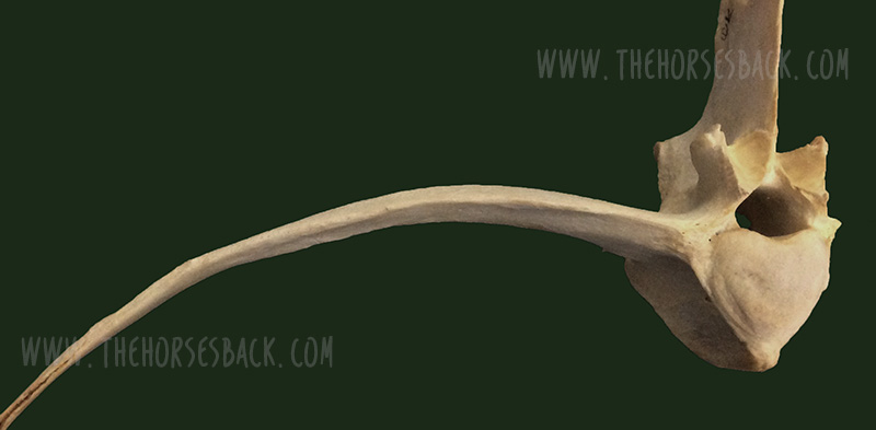
A transitional vertebra at L1. (c) J. Clothier

 The affected process or rib can hurt when the horse bends into it, as the abnormal rib/process is literally ‘stabbing’ into soft tissue.”


© All text copyright of the author, Jane Clothier, https://thehorsesback.com.


Thoracic and lumbar transitional vertebrae

Here are the three main types of variation, as shown in this diagram from one of the few research papers to mention this issue.

Here, we’re going to look at the first two – labeled A and B – which are the most common manifestations.
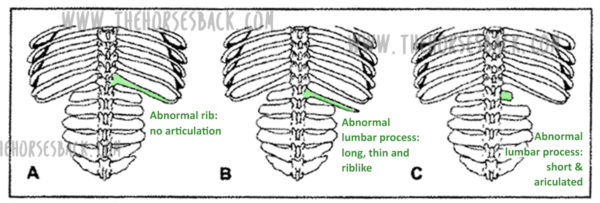
The three kinds of thoracolumbar transitional vertebrae. (c) American Journal of Veterinary Research. (Annotated in green by J. Clothier) Haussler, K.K., Stover, S.M., Willits, N.H. Developmental variation in lumbosacropelvic anatomy of Thoroughbred racehorses (1997); American Journal of Veterinary Research, 58 (10), pp. 1083-1091

A ‘process-like rib’ at T18

Labeled ‘A’ in the above diagram, this is a transitional vertebra at T18 (the last thoracic vertebrae) – a rib that thinks it might be a transverse process, lacking an articulation or joint with the vertebral body.
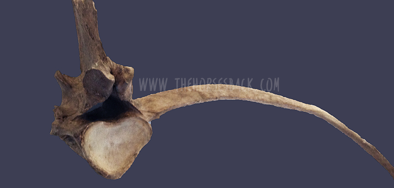
A normal facet on one side, a non-articulated process-like rib on the other (c) J. Clothier

Instead, the process-like rib is solidly attached, meaning there is no independent movement whatsoever. At its end point, it’s joined by costal cartilage to the preceding rib, partially restricting that rib’s movement, too.

This is a problem, as the caudal ribs are not directly attached to the sternum because they need to move more.

The abnormality can be on one or both sides of the vertebra, although single side is most common.

A ‘rib-like process’ at L1

Labeled ‘B’ in the above diagram, this is a transitional vertebra at L1 (the first lumbar vertebrae). Again, it’s usually one-sided, although two sides also occur.

Here, we’re looking at a transverse process that rather than being fairly short, wide and flat, instead extends outwards like a misshapen rib. There’s no articulation with the vertebral body.
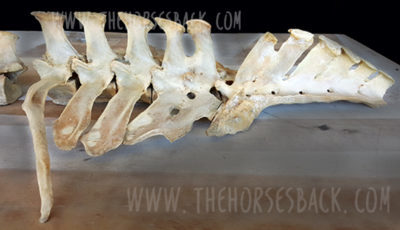
The first lumbar vertebrae (L1) of this Quarter Horse mare is a transitional vertebra. (c) Melissa Longhurst, www.equinebodybalance.com.au 

The above image shows an abnormal L1 found in a Quarter Horse mare. This mare was asymmetric throughout her body, and had a history of unsoundness both fore and rear throughout her lifetime.

Effect on the horse

Scoliosis is the major effect of transitional vertebrae. It’s an asymmetry that in these cases can be lifelong and permanent.

I’ve seen it a few times now in skeletons and on horses that have subsequently been euthanized for unrelated reasons – the spine curves in the affected direction, ie. the horse’s ‘short side’ is the same as the abnormal rib/process that is causing restriction.
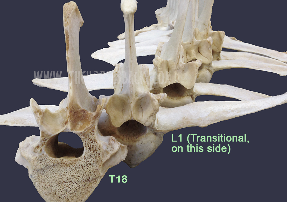
The above bones were from a TB gelding who was in his late teens. Over his lifetime, the additional pressure on the side of the abnormal L1 had caused greater bone development in the vertebra further forward. In this photo, T18, the last thoracic vertebra, has been cut to show this impact.

Cases are highly individual and the degree of impact depends on how abnormal the vertebra is, plus other factors affecting the horse’s musculoskeletal balance – including tack and riders. However, we can consider the following points.

There can be an obvious localized effect:

  • The affected process or rib can hurt when the horse bends into it, as it is literally ‘stabbing’ into soft tissue.
  • The attachments of the deep, short muscles involved in segmental stabilization at L1 and T18 are affected, also affecting proprioception and posture.
  • The abdominal muscles involved in breathing and flexion during locomotion are restricted over an affected T18.
  • The diaphragm inserts onto T18, meaning its function is also affected.
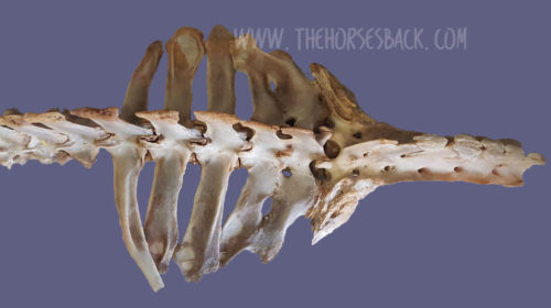
L1 transitional vertebra on the left side causing scoliosis along the spine, including the sacrum. (c) Melissa Longhurst, www.equinebodybalance.com.au


This can affect overall spinal health and biomechanics:

  • Scoliosis means that bending to the affected side can be uncomfortable, while bending to the opposite side can be highly limited.
  • Achieving straightness may be impossible. Scoliosis can extend through the withers and into the neck.
  • Impinging transverse processes and vertebral arthrosis at other vertebral joints further limit movement.
  • These restrictions make lifting the back problematic. 

And then there can be a host of secondary effects:

  • In the heavily pregnant mare, existing discomfort due to a T18 may worsen.
  • Achieving saddle fit is difficult on an asymmetric horse with scoliosis.
  • Abnormal loading can lead to recurrent lameness and persistent hoof issues.
  • Unrelated pathologies can scale up uncontrollably, as the horse cannot compensate effectively.


More on this Topic

Take a closer look at the vertebrae featured in this article (Equine Healthworks is my practice page in NSW, Australia – also on Facebook.)


Questions, thoughts or comments? Join us at The Horse’s Back Facebook Group. 


Can we spot transitional vertebrae in the living horse?

Yes, sometimes.
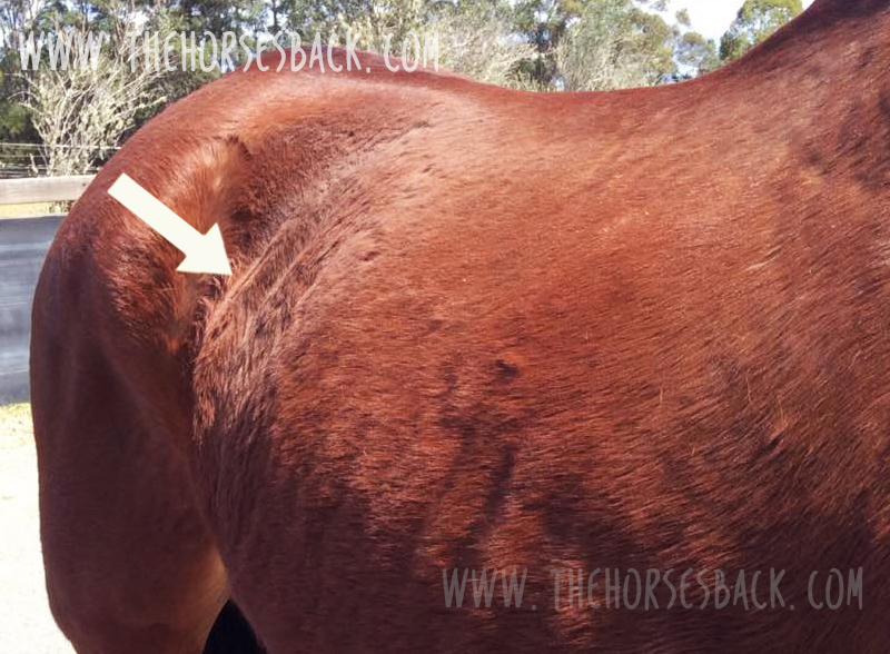
Unilateral transitional vertebra at T18. (c) J. Clothier

As this TB mare (above and below) was unable to maintain weight due to the physical stresses she was experiencing, her rib outline was fairly clear.

In her case, the last rib felt wider and flatter than the other ribs. The space between the rib and the point of hip was also noticeably narrower on the affected side (although this would be true of any horse with scoliosis, it’s a matter of putting the picture together, sign by sign).
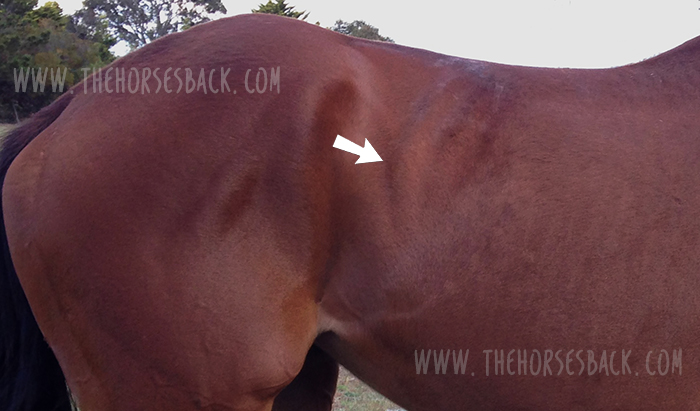
The problem is visible here. This mare’s body condition and tension reflects the stresses caused by the T18 transitional vertebra, which was later confirmed at necropsy. (c) J. Clothier

There were other reasons for suspicion. Even when all the surrounding tissue was relaxed, there was no ‘spring’ when the rib was palpated with a flat hand. That’s not definitive, but it’s a cause for concern.

Do something that most people never do – stand on a fence or mounting block and take a photo down the horse’s spine, when it’s standing square…”

This veteran grey Arabian, below, is one I’d also consider a suspect. Again, we can see a very obvious protruding last rib on the offside and a lack of straightness. Even with musculoskeletal bodywork and spinal mobilization, the rib remained just as pronounced.
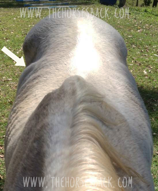
Arabian mare with a suspect rib. Photo: J. Clothier

Incidentally, I’ve also worked on this horse’s offspring, and the younger horse has the same profile to the ribs, on the same side, accompanied by a history of inexplicable back pain – and lack of straightness. 

Note: It’s important to eliminate other causes first, as horses will often have this appearance at the last rib, without it being caused by a transitional vertebra. What’s happening is that the rib is protruding because the vertebra is immobilised in a rotated position. When chiropractic, osteopathy or bodywork restores mobility to the spine, the rib returns to its normal position. 


Ongoing hoof issues

In the bay TB mare, spinal asymmetry (scoliosis, with bend to the right) had led to excessive loading of the near fore. This was no doubt compounded by constantly training and racing in a clockwise direction, plus the classic long toe/low heel frequently found in ex-racehorses.

As a result, her near fore had constant hoof wall separation, bacterial infection (seedy toe / white line disease) and a deep P3 problem that would never come right.

Here’s the hoof capsule and P3. Yes, the poor girl suffered, despite extensive efforts to reconstruct that hoof.
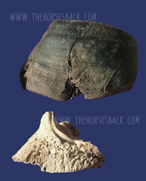
P3 and hoof capsule, near fore, TB mare. Photo: J. Clothier


Patreon members can view videos of this mare and further photos. Go to: www.patreon.com/thehorsesback for more details.


The TB gelding mentioned earlier also had chronic issues in the opposing fore hoof, with wall separation, damage to P3 and evidence of earlier laminitis.


How many horses are affected?

Who knows? The study mentioned earlier (Haussler et al, 1997) found that 22% of Thoroughbreds examined at necropsy, having died or been euthanized at the racetrack, had thoracolumbar transitional vertebrae.
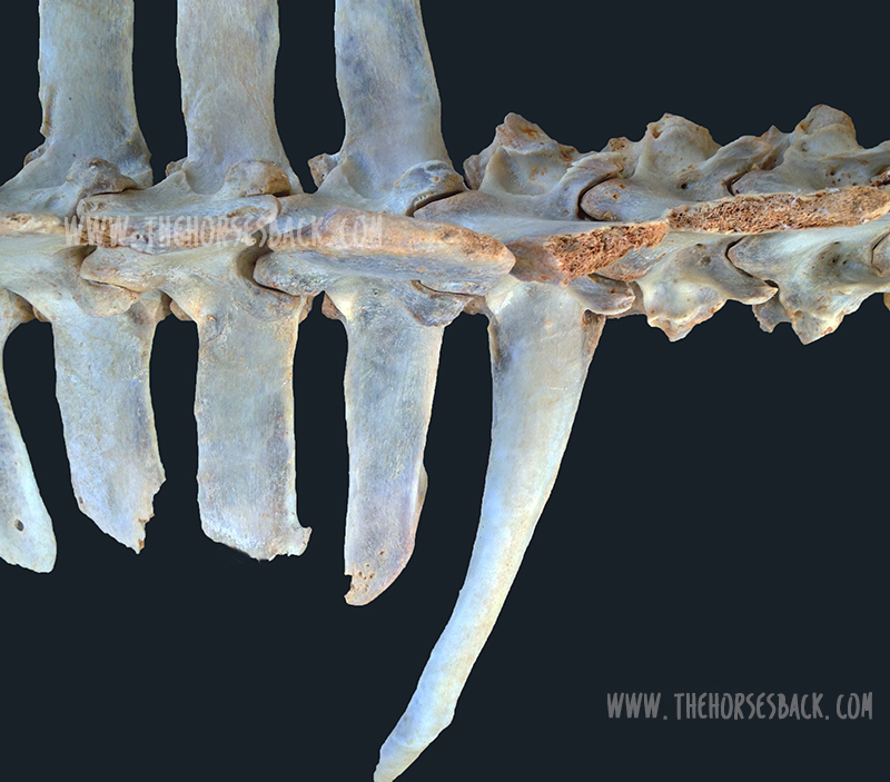
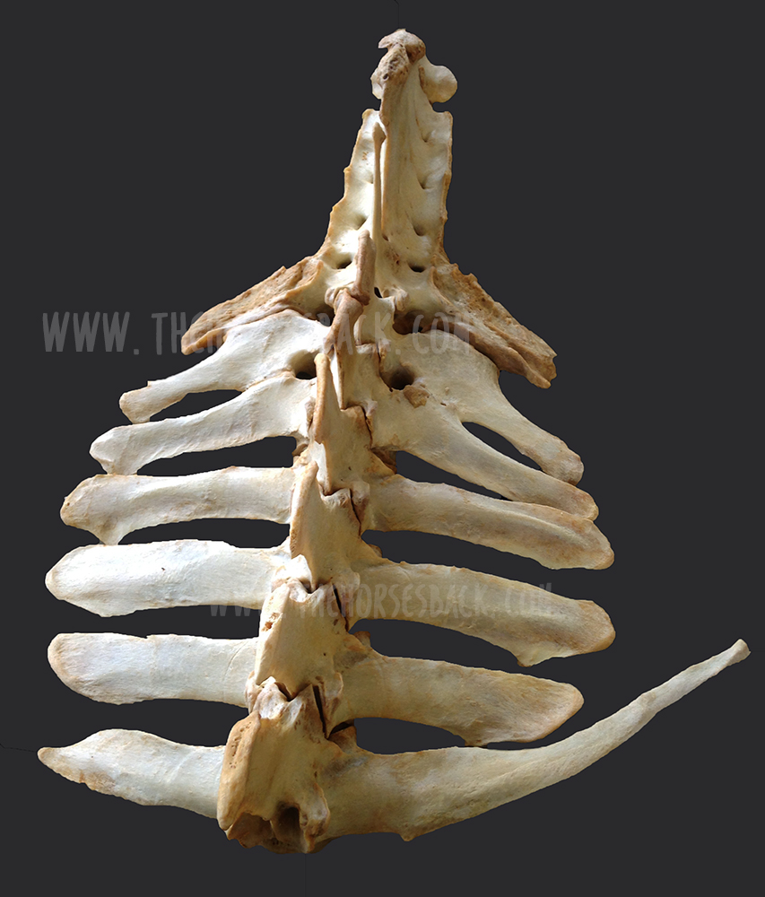
Transitional vertebra at T18 (above ground skeleton, damaged by scavengers)    (c) J. Clothier


To date, I’ve come across 3 in above-ground skeletons (2 x T18, 1 x L1), plus one in a horse later euthanized (1 x T18). These were TBs and Australian Stock Horses.

And as mentioned, I’ve suspected the T18 issue here and there amongst clients’ horses.

Although found mostly in Thoroughbreds, transitional vertebrae are seen across a range of breeds. And certainly, with equine dissection having taken off in quite a big way in the equine care industry, more and more of these anomalies are being observed.


Questions, thoughts or comments? Join us at The Horse’s Back Facebook Group. 


Should we be concerned?

The answer is, inevitably, both yes and no.

On the positive side, if the numbers harbouring this problem are as high as it seems, we have to assume that many horses are coping just fine. 

For as with any musculoskeletal anomaly, horses can compensate very well.

However, when another problem is added to the mix, things can head south very quickly indeed.

And it can all happen without us ever knowing that a skeletal anomaly is an underlying factor. When this happens, owners often have a lot of unanswered questions about their horses – and often large vets bills.
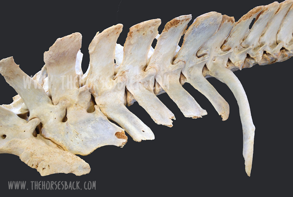
Transitional vertebrae at T18. (c) J. Clothier

It’s the TB or TB-derived breed horse that is most likely to present this (although not exclusively). If you’re buying one and you view a horse with an obvious T18 that really stands out, you might want to get that checked.

At the very least, do something that most people never do – stand on a fence or mounting block and take a photo down the horse’s spine, when it’s standing square or close to square.

If there’s a clear scoliosis along the spine, be cautious (this is a good rule of thumb anyway, no matter what the cause is). If you see an overly pronounced rib on the concave side, be doubly cautious.

And if you believe your horse may have one, the answer is the same as always: be aware, take a 360 degree approach in ensuring that hooves, tack, training and riding are as good as they can be, and your horse will have the best possible chance of functioning well without cause for concern.
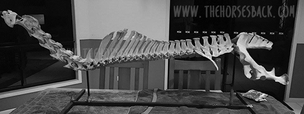
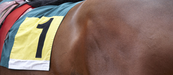
(c) Melissa Longhurst, www.equinebodybalance.com.au


Filed Under: Bodywork Tagged With: equine anatomy, equine bodywork, equine vertebrae, GA, Thoroughbred, transitional vertebrae

By Popular Demand: Here’s How You X-Ray for the C6-C7 Malformation

August 16, 2017 by Jane @ THB 2 Comments

News about the malformation of the C6 and C7 vertebrae has understandably raised a lot of concern. There are major implications for both buyers and breeders of TB and TB-derived breed horses, as outlined in the original article, All You Need to Know About the Hidden C6-C7 Malformation That’s Bringing Horses Down. In response, Sharon May-Davis has very kindly provided us with the English version of a paper she published in the Journal of Japanese Local Government Racing, outlining a preliminary protocol for radiographing this area of the neck and identifying the malformation. (Yes, a single racing authority has taken this issue on board!)

So without further ado, here is the paper in its entirety, the only adaptations being the positioning of images to better fit this site’s blog format.

© All text copyright of the published authors. No reproduction of partial or entire text without permission. Sharing the link back to this page is fine. Please contact me for more information. Thank you!


Preliminary Radiographic Protocols for Identifying Congenital Malformations of the Caudal Cervical Vertebrae

Authors: May-Davis S.E.R., Minowa F., Monoe S.


Abstract

In 2014, a published study based on dissections and skeletal examinations noted that 19:50 Thoroughbred horses had a congenital malformation of the 6th cervical vertebra (C6). In addition, it was found that in those 19 Thoroughbred horses expressing a congenital malformation of C6, 9 displayed a concurrent congenital alformation of the 7th cervical vertebra (C7). In this study, 3 Thoroughbred horses and 1 Thoroughbred type were clinically examined; 3 were radiographed for limb abnormalities and 2:3 radiographed for the congenital malformation of C6 and C7 prior to euthanasia. Upon dissection, 3:4 expressed a congenital malformation of C6 with 2:3 displaying a concurrent CM of C7. These 2 horses were positively radiographed for the CM of C6 and C7 prior to dissection. The radiographs of C6 were taken in direct lateral orientation with 0 degree of elevation and revealed the absence of the caudal ventral tubercle (CVT) of C6. Re-positioning the horse’s forelimbs caudally with an outstretched neck, C7 was radiographed at a 30° oblique lateral angle in a cranial to caudal direction with 0 degree of elevation; the transverse process of C5 remained cranial to the beam. The caudal aspect of the plate was positioned medial the Cranial deep pectoral and rotated vertically to expand the field of view of the cervical vertebrae, whilst remaining perpendicular to the beam.


Introduction

In recent times, 3 Australian studies reported congenital malformations of C6, C7, the 1st sternal rib, along with their associative soft tissue structures. Furthermore, it was noted that these congenital malformations were predominantly breed related and more specifically, to Thoroughbred horses or derivatives thereof. In addition, reports of forelimb proprioceptive dysfunction, neurological impediment and gait deficits were concurrently noted (May-Davis [a,b], May-Davis and Walker). The congenital malformations of C6 appeared as either a left or right unilateral absence of the caudal ventral tubercle (CVT) or as a bilateral absence of the CVT (Figure 1).


Left: Normal. Centre: Absent right CVT. Right: Bilaterally absent CVT

Figure 1. View of the ventral aspect of C6, cranial aspect top. Photo credit: Sharon May-Davis


The congenital malformation of C7 appeared with either a fully transposed CVT from C6 onto the ventral surface of C7 or a partial transposition of the CVT from C6 onto the ventral surface of C7 (Figure 2) (May-Davis [a]).


Left: Normal. Centre: Transposed right CVT. Right: Bilaterally transposed CVT

Figure 2. View of the ventral aspect of C7, cranial aspect top. Photo credit: Sharon May-Davis


The congenital malformation of the 1st sternal rib appeared in multiple presentations with gross anatomic variations including; an absent 1st sternal rib; bifid Tuberculum costae (Figure 3); bifid Sternochondral articulation onto the sternum; flared shaft; normal 1st sternal rib inserting onto the cranial branch of a bifid Sternochondral articulating 2nd sternal rib; straight costal shaft and an articulating rudimentary Tuberculum costae with a ligamentous extension replacing the bony shaft and attaching to a rudimentary Sternochondral articulation onto the sternum (May-Davis [b]).

Figure 3. Left costal bifid Tuberculum costae. Photo credit: Sharon May-Davis

Associative soft tissue structures varied according to the presentation of the congenital malformation with the most noted being, the Longus colli muscle, Scalene muscles and neural vessels such as the Phrenic nerve and Brachial plexus (May-Davis [b], May-Davis and Walker).

The congenital malformations noted in these 3 studies were determined via dissection and skeletal examination. They were based on 2 specific skeletal variations in C6 and C7 plus any variation to normal of the 1st sternal rib.

The congenital malformations could appear in C6 as a singular expression with no other anomalies, however, the congenital malformation of C7 only occurred when the CVT was absent on C6. The congenital malformations of the 1st sternal rib only occurred in the presence of the congenital malformation of C6 and C7 (Table 1) (May-Davis [b]).

With this in mind, the premise that if a congenital malformation exists in C6 then there is a 50% chance that transposition will occur on C7. In this format, it would stand to reason that there could also be an anomalous rib. Therefore, this study is designed to identify the absent CVT on C6 and investigate its transposition onto C7.


Table 1. Noted observations of 151 horses of mixed gender aged between 0 (stillborn) and 30, exhibiting a congenital malformation of C6, C7 and the 1st sternal rib.


Materials and Methods

After evaluation and clinical examination, the 2 horses (1 Thoroughbred and 1 Thoroughbred type) were restrained in cross ties. Detomidine Hydrochloride (Detomovet CEVA Animal Health Pty Ltd) was administered via left jugular vein venepuncture. The dosage was determined by the clinician’s experience and in consideration of the individual’s current health status.

The radiographs were obtained by utilising a Porta 100 HF High Frequency portable x-ray unit (7.8kgs). The kV range was 40-100kV with an mA range of 20-30 mA and a mAs range of 0.3 – 20 mAs. The images were captured on a wireless Rayence 1012WCA Medical Image processing unit (26cm x 32.5cm) and computerised with Vetview Digital Diagnostic Imaging software.

The radiographic angles were obtained with the horse’s neck in full extension and the forelimbs behind the vertical in standing position. With the clinician standing with the x-ray unit on the left side of the neck and the plate positioned on the right side of the neck, the distance between the x-ray unit and plate was 80cms. The x-ray unit and plate remained lateral to the cervical vertebrae determined by the transverse processes of C5.

The first view is a direct lateral at 90 degrees, with 0 degree of elevation whilst maintaining the transverse process of C5 to the left of the beam. 

Remaining lateral to the cervical vertebrae with a 0 degree of elevation, the second view is taken with the x-ray unit positioned at a 30° oblique lateral angle in a cranial to caudal direction with the transverse process of C5 cranial to the beam. The caudal aspect of the plate is positioned medial the Cranial deep pectoral and rotated vertically to expand the field of view of the cervical vertebrae, whilst remaining perpendicular to the beam.

Upon the radiographic evaluation of C6, radiographs of C7 are obtained. The 4 horses were then euthanized and dissected so to verify the radiographic findings. Only those pertaining to the congenital malformation of C6 and C7 are reported in this study.


Results

Figure 4. A compressed trachea (white arrow) in Thoroughbred No. 2 at C6 / C7. Photo credit: Sharon May-Davis

Upon euthanasia of the 4 horses, Thoroughbred No.1 was normal. Thoroughbred No.2 presented with an absent CVT in C6 and trachea compression (Figure 4).

Thoroughbred No. 3, presented with a C6 and C7 congenital malformation and a malformed 1st sternal rib. The Thoroughbred type also presented with a congenital malformation of C6 and C7 (Table 2).


Table 2. Noted observations via dissection of the congenital malformation of C6, C7 and the 1st sternal rib.


Left: Radiographic view of C6. Centre: The white line denotes a normal CVT. Right: The white denotes an absent CVT.

Figure 5. The radiographic lateral view of C6 depicting a unilaterally absent CVT in C6. Image credit: Sharon May-Davis


Thoroughbred No. 3 C7 radiograph (Figure 6) clearly presents the observer with a transposition of the CVT from C6 onto the ventral surface of C7, furthermore it denotes a reciprocal deviation in the trachea at the point of this transposition.


Left: Radiographic view of C7. Centre: The white line denotes the unilateral transposition of the CVT from C6 onto the ventral surface of C7. Right: The white line denotes the deviation in the trachea.

Figure 6. The radiographic view of C7 depicting a unilaterally transposition of the CVT from C6. Image credit: Sharon May-Davis


The Thoroughbred type (Figure 7) exhibits the absent CVT in C6. The radiograph clearly indicates an absent CVT that is unilateral in its presentation.


Left: Radiographic view of C6. Centre: The white line denotes a normal CVT. Right: The white denotes an absent CVT.

Figure 7. The radiographic lateral view of C6 depicting a unilaterally absent CVT in C6. Photo credit: Sharon May-Davis.


The Thoroughbred type C7 radiograph (Figure 8) clearly presents the observer with a transposition of the CVT from C6 onto the ventral surface of C7, furthermore it denotes a reciprocal deviation in the trachea at the point of this transposition.


Left: Radiographic view of C7. Centre: The white line denotes the unilateral transposition of the CVT from C6 onto the ventral surface of C7. Right: The white line denotes the deviation in the trachea.

Figure 8. The radiographic view of C7 depicting a unilaterally transposition of the CVT from C6. Photo credit: Sharon May-Davis.


Figure 9. Right: Absent CVT on C6 (white arrow) and transposed CVT from C6 onto the ventral surface of C7. Photo credit: Sharon May-Davis

Upon dissection, the Thoroughbred type displayed identical congenital malformations of C6 and C7 as depicted in the radiographs (Figure 9).


Discussion

This study was conducted with the view to obtain specific radiographic evidence of the congenital malformation of C6 and C7. Radiographic views of the 1st sternal rib were not possible due to thick and dense musculature in the region. The views obtained of C6 and C7 with exact radiographic angles proved that this region can be radiographed with precision so that the practitioner can make a clear diagnosis should a horse present with a neurological deficit, as previously published (May-Davis

[a, b], May-Davis and Walker]). Furthermore, the asymmetry of the cervical vertebra could exacerbate arthritis in the articular process joints as previously noted (May-Davis [a]). In addition, the incidental finding of the compression of the trachea is an important factor to consider when Thoroughbred racehorses present with compromised airways. An explanation for this occurring is that the Longus colli muscle hypertrophies in support of the weakened structures and due to its proximity to the Trachea, it impinges upon its dorsal surface. This impingement is further exacerbated by the ventral projection from C7 of the transposed CVT from C6.

Since the first publication in 2014, retrospective studies were conducted that added another dimension to the current research. Italian and American studies noted that Warmbloods, Quarter Horses and Arabs were also afflicted with this condition (Santinelli et. al 2016 and DeRouen et.al 2016). It is significant to note that these breeds present the majority of the ridden horse population in Japan extending from Racing, Showjumping, Dressage to Eventing and Endurance. However, a specific Dutch Warmblood study was conducted with fresh cadaver’s that were portioned and CT scanned (Veraa et.al 2016), this also included Oldenburg horses. The congenital malformation of C6 and C7 was present in both breeds with a noted malformed 1st sternal rib in a Dutch Warmblood. Combined percentages of these 3 studies in conjunction with the 3 Australian studies and the current study has the congenital malformation of C6 at; Warmbloods 30%, Quarter Horses 16% and Thoroughbred horses over 40%. 

Aside from the Arabian who is an ancestor to the Thoroughbred, the Warmblood breeds and Quarter Horses all have Thoroughbred lineage in the back line of their breeding. Thus, implying that this condition is heritable as noted by May-Davis [b] and more systemic than just one breed. With this in mind, all critical events should be assessed with the knowledge that a potential congenital malformation could exist in C6, C7 and the 1st sternal rib, as previously noted. With the largest population of horses in Japan being Thoroughbreds, it would be a recommendation to note the studies of several countries including Japan and the severity in percentage of this systemic congenital malformation. Especially in relation to Thoroughbred’ racehorses racing and cornering at speed.


Conclusion

This study showed that of the 4 horses investigated, 3:4 horses displayed a CM of C6, 2:3 displayed the congenital malformation in C6 and C7, and 1:2 a congenital malformation of the 1st sternal rib. Even with a small sample of Thoroughbreds, studies from other countries must be measured and it would therefore be a recommendation to radiograph for this condition in the caudal cervical vertebrae in a pre-purchase examination. The purpose of which would be to eliminate this condition so that riders and handlers are not put at risk.


Author contributions

Sharon May-Davis, Fumiko Minowa and Sadae Monoe wrote and reviewed this article.


Conflict of Interest

The authors have no conflict of interest in the preparation or presentation of this original research article.


Acknowledgement

The author wishes to thank Christine Gee for her professional advice on the manuscript. The Nippon Veterinary and Life Sciences University for the use of their facilities and to those authors / editors/ publishers of those articles, journals and books cited in this manuscript.


References

Derouen A, Spriet M, Aleman M. Prevalance of anatomical of the sixth cervical vertebra and association with vertebral canal stenosis and articular process osteoarthritis in the horse. Vet Radiol Ultrasound, Vol. 00, 2016, pp 1–5.

May-Davis SER. The Occurrence of a Congenital Malformation in the Sixth and Seventh Cervical Vertebrae Predominantly Observed in Thoroughbred Horses. J Equine Vet Sc 2014; 34:1313-17.

May-Davis SER. The Congenital Malformation of the 1st Sternal Rib. J Equine Vet Sc 2014; 34:1313-17.

May-Davis SER, Walker C. Variations and implications of the gross morphology in the Longus colli muscle in Thoroughbred and Thoroughbred derivative horses presenting with a congenital malformation of the sixth and seventh cervical vertebrae. J Equine Vet Sc 2015; 35:560-8.

Santinelli I, Beccati F, Pepe M. Anatomical variation of the spinous and transverse processes in the caudal cervical vertebrae and the first thoracic vertebra in horses. EVJ 48 (2016) 45–49.

Veraa S, Bergmann W, van den Belt A-J, Wijnberg I, Back W. Ex vivo computed tomographic evaluation of morphology variations in equine cervical vertebrae. Vet Radiol Ultrasound, Vol. 00, 1–7.


Filed Under: Bodywork, Sharon May-Davis Tagged With: C6, C7, cervical vertebrae, GA, homepage, TBs, Thoroughbred

8 Golden Rules For Helping Your Thoroughbred Get Right Off The Track

August 2, 2014 by Jane @ THB 38 Comments

OTTB Header

In this guest post, Kerry Warren Couch responds to the article on this site about ex-racehorses by stating, clearly and simply, how she has helped her OTTBs over many years, mainly by initially doing very little at all.

(c) All text copyright of the author at www.thehorsesback.com. No reproduction of partial or entire text without permission. Sharing the link back to this page is fine. Please contact jane@thehorsesback.com for more information. Thank you!

Kerry writes:

Sometimes, you can achieve more by doing very little. There’s a lot advice around about what you can do to help ex-racehorses, or OTTBs (Off the Track Thoroughbreds). Much of it focuses on what can be done through retraining, while some of it focuses on dealing with physical issues.

I have ridden many OTTBs over the decades of my riding career, professionally and for sheer pleasure. I just adore them – they have so much heart, and they give  everything if the rider is willing to be extraordinarily patient, partnering with their horse and never forcing them.

I loved the Buying an Ex-Racehorse: Can You Spot the Major Physical Issues? article – it’s absolutely on target. I have a few suggestions for folks to consider when they bring their OTTBs home. These come from my personal experience – they may not apply to everyone and all cases, so do keep that in mind.

The author's new OTTB
The author’s new OTTB

Seven months ago, I purchased yet another OTTB. He came from a top trainer/track and we knew his entire medical history, including x-rays. This is NOT typical – I was very fortunate to have access and for my vet to be able to talk with the stable and their vets. Very lucky me.

Despite coming from a top trainer, my new OTTB does have some issues – he IS an ex-race horse, so this is to be expected.

He has benefited from my usual approach, which helps the horse long before we start work.

What follows is my approach to helping every OTTB that comes to live at my farm.


1. Allow the OTTB plenty of downtime

My OTTBs have always needed time to ‘defuse’ from the training barn life, so I turn them out, slowly to acclimatize.

But then as they come to understand turn out, I find them pasture mates and allow turn-out at will, so they can go in their stalls or walk out to the field as they wish.

2. Give the OTTB a full check-up

I have a complete chiro assessment done before ANY work is begun – and that includes groundwork.

We start working on the serious physical issues right away, but leave simple ones for a little later, so we don’t overwhelm the horse in his transition to a non-track life.


3. Use paddock time as self-help time

I am lucky that my farm has hills, so I can help my OTTBs work on symmetrical muscle building long before I begin groundwork.

I purposefully keep their water in their stalls so they must walk the hills every day, from pasture to barn and back.

Their attitudes really change with this time to ‘reprogram’.

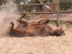
OTTB SandI also have a designated ‘rolling area’ near the paddock, filled with super soft sand.  I have been amazed at how smaller, simpler issues can be naturally worked out by allowing horses to relax and roll in lovely sand.

My region has hard clay soil, so I had the sand area made, because I know that rolling on hard clay can actually cause or exacerbate injuries. It’s not that expensive and I’ve found that horses LOVE it.

Do consider putting it in a well-drained area with a little bit of full sun, as warm sand is wonderful!

4. Use grooming as therapy time

I groom them every day ­– slowly and methodically.  I fully believe this has wonderful therapeutic benefits.

It helps with circulation and muscle tone, and helps me partner with my horses without asking anything of them.

5. Work out an individual nutritional plan

Where feed’s concerned, I have full blood panels done and put a nutrition program together based on each horse’s needs. These vary from horse to horse.

I have been fortunate to acquire OTTBs from known training barns, so my horses typically have few deficiencies. This may NOT be the case for so many people who are purchasing OTTBs from a TB rescue operation or general sale.

By the way, I am NOT implying that rescue groups do not take care of the horses. It’s just that sometimes, the rescue folks aren’t told about the horse’s medical history, so they simply don’t know. It is not due to any lack of care or concern on their part.

Hills at the author's place
Hills at the author’s place

We some times see minor anemia, and imbalance in the micro-minerals.  Many OTTBs do have some signs and symptoms of gastric discomfort. This may reflect fore or hind gut ulcers.

I don’t like to have an endoscopy performed, as the pre and post procedures often exacerbate the ulcers, if they do exist.

I actually prefer to treat this area empirically. So, if my vet and I both feel the horse is exhibiting signs consistent with ulcers, we treat them without the instrumental diagnostics.

That said, if there IS a serious issue, one certainly may wish to pursue the endoscopy.

Diet is really individualized to the horse. It is not advisable to suddenly change a horse’s diet. I like to implement a tapering program. I keep the horse on whatever he was being fed at the training barn for two weeks, with a very slow changeover to whatever program we have determined for the new horse.

I typically have acquired very young OTTBs, ie, 3-4 year olds. So I know my horses are still growing and will require nutrition appropriate to a growing horse. We do feed a LOW carbohydrate feed mix and will supplement as warranted.

If one feeds the proper amount as recommended by the feed company and one’s vet, the horse ought to receive the right blend of vitamins and minerals.

I do like to add flax seed meal, not the seeds. If a horse has really thin walls or poor feet, I add a hoof supplement with biotin and trace minerals. If a horse requires, we will add probiotics.

If a horse is a poor drinker, I may add some soaked, drained, no-molasses-added beet pulp.

I think the main thing here is that no two horses are exactly the same.  There is NO cookie cutter approach to caring for horses. There are only general principles:

  • Fresh, clean water, daily (scrub out those water buckets).
  • Plenty of quality hay and forage at will, so the horse does not stand for hours without roughage (the average 1100 lbs horse requires approx 22 lbs of total quality forage a day, which can be a combination of pasture and hay, all hay or all pasture).
  • Get the horse out, moving at will in a pasture so the fore and hind gut can work effectively.
  • Make sure a dentist has assessed their teeth.
  • A fecal test is also a good idea, especially for OTTBs coming from a rescue or general sale.  Good training barns have a worming routine.  This may not be the case with a rescue operation or at a general sale when one doesn’t know much about the OTTB’s origin.
  • I also know the vaccination history of my newly acquired horses, but this may not always be the case for others.  I’d advise anyone to discuss this with the veterinarian.


6. Improve poor hoof balance slowly

I get the farrier in from the beginning to slowly – NOT rapidly – change those hoof angles over several months of trims.

Most OTTBs have under run heels, long toes and flat soles.

A slow correction  allows longer term adaptation of the hoof structures and avoids short term soreness. It also avoids dramatic changes that affect posture

OTTBOne MUST take it very slowly.  I believe this holds true for any horse, but particularly for the OTTBs.

Their muscles, tendons and ligaments are put at risk if angle changes are made too rapidly. It leads to transitory lameness, soreness and increased risk of injury, even in the pasture.

I offer the analogy of a person wearing barn boots, day-in, day-out.

Then, suddenly switching to three inch high heels and attempting to carry out the same daily chores. OUCH!

I think it is SO important to have all the professionals involved in the horse’s care/transition on the same page and are informed of each other’s interventions with the horse.

It’s really important to have the big players on the ‘team’ communicating with one another:  the chiropractor, the farrier, the vet/nutritionist, hay and feed dealer.

7. Allow time for slow and steady progress

I have also found that my OTTBs have really needed to learn how to enjoy other horses and life before I start any type of training program.

We humans seem to be in such a hurry.  It’s so helpful to let their bodies and minds defuse.

I have noted that less experienced horse owners would do well to remember that these horses are NOT, at this stage, pets. They have frequently not been cuddled or hand fed carrots, or hugged or loved.

These horses were working horses. They had a job to do.

I have observed some people new to horses being so enthralled with the idea of adopting an OTTB that they immediately want to love them, pet them, stand very close to them and even throw their arms around their necks.

This is a lovely sentiment but oh, so dangerous. Ex-track horses are often more accustomed to varied stall muckers coming in, varied groomers, different hot walkers, exercise riders, jockeys, etc.

So many OTTBs are simply NOT accustomed to being fussed. They may pin their ears and toss their heads to even bite or nip at the new owner, raise a hind foot, or wring the tail as a warning.

People new to horses who start with an OTTB really really need to be aware of this and put together a realistic plan to allow the horse to defuse, detox, rebuild and SLOWLY re-acclimatize to a life where his new people will show affection.

This happens over weeks or months – NOT days. Take it very slowly and give the horse his personal space and time to come to you – don’t force your eager affection on the horse too soon. It may likely backfire.

In time, with patience and understanding, you WILL have a lovely, affectionate horse. But not right away, as a general rule.

Talking to your horses daily, or singing, is good – I sing a lot to my horses so that they learn my voice and intonations.  I do this over a long period, way before I ever attempt to be affectionate with my new OTTB. And even then, I wait for my horse to show me he is ready for me to rub his ears, etc.

Grooming helps this along, too. I often sing while I groom.

8. Enjoy starting work with your OTTB

(c) retiredracehorseblog.com

With all this, they are so much easier to work with – even for their rehab therapies, and physical therapy is always part of the program.

They ARE off the track and there WILL be issues, almost always hips, pelvis, back and others…

I give my OTTBs six full months, and sometimes up to a year, as each horse is different.

I know that may not be reasonable for many people, but even so, at least three months would yield a big benefit.


I do hope others have suggestions to add in the Comments section, below. Horses are such magnificent and amazing creatures.  I have spent my lifetime, to date, with them, and I feel I have merely scratched the surface of understanding. I learn something new every single day.  They are wonderful teachers, if we simply have the heart and open mind to ‘listen’ to their voices. 


Filed Under: Guest Posts, Thoroughbreds Tagged With: ex-racehorse, ex-racehorses, Flat racers, GA, off the track, off the track thoroughbreds, OTTB, OTTBs, Thoroughbred, thoroughbreds

Footer

Contact Me

Send your questions this way…

Get in Touch

  • Store Support
  • Shipping & Returns
  • Contact

Copyright © 2025 · Parallax Pro on Genesis Framework · WordPress · Log in

MAY Red Light Products Now In Stock! Dismiss