• Skip to main content
  • Skip to footer

The Horse's Back

Body Talk for Thinking Owners

  • Home
  • Articles
  • BEMER
  • Store
    • Store
    • Shipping & Returns
    • Support
  • Equine Healthworks
    • Bodywork for Horses
    • Reviews
    • Leave a Review
  • About & Contact
    • About
    • Contact

horse anatomy

Here’s a Round Up of My Premature and Dysmature Foal Research

May 26, 2022 by Jane @ THB Leave a Comment

Here are abstracts, downloads and links for my research into the ongoing effects or premature or dysmature birth in horses.

These are the publicly available details of my thesis (full download) and published, peer-reviewed journal articles. The articles aren’t open access, but if you really want to read something, please contact me.

As always, huge thanks are due to the breeder owners who so very kindly allowed me to study their horses, and who provided such valuable images. Together, you’ve helped me to learn a lot and reach initial findings that I now hope to pass on.


Beyond the Miracle Foal: A Study into the Persistent Effects of Gestational Immaturity in Horses 

PhD Thesis, University of New England and CSIRO

Abstract
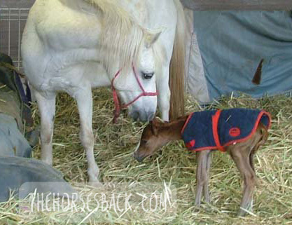
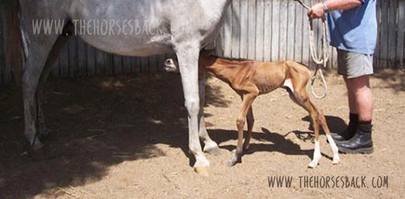
Breeding horses can be a financially and emotionally expensive undertaking, particularly when a foal is born prematurely, or full term but dysmature, showing signs normally associated with prematurity. In humans, a syndrome of gestational immaturity is now emerging, with associated long-term sequelae, including metabolic syndrome, growth abnormalities and behavioural problems.

If a similar syndrome exists in the equine and can be characterised, opportunities for early identification of at-risk individuals emerge, and early intervention strategies can be developed. This thesis explores the persistent effects of gestational immaturity manifest as adrenocortical, orthopaedic and behavioural adaptation in the horse.

Basal diurnal cortisol levels do not differ from healthy, term controls, but when subjected to a low dose ACTH challenge, gestationally immature horses presented a depressed or elevated salivary cortisol response, suggesting bilateral adaptation of the adrenocortical response. This may be reflected in behavioural reactivity, but the outcomes from a startle test were inconclusive.

A survey of horse owners indicated that gestationally immature horses tended to be more aggressive and active than controls, aggression being displayed mostly in families of Arabian horses. Case horses also tended to be more active, intolerant, and untrusting.

Gestationally immature horses have restricted growth distal to the carpal and tarsal joints, and this results in a more ‘rectangular’ conformation in adulthood compared to controls. They also often present with angular limb deformities that adversely affect lying behaviour and recumbent rest. This, however, can be mitigated using analgesic therapy, suggesting chronic discomfort.

Based on these findings, it is reasonable to postulate that a syndrome of gestational immaturity may persist, both clinically and sub-clinically, in affected adult horses. Further work is required to fully characterise this syndrome and validate the outcomes in larger populations, thereby providing a foundation for interventions applicable in the equine breeding industry.

The entire PhD thesis can be downloaded here. This is a 236-page PDF.

Clothier, Jane  (author); Brown, Wendy  (supervisor); Small, Alison (supervisor); Hinch, Geoff  (supervisor)


Equine Gestational Length and Location: Is There More That The Research Could Be Telling Us?

Australian Veterinary Journal

Abstract
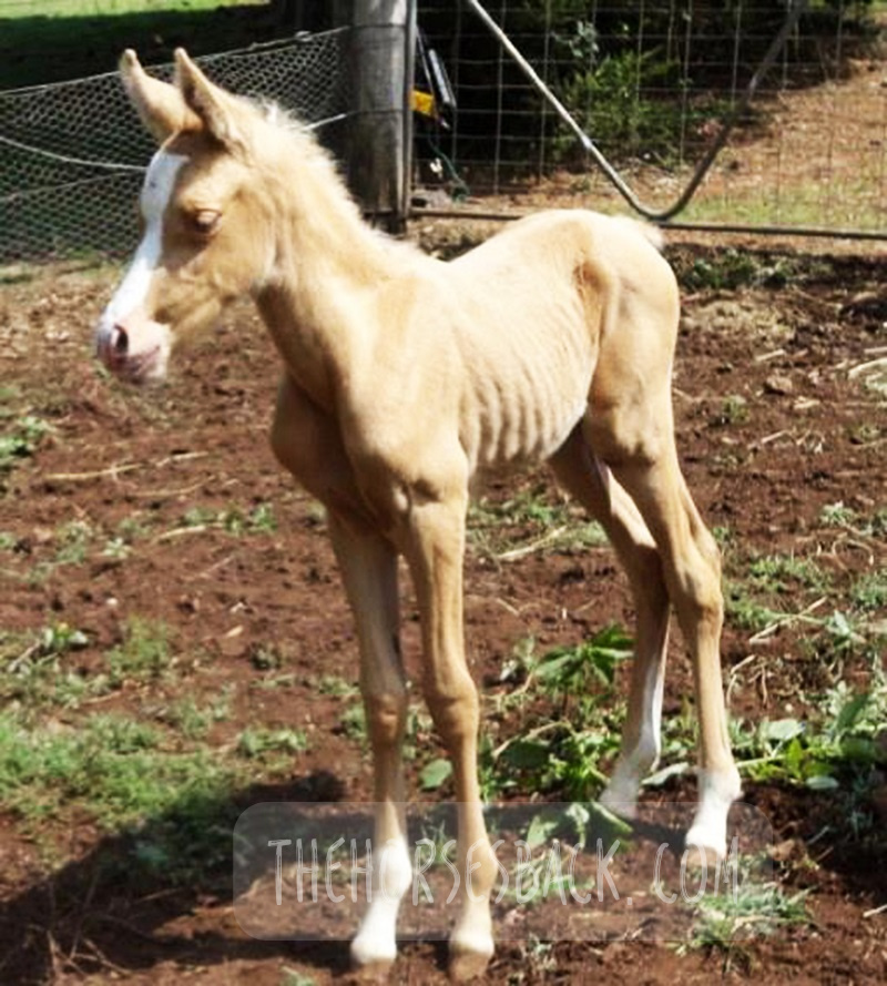
Clear definitions of ‘normal’ equine gestation length (GL) are elusive, with GL being subject to a considerable number of internal and external variables that have confounded interpretation and estimation of GL for over 50 years. Consequently, the mean GL of 340 days first established by Rossdale in 1967 for Thoroughbred horses in northern Europe continues to be the benchmark value referenced by veterinarians, breeders and researchers worldwide. Application of a 95% confidence limit to reported GL range values indicates a possible connection between geographic location and GL.

Improved knowledge of this variable may help in assessing the degree of the neonate’s prematurity and dysmaturity at or soon after birth, and identification of conditions such as incomplete ossification of the carpal and tarsal bones. Associated pathologies such as bone malformation and fracture, angular limb deformity and degenerative joint disease can cause chronic unsoundness, rendering horses unsuitable for athletic purpose and shortening ridden careers.

This review will examine both the factors contributing to GL variation and the published data to determine whether there is potential to refine our understanding of GL by establishing a more accurate and regionally relevant GL range based on a 95% confidence limit. This may benefit both equine industry economics and equine welfare by improving early identification of skeletally immature neonates, so that appropriate intervention may be considered.

The paper can be accessed here.

Clothier, J., Hinch, G., Brown, W. and Small, A. (2017), Equine gestational length and location: is there more that the research could be telling us?. Aust Vet J, 95: 454-461. https://doi.org/10.1111/avj.12653

Using Movement Sensors to Assess Lying Time in Horses With and Without Angular Limb Deformities 

Journal of Equine Veterinary Science

Abstract
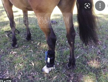
Chronic musculoskeletal pathologies are common in horses, however, identifying related effects can be challenging. This study tested the hypothesis that movement sensors and analgesics could be used in combination to confirm the presence of restrictive pathologies by assessing lying time. Four horses presenting a range of angular limb deformities (ALDs) and four non-affected controls were used.

The study comprised two trials at separate paddock locations. Trial A consisted of a 3-day baseline phase and 2 × 3-day treatment phases, during which two analgesics were administered to two ALD horses and two controls in a standard crossover design. Trial B replicated trial A, except that as no difference between analgesics had been evident in trial A, only one analgesic was tested. Movement sensors were used to measure the horses’ lying time and lying bouts.

In trial A, ALD horses’ basal mean lying time was significantly less than controls (means ± SD for ALD horses 213 ± 1.4 minutes and for controls 408 ± 46.7 minutes, P = .007); with analgesic administration, the difference became nonsignificant. In trial B, ALD horses’ basal mean lying time was also significantly less than controls (ALD horses 179 ± 110.3 minutes; controls 422.5 ± 40.3 minutes, P < .001), again becoming nonsignificant with analgesic administration. Given the increases in ALD horses’ lying time with analgesic administration, it is possible that their shorter basal lying time is associated with musculoskeletal discomfort. Despite the small sample size, movement sensors effectively measured this behavior change, indicating that they could be a useful tool to indirectly assess the impact of chronic musculoskeletal pathologies in horses.

The paper can be accessed here.

Clothier J, Small A, Hinch G, Barwick J, Brown WY. Using Movement Sensors to Assess Lying Time in Horses With and Without Angular Limb Deformities. J Equine Vet Sci. 2019; 75:5559. doi: 10.1016/j.jevs.2019.01.011


Prematurity and Dysmaturity Are Associated With Reduced Height and Shorter Distal Limb Length in Horses 

Journal of Equine Veterinary Science

Abstract
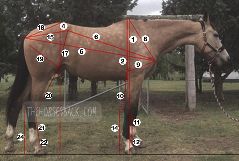
The long-term effects of gestational immaturity in the premature (defined as < 320 days gestation) and dysmature (normal term but showing some signs of prematurity) foal have not been thoroughly investigated. Studies have reported that a high percentage of gestationally immature foals with related orthopedic issues such as incomplete ossification may fail to fulfill their intended athletic purpose, particularly in Thoroughbred racing. In humans, premature birth is associated with shorter stature at maturity and variations in anatomical ratios, linked to alterations in metabolism and timing of physeal closure in the long bones.

We hypothesized that gestational immaturity in horses might similarly be associated with reduced height and different anatomical ratios at maturity. In this preliminary study, the skeletal ratios of horses with a history of gestational immaturity, identified through veterinary and breeder records, were compared with those of unaffected, closely related horses (i.e., sire, dam, sibling).

External measurements were taken from conformation photographs of cases (n = 19) and related horses (n = 28), and these were then combined into indices to evaluate and compare metric properties of conformation. A principal component analysis showed that the first two principal components account for 43.8% of the total conformational variation of the horses’ external features, separating horses with a rectangular conformation (body length > height at the withers), from those that are more square (body length = height at the withers). Varimax rotation of PC1 and analysis of different gestational groups showed a significant effect of gestational immaturity (P = .001), with the premature group being more affected than the dysmature group (P = .009, P = .012). Mean values for the four dominant indices showed that these groups have significantly lower distal limb to body length relationships than controls. The observed differences suggest that gestational immaturity may affect anatomical ratios at maturity, which, in combination with orthopedic issues arising from incomplete ossification, may have a further impact on long-term athletic potential.

The paper can be accessed here.

Clothier J, Small A, Hinch G, Brown WY. Prematurity and Dysmaturity Are Associated With Reduced Height and Shorter Distal Limb Length in Horses. J Equine Vet Sci. 2020 Aug;91:103129. doi: 10.1016/j.jevs.2020.103129. Epub 2020 May 22. PMID: 32684267.


Perinatal Stress in Immature Foals May Lead to Subclinical Adrenocortical Dysregulation in Adult Horses: Pilot Study 

Journal of Equine Veterinary Science

Abstract
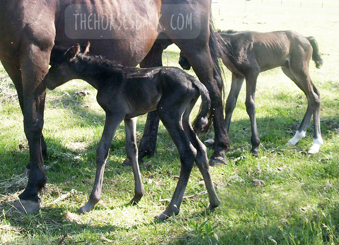
The persistent endocrinological effects of perinatal stress due to gestational immaturity in horses are unknown, although effects have been reported in other livestock species. This pilot study tested the hypothesis that persistent adrenocortical dysregulation is present in horses that were gestationally immature at birth by assessing the salivary cortisol response to exogenous ACTH.Case horses (n = 10) were recruited with histories of gestation length < 315 d or dysmaturity observable through neonatal signs. Positive controls (n = 7) and negative controls (n = 5) were recruited where possible from related horses at the same locations.

Cases and positive controls received an intramuscular, low-dose (0.1 ug/kg) of synthetic ACTH (Tetracosactrin 250 mg/mL, Synacthen); negative controls received no ACTH. Saliva samples were collected from all horses at baseline T = 0 and at 30 min intervals post injection from T = 30 to T = 150. These were assayed for salivary cortisol concentration (SCC) using a commercially available ELISA kit (Salimetrics).All baseline values (T = 0) were within normal published ranges. Peak and AUC values (corrected for baseline) for case horses were significantly different (ANOVA P < .001) to positive controls, with either higher (H-cases) or lower (L-cases) SCC values, outside the 95% Confidence Interval of the reference population.

There was no significant effect of breed, age, sex, test month, or location on results. The results suggest that gestational immaturity may lead to subclinical adrenocortical dysregulation, with affected horses presenting an elevated or blunted response to a low-dose ACTH stimulation, despite normal basal levels.

The paper can be accessed here.

Clothier J, Small A, Hinch G, Brown WY. Perinatal Stress in Immature Foals May Lead to Subclinical Adrenocortical Dysregulation in Adult Horses: Pilot Study. J Equine Vet Sci. 2022 Apr;111:103869. doi: 10.1016/j.jevs.2022.103869. Epub 2022 Jan 21. PMID: 35074402.

Filed Under: Bodywork, Foals Tagged With: dysmature foals, equine anatomy, equine bodywork, equine dysmaturity, equine prematurity, GA, horse anatomy, immature foals, Premature foals

Yes, We Can Image for Transitional Vertebrae in Horses

September 23, 2021 by Jane @ THB 7 Comments

It’s been a question of mine for a while. Can diagnostic imaging show the presence of transitional vertebrae?

We’re seeing many bone samples from dissections, as shown in my previous article on transitional vertebrae.

But if we’re to help our horses that live with this issue, we need to identify it before they’re dead. (Yes, right?!)

Allow me to introduce a practicing vet and educator who is doing just that.


Imaging for Transitional Vertebrae 

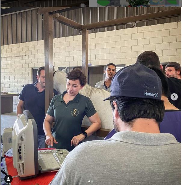
Meet Dr Brunna Fonseca, Associate Professor, educator and specialist in equine orthopedics, focusing on the spine and nervous system. She’s based in São Paulo, Brazil.
I’ve been following her Instagram for a while, because she posts brilliant videos and photos explaining what she does, and how, and why.

I was delighted to see a recent post on imaging for a transitional vertebra, which included fantastic visuals. Such a great communicator!

Dr Brunna has kindly given me permission to repost her images and descriptions here. So without further ado…

  • All images copyright of Axial Vet

Ultrasonograms

Ultrasonography for transitional vertebrae
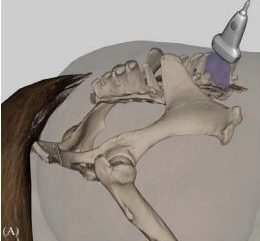
Angle of transducer. Image: Equine Neck and Back Pathology: Diagnosis and Treatment, 2nd Edn. Ed. Frances M.D. Henson. © 2018 John Wiley & Sons, Ltd.

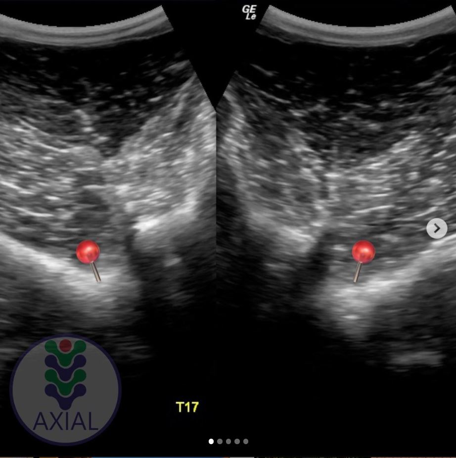
The following ultronographic images are each a composite of two images, one showing the left side and the other the right.

This textbook illustration helps to show the angle the image is taken at. This angle is usually used for imaging the articular facets of the vertebrae.

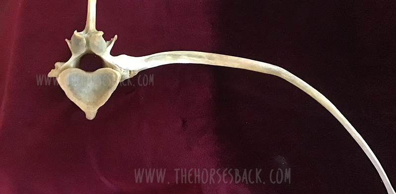
Additionally, the image at the top of this article shows a transitional vertebra at T18, like the mare being diagnosed by Dr Brunna.


1.  Can we recognise transitional vertebrae?

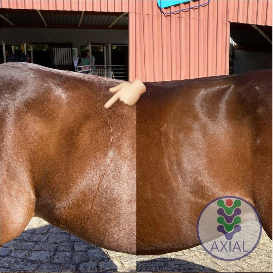
The first image shows two sides of a mare’s body. The hand icon gives us a strong hint of where to look… This appearance is very similar to that of the TB mare in my previous post.

Dr Brunna writes, “This mare has the T18 transitional vertebra, presenting a transverse process similar to the lumbar vertebrae on the right side, which causes the appearance of the horse to have the most visible rib on that side.

The occurrence of transactional vertebrae in the horse is not uncommon, especially in the thoracolumbar transition, which can occur in T18 or L1.”


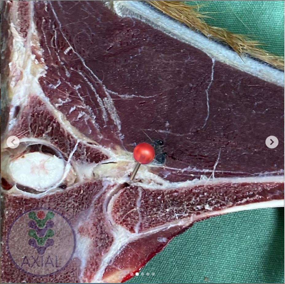
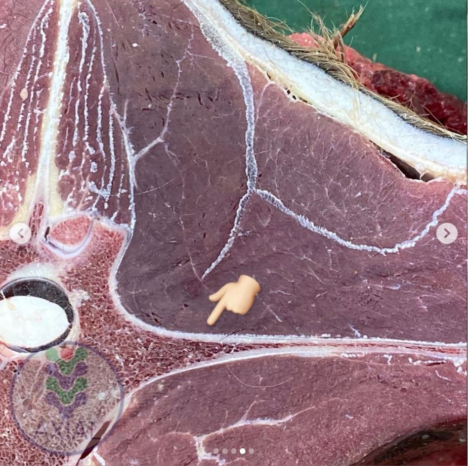
2. Section of a thoracic vertrebra

This image is from a different horse showing a normal rib head and its joint with the vertebra.

Dr Brunna writes, “This is the image of a thoracic vertebra, showing the costotransverse joint.”


3. Image of a normal vertebra

Dr Brunna writes, “This is a T17 ultrasound image, where we can see the image of the normal costotransverse joints.”

This is the bay mare again.

As with the previous cross section, the red pins which show the facet joint between rib head and vertebra.


4. Section of a lumbar vertebra

This is cross section is of a normal lumbar vertebra from a different horse.

As you can see,  there is no joint between the  transverse process and the vertebral body.

The process is wide and flat, and integral to the vertebra.


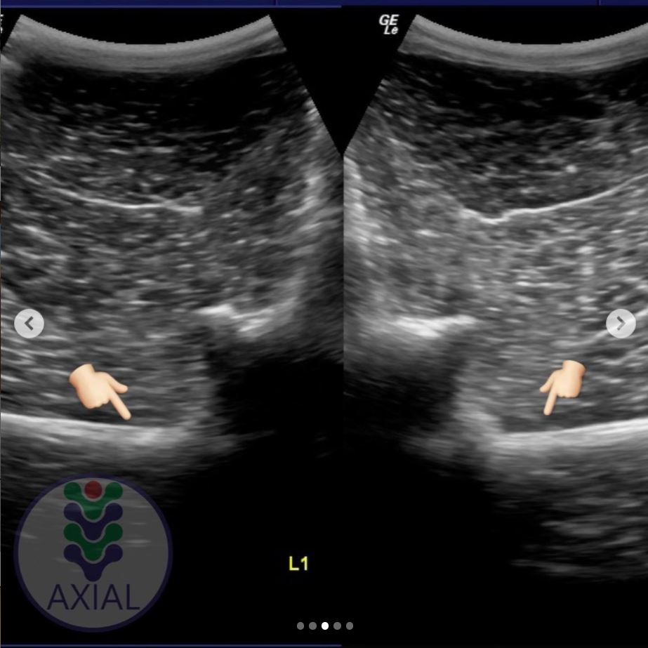
5. Image of a lumbar vertebra

Here’s an ultrasound of the first lumbar vertebra (L1) in the bay mare.

As in the above cross section (picture 4), there is no joint between the transverse processes and the vertebral body.

We now have ultrasound images of the normal T17 and normal L1. As we will see, the transitional vertebra mixes elements from both.


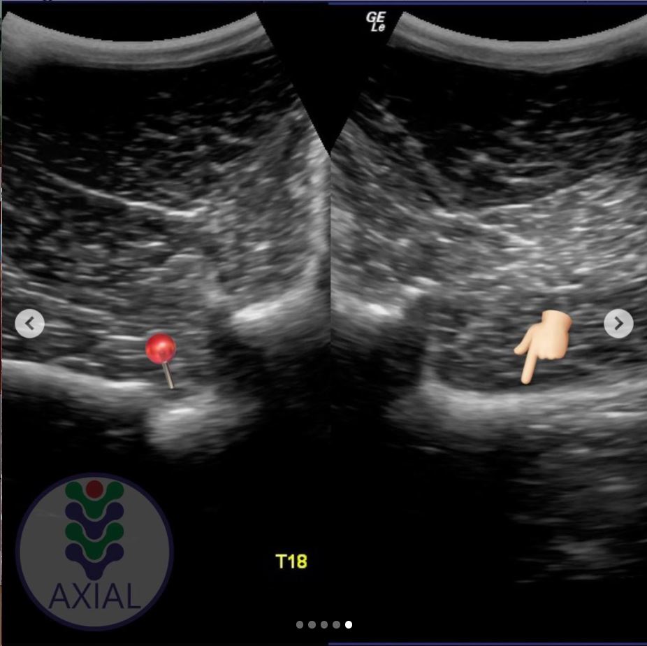
6. Imaging transitional vertebrae

“This is an ultrasound image of T18, where we can see the image of the costotransverse joint on the left side (red pin) and image of the transverse process on the right side.”

So here’s the underlying skeletal issue in the bay mare.

The left side is a normal joint, being the same as the T17 thoracic vertebra (picture 3).

The right side is similar to the previous image of the lumbar vertebra (picture 5).

It is not identical, for while the process-like rib is joined to the vertebra, it is not the same shape and does not lie as flat as the lumbar process.


Want to Hear More From Dr Brunna Fonesca?

You can follow her Axial Vet Instagram page to see examples of her equine cases and their assessment, in images and videos.

An increasing number of captions are now translated into English.


Filed Under: Bodywork Tagged With: Anatomy, equine anatomy, equine bodywork, equine malformation, equine skeleton, GA, horse anatomy, transitional vertebra, transitional vertebrae

‘The Size of a Walnut’ – Does Equine Brain Size Matter?

November 5, 2019 by Jane @ THB 7 Comments

There seem to be quite a few social media posts about the equine brain of late – and that’s no bad thing. 

In some ways, the brain is simply the latest part of the equine anatomy to come under the spot light. It’s being subject to statements about welfare, training and psychology – and that’s definitely a good thing (here’s one from Hippologic.)

However, I want to add something to this equine brain discussion. I just happened to run in to it when I went down a research rabbit hole a couple of years back.

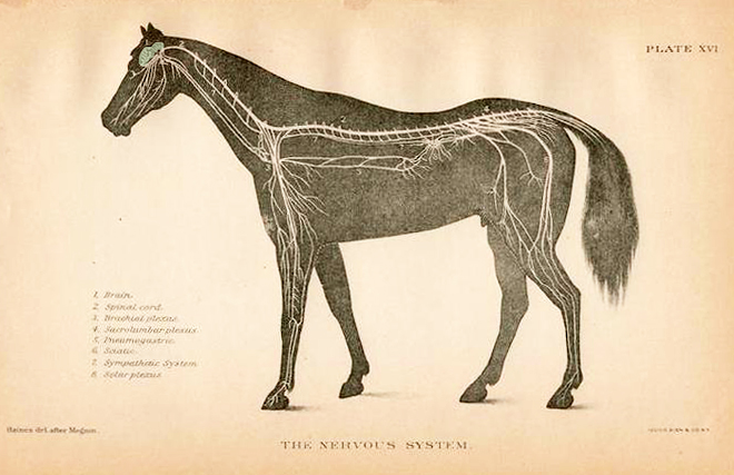
We often hear how brain size is not directly linked to an individual’s intelligence. At the same time, a relatively large brain is said to signify intelligence in humans, while that of the horse, popularly said to be the size of a (large) walnut, is said to account for their lack of intelligence.

Vintage anatomy print showing relatively small size of equine brain to body size.

This falls down once we look at elephants, which have relatively small brains yet are pretty cluey.

In horses, innovative behaviors without evolutionary basis are often used as a measure of intelligence (read more here about unlatching gates). Leaving behaviour-based measurement methods aside for the moment, let’s ask: how do we figure out if there’s an association between different brain sizes and intelligence levels?

(Note: if you’re a neuroscientist of any description, look away now. What follows is a highly simplistic overview of this incredibly complex subject area.)

© All text copyright of the author, Jane Clothier, www.thehorsesback.com. No reproduction of partial or entire text without permission. Sharing the link back to this page is fine. Please contact me for more information. Thank you!


Measuring Equine Brain Mass and Body Mass 

In zoology, the starting point isn’t about brain size, but brain mass compared with body mass or weight.

Even then, it’s not a matter of separating the brain from the body and then weighing both. The most accurate way of measuring this accounts for several anatomical, physiological factors, including the amount of water in the brain.

The result is a single figure that is called the encephalization quotient (EQ). The EQ for a species is arrived at after researchers have performed the calculation for dozens of animals.

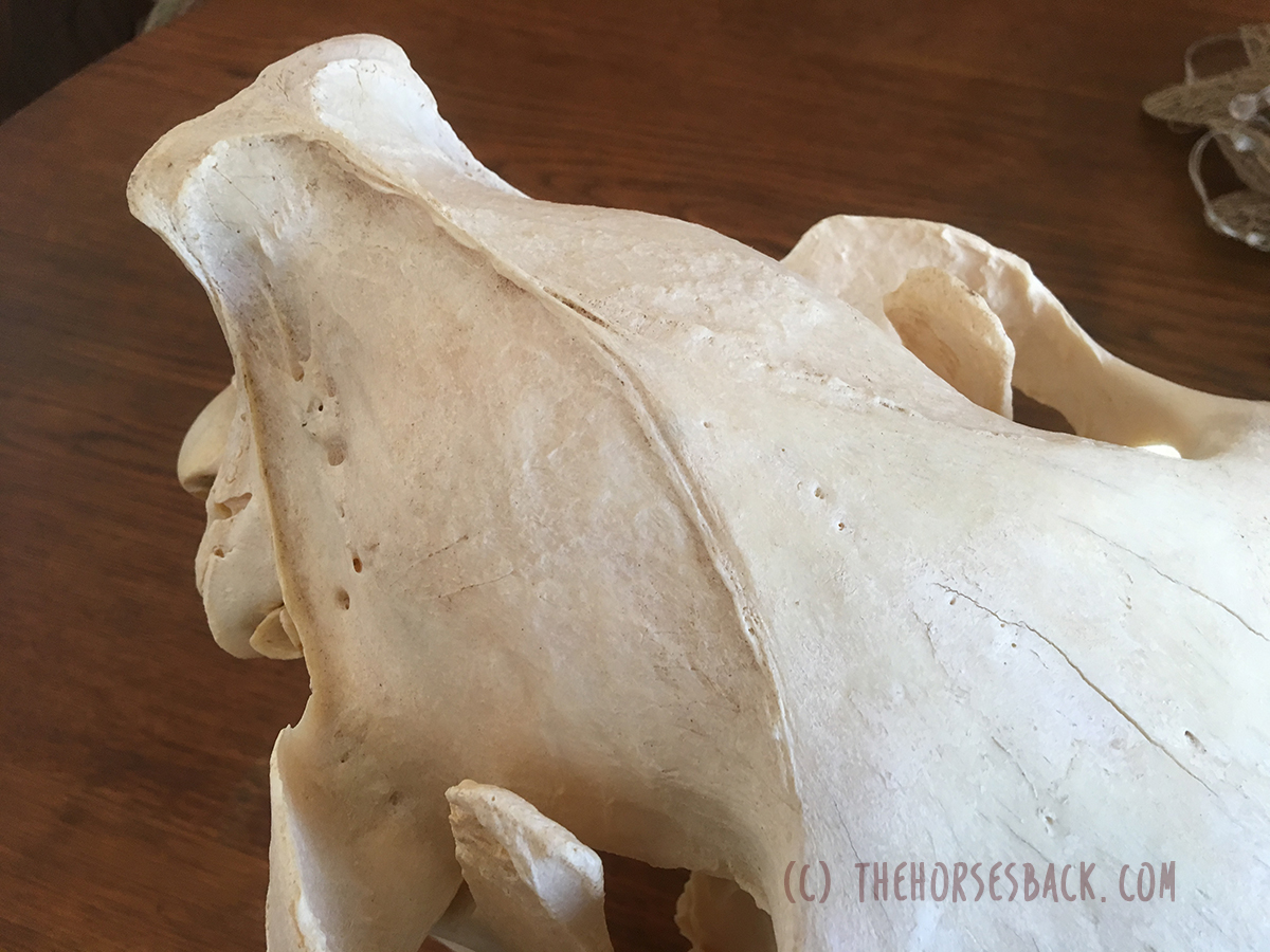
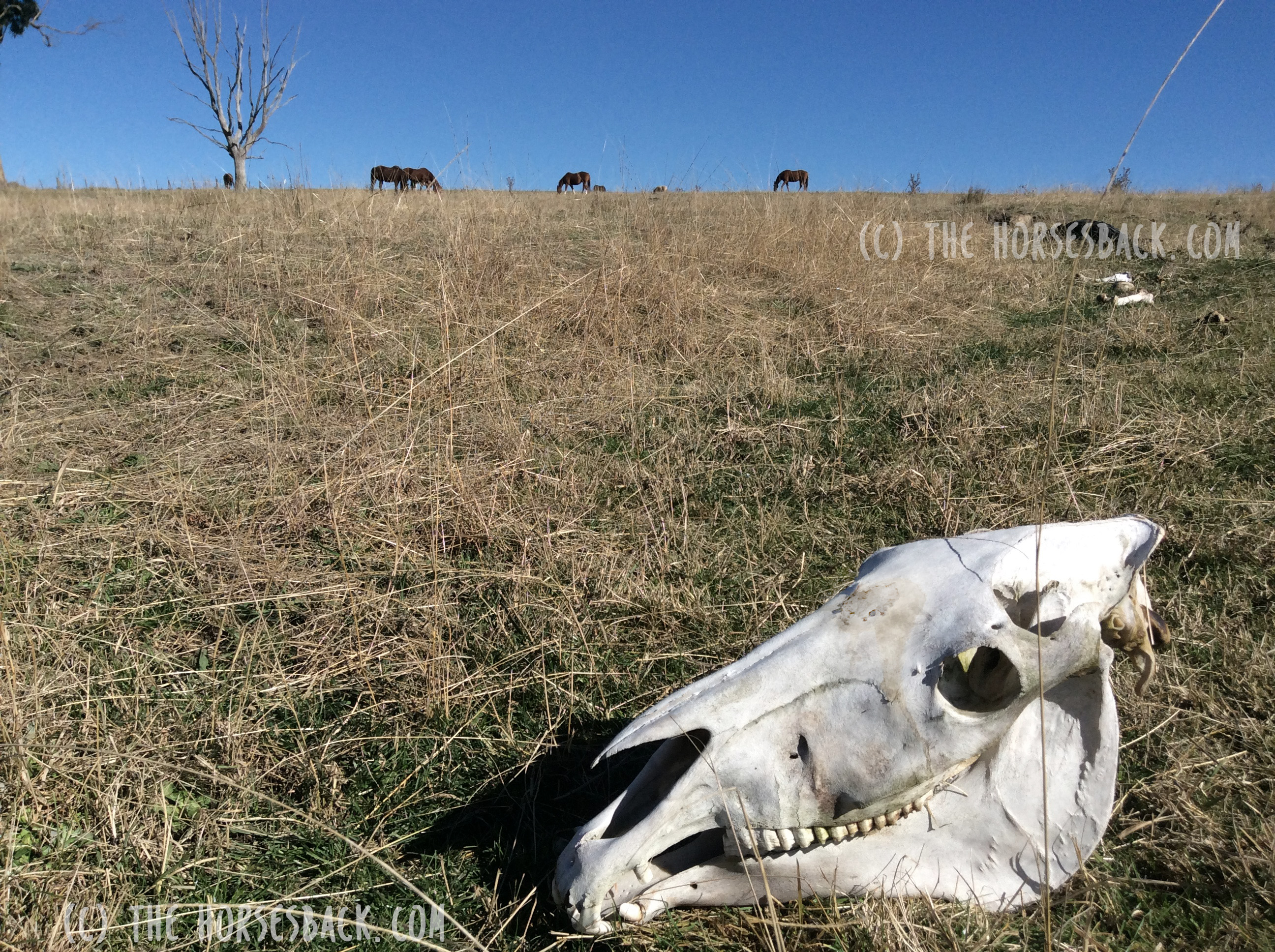
The parietal bones form the domed ‘cranial vault’ of the skull.


So How Does This Look for the Equine Brain?

Only a handful of equine researchers have delved into EQs, as this is mostly an area of zoological neuroanatomy.

In this study by Cozzi et al (2014), the brains of 131 mixed breed adult horses (no ponies) were collected and weighed.[1] Researchers found first that the adult horse’s brain weighs 600 – 700 g. The average brain weight for horses aged 2 years and over was 606 g, while the average bodyweight was 535.22 kg.

This meant the horses in this study had an EQ of 0.78.

Here are the EQs for some of the large mammals: Cow – 0.55, Pig – 0.6, Camel – 0.61, Horse – 0.78, Goat – 0.8, Wolf – 0.9, Domestic Cat – 1.00, African Elephant – 1.67, Gorilla – 1.76, Human – 6.62.

And if you’re really interested, here’s the calculation used in the equine paper. Other scientists use different calculations – there is no standard approach.

EQ = E i / 0.12 P2/3Ea/Ee


So, Are Horses Intelligent – or Not?

A larger brain mass compared with body mass is often associated with better cognitive functioning, but that does not mean it causes it.

Brain size is therefore a very general measure for intelligence. What actually matters are the specific areas of the brain and their relative sizes.

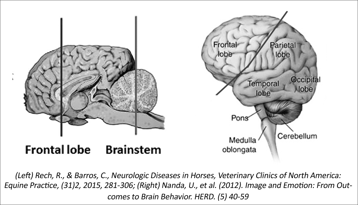
The bigger the frontal lobes, the more capable the species is of ‘goal directed’ behaviors – that is, the ability to analyse information and act accordingly, planning ahead. [2]

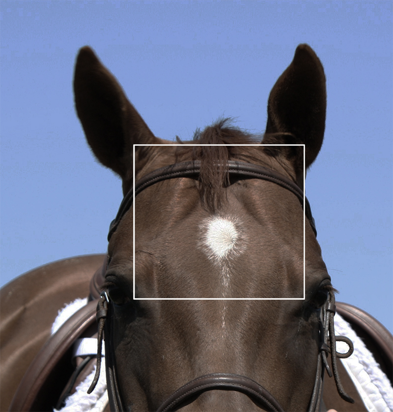
Here we hit an issue. The frontal lobes are either relatively small in the horse, or non-existent – and this is a matter of contention. Some published veterinary researchers maintain that they do, as shown below.

Rough comparison of the frontal lobes of the horse (left) and human brains.

However, researcher and author of Horse Brain, Human Brain Janet Jones PhD writes, “Basic anatomy shows that horses have no frontal lobes and no prefrontal cortex. No qualified PhD trained in neuroscience disputes this anatomy.”[3]

Whichever is true, the take home for both is that the horse is more likely to react in the moment. This is not to say that horses lack intelligence, but that they think and respond differently.


The brain’s fissures are also important. These are the wrinkles and grooves, known as sulci (sunken inwards) and gyri (protrude outwards). They’re standard within species, although the brains of some species have more complex surfaces than others.

Rats, considered to be on the lower end of the intelligence scale of mammals (although rat owners will surely disagree), have smoother brain surfaces than horses. In turn, horses have fewer fissures in their brains than primates.

The area contained within the cranium is the ‘cranial vault’. Its inner surface perfectly matches the outer surface of the brain, as they develop together as the animal grows.  If you could look inside this part of the skull, you would see a perfect mould of the fissures.

More recent research also links the organization of neurons (nerve cells) and synapses in the brain to intelligence.


Surely There’s a Difference Between Breeds?

Different breeds of horses certainly have differences in the shapes of their heads.

However, these differences are slight overall. In a study of TBs, STBs and Arabians, the relative proportions of the ‘neurocranium’ – the area above the frontonasal suture, including the cranium – were reasonably similar between breeds.

It was the lower part of the skull, primarily the nasal bones and the maxilla, that varied most and gave the breeds their different looks [4]. The study did not measure the cranium itself.

The neurocranium aligns with the ends of the frontonasal suture and includes the temporal and parietal bones, ending at the occiput.

This suggests that while some breeds may look extremely different – take the Welsh Cob and the TB, for instance – the neurocranium may be nearly square in all, at least when viewed from the front.

And even though some breeds may have proportionately larger heads, all (excluding ponies) will have EQs grouped around the average score of 0.78 mentioned earlier.

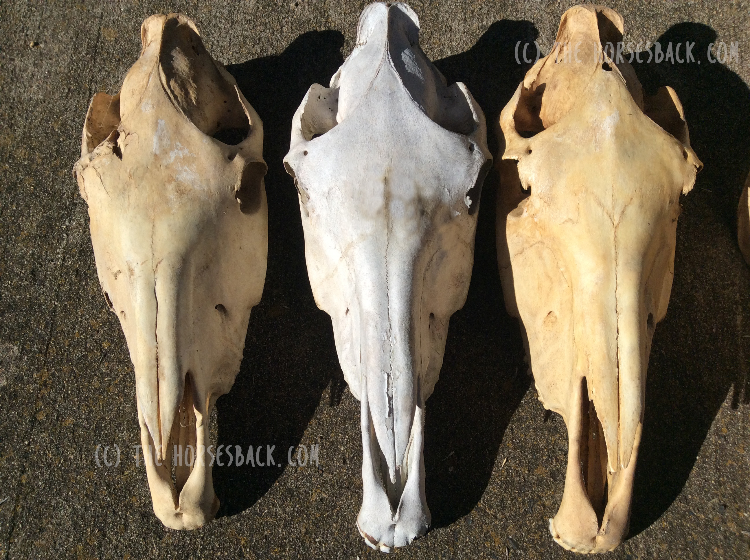
Small and wide ponies, incidentally, often have quite large parietal domes (or tuberosities, as they should be known), but the jury is out as to whether this makes them more intelligent… The fact is that we don’t know.


A Little More on Equine Brain Size

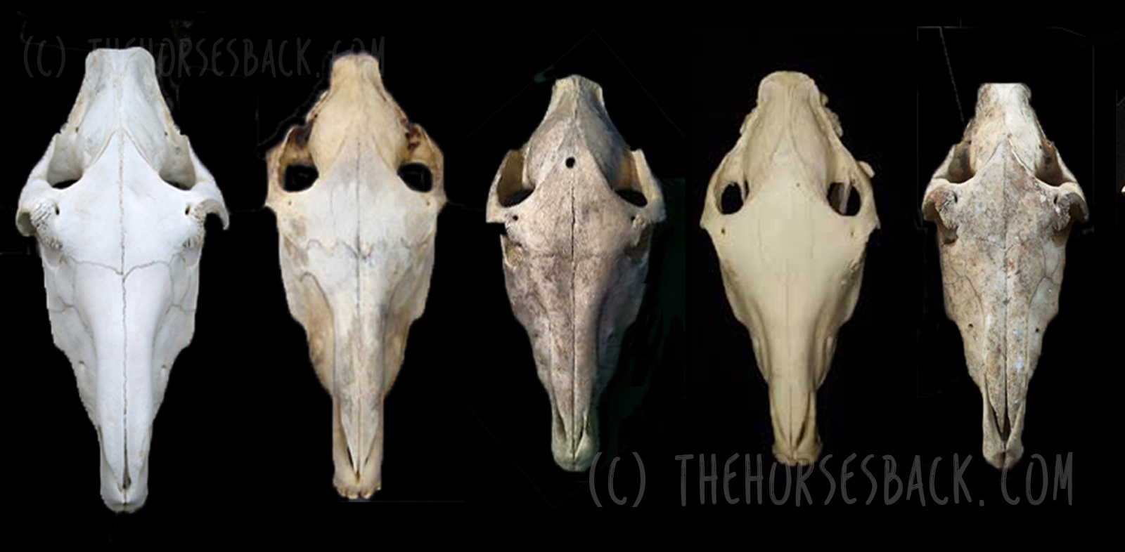
There are a few other differences that aren’t documented. Comparing horse skulls, we can see that some have a cranium that is narrower in relation to overall skull width than others. They also vary in shape: some are very full and round, while others are more teardrop shaped.

You can see this when you look at the spaces to either side of the parietal ‘dome’ and temporal bones, where the coronoid processes (tips) of the mandible protrude behind the zygomatic arch.

This may be due to breed or it may be individual. Our own skulls vary from person to person, with some aspects being just how we are, while others may be more developmental.

We can see this in horses too. Dwarf horses can have domed heads, as can horses that have been born prematurely.

This can affect intelligence – researchers have found that in humans, when the brain is smaller due to development delays, the intelligence can be lower. If it is smaller without any developmental delay, it makes no difference at all. [5]

Interestingly, a new study in humans shows that the longer the time Romanian orphans spent in the institutions as babies, the smaller their total brain volume, with these changes being associated with a lower IQ. You can read more about that study here. [6]

Personally, I would love to know more about this, as I’ve been researching the developmental effects of gestational problems in horses, including the effects of premature birth (my PhD thesis lives here). It’s the same old problem though: once a horse is at the stage where we can examine its skull, its early history is usually lost in the mists of time.

Ultimately, as with humans, what is going to make the most difference to us as horse owners is the individual’s learning experiences at different stages of its life. This is also where equine personality comes in, and the methods of training used, but those are different subject areas altogether.


[1] Cozzi et al., The Brain of the Horse: Weight and Cephalization Quotients, Brain Behav. Evol., 2014; 83:9-16

[2] McGreevy, P., Equine Behavior – A Guide for Veterinarians and Equine Scientists, Elsevier, 2012.

[3] Janet Jones – Horse Brains Facebook page

[4] Evans KE, McGreevy, PD., Conformation of the Equine Skull: a Morphometric Study, Anat. Histol. Embryol., 2006, 35(4): 221-7

[5] de Bie H. et al. Brain Development, Intelligence and Cognitive Outcome in Children Born Small for Gestational Age. Horm Res Paediatr 2010, (73)6-14.

[6] Mackes, NK. et al.,  on behalf of the E. Y. A. F. (2020). Early childhood deprivation is associated with alterations in adult brain structure despite subsequent environmental enrichment. Proceedings of the National Academy of Sciences.

Filed Under: Bodywork Tagged With: Anatomy, equine anatomy, equine brain, equine skull, GA, horse anatomy, horse brain, horse skull

How The Anatomy Books (Unintentionally) Fail Us Over The Nuchal Ligament

July 13, 2014 by Jane @ THB 5 Comments

nuchal-ligament-header-1

The nuchal ligament is a soft tissue structure that is widely discussed in dressage circles. Unsurprisingly, given its deep location, relatively few of us get to cast eyes on it or feel it directly under our hands.

It’s equally unsurprising, then, that most of us don’t realize that the image we hold in our heads is somewhat different to the reality of the ligament inside our horse.

 © All text copyright of the author, Jane Clothier, www.thehorsesback.com. No reproduction of images, partial or entire text without permission. Sharing the link back to this page is fine. Please contact me for more information. Thank you!

I have recently been fortunate enough to attend another dissection with renowned Australian gross anatomist (and she will point out repeatedly that despite this title, she is not gross – or, at least, not that often), Sharon May-Davis.

In this dissection workshop, Sharon had yet another opportunity to show us that an aspect of textbook anatomy is incorrect.

Yes, apparently there are many points where this is the case.

Where the nuchal ligament is and what it connects

The structure in question is the nuchal ligament, or the nuchal ligament lamellae to be exact.

George Stubbs illustration
George Stubbs, 1777, showed the NLL attaching from C2 to C7.

To quickly explain, the funicular part of the nuchal ligament is the cord-like part that runs from the withers to the occiput (back of skull). The lamellae is the fibrous sheet-like part that extends from the funicular part to the cervical (neck) vertebrae.

According to the majority of anatomy diagrams and textbooks, it extends down to attach to the cervical vertebrae, from C2 to C7.

According to Sharon, it doesn’t. And here’s why.

Findings on the nuchal ligament’s true location

In this study of 35 horses on the dissection table, Sharon found:

  • No cases where the attachments were from C2 to C7.
  • No horses where the attachments were from C2 to C6.
  • In all 35 horses, the attachments were from C2 to C5.
  • And in 9 of the 35, the attachments to C5 consisted of thin and feeble fibers.
  • The horses were of a mixture of identifiable breeds, aged 2 to 28 years old.

So, why do the majority of anatomical drawings of the deeper structures of the horse show something different?

When received knowledge can be a problem

Nuchal ligament, 5yo TB [click to enlarge]
Many of today’s illustrators are referring to illustrations that have themselves been amended from earlier illustrations.

(The header image for this site’s most viewed post, The Disturbing Truth About  Neck Threadworms and Your Itchy Horse, shows an inaccurate rendering of this ligament, as do most of the other illustrations I used. Dang!)

Inaccuracy is a recognized problem when it comes to received knowledge – was this anomaly due to an earlier artist’s error, or was it a characteristic of some 17th century horses that has been progressively bred out over subsequent centuries?

  • And this raises the question of which structure, exactly, is supporting the base of the neck of the horse in motion? Read more about m. Spinalis cervicis in this post, Meet Spinalis, the Forgotten Muscle in Saddle Fitting.
  • And how does this awareness inform current training approaches that require horses to raise themselves into self-carriage?

The findings from this study are in a peer-reviewed paper by Sharon May-Davis and Janeen Kleine currently in press with the Journal of Equine Veterinary Science. The paper includes a detailed review of illustrations in equine anatomy literature, an explanation of the study, and a thought-provoking discussion on the implications for our understanding of equine biomechanics.

Variations and implications of the gross anatomy in the equine nuchal ligament lamellae, Sharon May-Davis, Janeen Kleine, Journal of Equine Veterinary Science 30 June 2014 (Article in Press DOI: 10.1016/j.jevs.2014.06.018)

Have you read about Sharon’s findings on arthritis of the humeroradial (elbow) joint in all ridden or driven horses?


Questions, thoughts or comments? Join us at The Horse’s Back Facebook Group. 


Filed Under: Bodywork, Sharon May-Davis Tagged With: equine anatomy, equine bodywork, GA, horse anatomy, Nuchal ligament, Sharon May-Davis

Footer

Contact Me

Send your questions this way…

Get in Touch

  • Store Support
  • Shipping & Returns
  • Contact

Copyright © 2025 · Parallax Pro on Genesis Framework · WordPress · Log in

MAY Red Light Products Now In Stock! Dismiss