• Skip to main content
  • Skip to footer

The Horse's Back

Body Talk for Thinking Owners

  • Home
  • Articles
  • BEMER
  • Store
    • Store
    • Shipping & Returns
    • Support
  • Equine Healthworks
    • Bodywork for Horses
    • Reviews
    • Leave a Review
  • About & Contact
    • About
    • Contact

homepage

By Popular Demand: Here’s How You X-Ray for the C6-C7 Malformation

August 16, 2017 by Jane @ THB 2 Comments

News about the malformation of the C6 and C7 vertebrae has understandably raised a lot of concern. There are major implications for both buyers and breeders of TB and TB-derived breed horses, as outlined in the original article, All You Need to Know About the Hidden C6-C7 Malformation That’s Bringing Horses Down. In response, Sharon May-Davis has very kindly provided us with the English version of a paper she published in the Journal of Japanese Local Government Racing, outlining a preliminary protocol for radiographing this area of the neck and identifying the malformation. (Yes, a single racing authority has taken this issue on board!)

So without further ado, here is the paper in its entirety, the only adaptations being the positioning of images to better fit this site’s blog format.

© All text copyright of the published authors. No reproduction of partial or entire text without permission. Sharing the link back to this page is fine. Please contact me for more information. Thank you!


Preliminary Radiographic Protocols for Identifying Congenital Malformations of the Caudal Cervical Vertebrae

Authors: May-Davis S.E.R., Minowa F., Monoe S.


Abstract

In 2014, a published study based on dissections and skeletal examinations noted that 19:50 Thoroughbred horses had a congenital malformation of the 6th cervical vertebra (C6). In addition, it was found that in those 19 Thoroughbred horses expressing a congenital malformation of C6, 9 displayed a concurrent congenital alformation of the 7th cervical vertebra (C7). In this study, 3 Thoroughbred horses and 1 Thoroughbred type were clinically examined; 3 were radiographed for limb abnormalities and 2:3 radiographed for the congenital malformation of C6 and C7 prior to euthanasia. Upon dissection, 3:4 expressed a congenital malformation of C6 with 2:3 displaying a concurrent CM of C7. These 2 horses were positively radiographed for the CM of C6 and C7 prior to dissection. The radiographs of C6 were taken in direct lateral orientation with 0 degree of elevation and revealed the absence of the caudal ventral tubercle (CVT) of C6. Re-positioning the horse’s forelimbs caudally with an outstretched neck, C7 was radiographed at a 30° oblique lateral angle in a cranial to caudal direction with 0 degree of elevation; the transverse process of C5 remained cranial to the beam. The caudal aspect of the plate was positioned medial the Cranial deep pectoral and rotated vertically to expand the field of view of the cervical vertebrae, whilst remaining perpendicular to the beam.


Introduction

In recent times, 3 Australian studies reported congenital malformations of C6, C7, the 1st sternal rib, along with their associative soft tissue structures. Furthermore, it was noted that these congenital malformations were predominantly breed related and more specifically, to Thoroughbred horses or derivatives thereof. In addition, reports of forelimb proprioceptive dysfunction, neurological impediment and gait deficits were concurrently noted (May-Davis [a,b], May-Davis and Walker). The congenital malformations of C6 appeared as either a left or right unilateral absence of the caudal ventral tubercle (CVT) or as a bilateral absence of the CVT (Figure 1).


Left: Normal. Centre: Absent right CVT. Right: Bilaterally absent CVT

Figure 1. View of the ventral aspect of C6, cranial aspect top. Photo credit: Sharon May-Davis


The congenital malformation of C7 appeared with either a fully transposed CVT from C6 onto the ventral surface of C7 or a partial transposition of the CVT from C6 onto the ventral surface of C7 (Figure 2) (May-Davis [a]).


Left: Normal. Centre: Transposed right CVT. Right: Bilaterally transposed CVT

Figure 2. View of the ventral aspect of C7, cranial aspect top. Photo credit: Sharon May-Davis


The congenital malformation of the 1st sternal rib appeared in multiple presentations with gross anatomic variations including; an absent 1st sternal rib; bifid Tuberculum costae (Figure 3); bifid Sternochondral articulation onto the sternum; flared shaft; normal 1st sternal rib inserting onto the cranial branch of a bifid Sternochondral articulating 2nd sternal rib; straight costal shaft and an articulating rudimentary Tuberculum costae with a ligamentous extension replacing the bony shaft and attaching to a rudimentary Sternochondral articulation onto the sternum (May-Davis [b]).

Figure 3. Left costal bifid Tuberculum costae. Photo credit: Sharon May-Davis

Associative soft tissue structures varied according to the presentation of the congenital malformation with the most noted being, the Longus colli muscle, Scalene muscles and neural vessels such as the Phrenic nerve and Brachial plexus (May-Davis [b], May-Davis and Walker).

The congenital malformations noted in these 3 studies were determined via dissection and skeletal examination. They were based on 2 specific skeletal variations in C6 and C7 plus any variation to normal of the 1st sternal rib.

The congenital malformations could appear in C6 as a singular expression with no other anomalies, however, the congenital malformation of C7 only occurred when the CVT was absent on C6. The congenital malformations of the 1st sternal rib only occurred in the presence of the congenital malformation of C6 and C7 (Table 1) (May-Davis [b]).

With this in mind, the premise that if a congenital malformation exists in C6 then there is a 50% chance that transposition will occur on C7. In this format, it would stand to reason that there could also be an anomalous rib. Therefore, this study is designed to identify the absent CVT on C6 and investigate its transposition onto C7.


Table 1. Noted observations of 151 horses of mixed gender aged between 0 (stillborn) and 30, exhibiting a congenital malformation of C6, C7 and the 1st sternal rib.


Materials and Methods

After evaluation and clinical examination, the 2 horses (1 Thoroughbred and 1 Thoroughbred type) were restrained in cross ties. Detomidine Hydrochloride (Detomovet CEVA Animal Health Pty Ltd) was administered via left jugular vein venepuncture. The dosage was determined by the clinician’s experience and in consideration of the individual’s current health status.

The radiographs were obtained by utilising a Porta 100 HF High Frequency portable x-ray unit (7.8kgs). The kV range was 40-100kV with an mA range of 20-30 mA and a mAs range of 0.3 – 20 mAs. The images were captured on a wireless Rayence 1012WCA Medical Image processing unit (26cm x 32.5cm) and computerised with Vetview Digital Diagnostic Imaging software.

The radiographic angles were obtained with the horse’s neck in full extension and the forelimbs behind the vertical in standing position. With the clinician standing with the x-ray unit on the left side of the neck and the plate positioned on the right side of the neck, the distance between the x-ray unit and plate was 80cms. The x-ray unit and plate remained lateral to the cervical vertebrae determined by the transverse processes of C5.

The first view is a direct lateral at 90 degrees, with 0 degree of elevation whilst maintaining the transverse process of C5 to the left of the beam. 

Remaining lateral to the cervical vertebrae with a 0 degree of elevation, the second view is taken with the x-ray unit positioned at a 30° oblique lateral angle in a cranial to caudal direction with the transverse process of C5 cranial to the beam. The caudal aspect of the plate is positioned medial the Cranial deep pectoral and rotated vertically to expand the field of view of the cervical vertebrae, whilst remaining perpendicular to the beam.

Upon the radiographic evaluation of C6, radiographs of C7 are obtained. The 4 horses were then euthanized and dissected so to verify the radiographic findings. Only those pertaining to the congenital malformation of C6 and C7 are reported in this study.


Results

Figure 4. A compressed trachea (white arrow) in Thoroughbred No. 2 at C6 / C7. Photo credit: Sharon May-Davis

Upon euthanasia of the 4 horses, Thoroughbred No.1 was normal. Thoroughbred No.2 presented with an absent CVT in C6 and trachea compression (Figure 4).

Thoroughbred No. 3, presented with a C6 and C7 congenital malformation and a malformed 1st sternal rib. The Thoroughbred type also presented with a congenital malformation of C6 and C7 (Table 2).


Table 2. Noted observations via dissection of the congenital malformation of C6, C7 and the 1st sternal rib.


Left: Radiographic view of C6. Centre: The white line denotes a normal CVT. Right: The white denotes an absent CVT.

Figure 5. The radiographic lateral view of C6 depicting a unilaterally absent CVT in C6. Image credit: Sharon May-Davis


Thoroughbred No. 3 C7 radiograph (Figure 6) clearly presents the observer with a transposition of the CVT from C6 onto the ventral surface of C7, furthermore it denotes a reciprocal deviation in the trachea at the point of this transposition.


Left: Radiographic view of C7. Centre: The white line denotes the unilateral transposition of the CVT from C6 onto the ventral surface of C7. Right: The white line denotes the deviation in the trachea.

Figure 6. The radiographic view of C7 depicting a unilaterally transposition of the CVT from C6. Image credit: Sharon May-Davis


The Thoroughbred type (Figure 7) exhibits the absent CVT in C6. The radiograph clearly indicates an absent CVT that is unilateral in its presentation.


Left: Radiographic view of C6. Centre: The white line denotes a normal CVT. Right: The white denotes an absent CVT.

Figure 7. The radiographic lateral view of C6 depicting a unilaterally absent CVT in C6. Photo credit: Sharon May-Davis.


The Thoroughbred type C7 radiograph (Figure 8) clearly presents the observer with a transposition of the CVT from C6 onto the ventral surface of C7, furthermore it denotes a reciprocal deviation in the trachea at the point of this transposition.


Left: Radiographic view of C7. Centre: The white line denotes the unilateral transposition of the CVT from C6 onto the ventral surface of C7. Right: The white line denotes the deviation in the trachea.

Figure 8. The radiographic view of C7 depicting a unilaterally transposition of the CVT from C6. Photo credit: Sharon May-Davis.


Figure 9. Right: Absent CVT on C6 (white arrow) and transposed CVT from C6 onto the ventral surface of C7. Photo credit: Sharon May-Davis

Upon dissection, the Thoroughbred type displayed identical congenital malformations of C6 and C7 as depicted in the radiographs (Figure 9).


Discussion

This study was conducted with the view to obtain specific radiographic evidence of the congenital malformation of C6 and C7. Radiographic views of the 1st sternal rib were not possible due to thick and dense musculature in the region. The views obtained of C6 and C7 with exact radiographic angles proved that this region can be radiographed with precision so that the practitioner can make a clear diagnosis should a horse present with a neurological deficit, as previously published (May-Davis

[a, b], May-Davis and Walker]). Furthermore, the asymmetry of the cervical vertebra could exacerbate arthritis in the articular process joints as previously noted (May-Davis [a]). In addition, the incidental finding of the compression of the trachea is an important factor to consider when Thoroughbred racehorses present with compromised airways. An explanation for this occurring is that the Longus colli muscle hypertrophies in support of the weakened structures and due to its proximity to the Trachea, it impinges upon its dorsal surface. This impingement is further exacerbated by the ventral projection from C7 of the transposed CVT from C6.

Since the first publication in 2014, retrospective studies were conducted that added another dimension to the current research. Italian and American studies noted that Warmbloods, Quarter Horses and Arabs were also afflicted with this condition (Santinelli et. al 2016 and DeRouen et.al 2016). It is significant to note that these breeds present the majority of the ridden horse population in Japan extending from Racing, Showjumping, Dressage to Eventing and Endurance. However, a specific Dutch Warmblood study was conducted with fresh cadaver’s that were portioned and CT scanned (Veraa et.al 2016), this also included Oldenburg horses. The congenital malformation of C6 and C7 was present in both breeds with a noted malformed 1st sternal rib in a Dutch Warmblood. Combined percentages of these 3 studies in conjunction with the 3 Australian studies and the current study has the congenital malformation of C6 at; Warmbloods 30%, Quarter Horses 16% and Thoroughbred horses over 40%. 

Aside from the Arabian who is an ancestor to the Thoroughbred, the Warmblood breeds and Quarter Horses all have Thoroughbred lineage in the back line of their breeding. Thus, implying that this condition is heritable as noted by May-Davis [b] and more systemic than just one breed. With this in mind, all critical events should be assessed with the knowledge that a potential congenital malformation could exist in C6, C7 and the 1st sternal rib, as previously noted. With the largest population of horses in Japan being Thoroughbreds, it would be a recommendation to note the studies of several countries including Japan and the severity in percentage of this systemic congenital malformation. Especially in relation to Thoroughbred’ racehorses racing and cornering at speed.


Conclusion

This study showed that of the 4 horses investigated, 3:4 horses displayed a CM of C6, 2:3 displayed the congenital malformation in C6 and C7, and 1:2 a congenital malformation of the 1st sternal rib. Even with a small sample of Thoroughbreds, studies from other countries must be measured and it would therefore be a recommendation to radiograph for this condition in the caudal cervical vertebrae in a pre-purchase examination. The purpose of which would be to eliminate this condition so that riders and handlers are not put at risk.


Author contributions

Sharon May-Davis, Fumiko Minowa and Sadae Monoe wrote and reviewed this article.


Conflict of Interest

The authors have no conflict of interest in the preparation or presentation of this original research article.


Acknowledgement

The author wishes to thank Christine Gee for her professional advice on the manuscript. The Nippon Veterinary and Life Sciences University for the use of their facilities and to those authors / editors/ publishers of those articles, journals and books cited in this manuscript.


References

Derouen A, Spriet M, Aleman M. Prevalance of anatomical of the sixth cervical vertebra and association with vertebral canal stenosis and articular process osteoarthritis in the horse. Vet Radiol Ultrasound, Vol. 00, 2016, pp 1–5.

May-Davis SER. The Occurrence of a Congenital Malformation in the Sixth and Seventh Cervical Vertebrae Predominantly Observed in Thoroughbred Horses. J Equine Vet Sc 2014; 34:1313-17.

May-Davis SER. The Congenital Malformation of the 1st Sternal Rib. J Equine Vet Sc 2014; 34:1313-17.

May-Davis SER, Walker C. Variations and implications of the gross morphology in the Longus colli muscle in Thoroughbred and Thoroughbred derivative horses presenting with a congenital malformation of the sixth and seventh cervical vertebrae. J Equine Vet Sc 2015; 35:560-8.

Santinelli I, Beccati F, Pepe M. Anatomical variation of the spinous and transverse processes in the caudal cervical vertebrae and the first thoracic vertebra in horses. EVJ 48 (2016) 45–49.

Veraa S, Bergmann W, van den Belt A-J, Wijnberg I, Back W. Ex vivo computed tomographic evaluation of morphology variations in equine cervical vertebrae. Vet Radiol Ultrasound, Vol. 00, 1–7.


Filed Under: Bodywork, Sharon May-Davis Tagged With: C6, C7, cervical vertebrae, GA, homepage, TBs, Thoroughbred

All You Need to Know About the Hidden C6-C7 Malformation That’s Bringing Horses Down

July 16, 2017 by Jane @ THB 71 Comments


What if you were to learn that your horse is living with a hidden malformation? A skeletal abnormality that could be affecting it every day, changing the way it moves, creating a string of other physical problems, and possibly underlying the hard-to-pinpoint problems you’ve been noticing for months or even years ?

And that might even be causing a level of inherent instability that could be putting the rider in danger?

Sadly, this isn’t a hypothetical question. Instead it’s a reality that is only now being slowly uncovered.

And like the proverbial stone rolling down a mountain, the issue is gathering momentum as the equine industry, owners, breeders and researchers learn about it.

  • It’s a skeletal malformation and it can’t be corrected.
  • It’s congenital, ie inherited, so is present from birth.
  • It has been in some lines of TBs for hundreds of years.
  • It creates biomechanical issues due to asymmetry and lack of anchor points for key muscles.
  • At its worst, it can contribute to neurological issues such as Wobbler syndrome.
  • Some horses are so unstable, they are more prone to falling (not good news for jockeys).
  • It can cause constant pain and associated behavioural changes.
  • It’s primarily found in Thoroughbreds, Thoroughbred crosses and Warmbloods, but has also been identified in European breeds, Quarter Horses, Arabs and Australian Stock Horses.


Note: in the years since this post was written with direct support from Dr Sharon May-Davis, research has moved on. The body of literature now includes papers from Sharon and also from other researchers who do not reach similar conclusions about the issues caused by this malformation. I have included a list of journal papers and webinars on the topic at the end of the post, for reference if you wish to deepen your knowledge. You can also go view the full list here (opens in a fresh tab).


The problem behind this is ECVM, a congenital malformation of the C6 and C7 cervical vertebrae (ie, base of neck) – and it’s pretty nasty.

I’ve written about the work of Sharon May-Davis on this blog before and here I’m going to do so again. Through her many dissections per year, gross anatomist Sharon has become the first person to comprehensively document and quantify this problem.

In doing so, and publishing her findings in peer-reviewed journals, she has triggered a minor research avalanche as others take up the subject.

Update: since this article was published in July 2017, this malformation has been labelled Equine Complex Vertebral Malformation (ECVM). Some amendments have been made to include this term. To preserve existing links to the page, I’ve not updated the title and URL.

© All text copyright of the author, Jane Clothier, https://thehorsesback.com. No reproduction of partial or entire text without permission. Sharing the link back to this page is fine. Please contact me for more information. Thank you!


A hidden problem: this OTTB had the C6-C7 malformation but presented few outward signs.


Twenty years + of research into ECVM

Sharon May-Davis’s path with this research began some 20 years ago. In February 1996, a Thoroughbred called Presley came down unimpeded in a race in Grafton, NSW, fracturing his pelvis, a hock bone, and right front fetlock.

Three years later, Sharon examined his bones, and saw something strange in his last two cervical vertebrae and his first ribs.

Fast forward to 2014, when Sharon published the first of her four peer-reviewed papers in the Journal of Equine Veterinary Science, concerning a congenital malformation in the sixth and seventh cervical (neck) vertebrae.

Although the problem had been mentioned briefly in papers, this was the first time that a researcher had accurately described and quantified the problem in its various forms.

Sharon’s unique perspective, gained as an anatomist who dissects between 15 and 20 horses per year, had certainly placed her in a position to do so.


The horse’s seven cervical vertebrae – made simple

Horses have seven vertebrae in their necks, labelled C1 to C7. Of these, four have unique shapes. Most horse people are familiar with C1, the first vertebrae known as the atlas, as it can be both seen and felt by hand with its distinctive ‘wing’ at the top of the neck.

Cervical vertebrae from C1 (top) to C7 (bottom), view from above (L) and below (R).

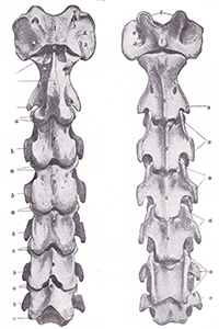
Almost as well-known is C2, the second vertebrae, known as the axis.

Both atlas and axis have unique shapes for a special reason: they support the heavy skull and anchor the muscles that control the head’s movement.

Heading down the neck, C3, C4 and C5 are broadly similar in shape, with each being a bit shorter and blockier than the one above.

However, C6 and C7 are both slightly different on the ventral (lower) side, for here they provide insertion points for muscles arising from the chest.

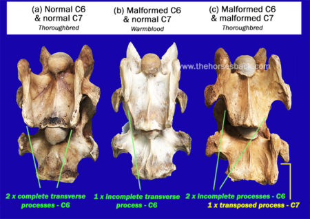
  • C6 has transverse processes (the protrusions extending outwards) that are different to those of neighbouring bones, with two distinctive ridges running the vertebrae’s length. C6 also has two large transverse foramen, the openings that the arteries pass through.
  • C7 is the shortest and squattest cervical vertebrae of all. Its transverse processes are shorter, while there are also two facets that articulate with the first ribs. C7 has no transverse foramen.

At least, that’s how the vertebrae should be in a normal horse.


So, what is wrong with the malformed C6 and C7 vertebrae in ECVM?

In certain horses, these last two vertebrae are rather different, being malformed.

Sharon has identified the manifestations of this problem as a congenital (inherited) malformation affecting some Thoroughbred horses, and horses with Thoroughbred blood in their ancestry.

In C6, there is a problem with the two ridges of the transverse processes, as one or both can be partially absent.

When both are partially missing, it is common for one or two ridges (ie, parts of the transverse processes) to appear on C7 instead.

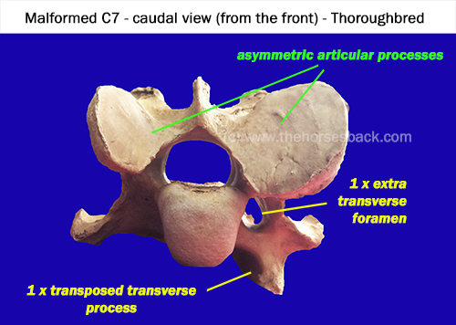
Also, the articular processes (the oval surfaces on the upper side, where each vertebrae links to its neighbours) can be radically different sizes. There can also be an additional arterial foramen or two.

The level of asymmetry can be radical.

The secondary problems the ECVM malformation causes

Being at the base of the neck, the asymmetry of C6 and C7 can cause alignment problems all the way up the vertebral column, leading to osteoarthritis of the articular facets.

It can also contribute to Wobbler Syndrome (Cervical Vertebral Stenotic Myelopathy), due to narrowing and/or malalignment of the vertebral foramen/canal, the opening through which the spinal cord passes. Not all Wobbler cases have this particular malformation, though.

A further problem is that the lower part of the longus colli muscle, which is involved in flexing the neck, would normally insert on the transverse processes of C6 and C7. When these processes are malformed, the normal insertions are not possible.

This means there is a serious symmetry problem in the junction of the thorax and neck, which can have a deeper effect on the horse’s neurology and proprioception, as well as respiration.

Asymmetry and narrowed vertebral foramen (canal) contribute to DJD and Wobbler Syndrome.


In a few cases, horses with both the C6 and C7 problem also have malformations of the first sternal rib, on one or both sides. This can cause problems beneath the scapular and further issues with muscular attachments.

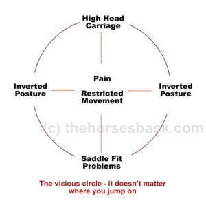
Associated stability problems can have far-reaching consequences for the horse, not to mention some serious safety issues for the rider. The safety issue can’t be stated often enough.


Why isn’t ECVM – the C6-C7 problem – more widely known ?

Why hasn’t this problem been noticed in regular veterinary interventions?

The answer is quite simple. While neurological issues may have been diagnosed, the exact cause has often remained hidden.

Both Thoroughbred horses and Warmbloods are known to have higher incidences of Wobbler Syndrome than other breeds, and while this is certainly not always due to C6-C7 malformation, the malformation has been found in some when dissected.

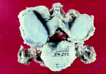
For example, the following dissection image appears in a veterinary account of large animal spinal cord diseases. It clearly shows a malformed C7 vertebrae, very similar to the one in the above image, but without giving any further categorisation.

The difficulty lies in the deep location of the lower cervical vertebrae. While normal radiographs can show all or some of C6, they are unable to penetrate the deeper tissues beneath the shoulder to image C7.

Photo from another online article: the familiar asymmetry of a malformed C7 is clearly visible in a dissected set of vertebrae.  (c) veteriankey.com (click image to access full article.)


Nevertheless, the malformation can be identified in radiographs of C6, once you know where to look.

Since Sharon’s first paper appeared, the School of Veterinary Medicine, University of California, Davis, has reviewed its history of radiographs from horses with Wobbler Syndrome.

Researchers found that 24 cases out of 100 (close to 25%) showed malformation of one or both C6 transverse processes. This study also clarified how to identify the problem on standard radiographs of C6.

In another study, the Faculty of Veterinary Medicine, Utrecht University, completed CT scans on horses’ necks and found the various forms of the malformation in 26 horses out of 78 (33%). Unlike radiographs, the CT scans enabled identification of the C7 and first rib issues, although of course this imaging was conducted post mortem.

Is this rare, or are many horses affected?

While the problem has been identified primarily in TBs, it affects most breeds with TB blood in the ancestry to some degree.

Sharon May-Davis reports that to date, published, peer-reviewed journal papers have tallied 136 out of 471 horses as exhibiting congenital malformation of C6.

These have been in a range of breeds including Thoroughbreds (39%), Thoroughbred crosses (27%), Warmbloods and European breeds (30%), Quarter Horses (11%), and Arabs (11%). Standardbreds have also shown the problem, although the numbers included in studies are very small.

A common question is whether it’s known which TB lines predominantly carry this problem. The answer is: Yes. However, it is now so disseminated amongst the modern equine population beyond TBs, that it is of little help to identify them.

It must be remembered that these horses are those already brought to veterinary attention and/or euthanized for a related or unrelated reason, so the percentages may be higher than those for the general horse population. At the same time, the malformation might have played a major part in the horses’ decline, due to the many locomotory and postural problems it can lead to.


How do we identify these horses in life?

It’s all very well looking at these bones post mortem, you might say. Yet how can I tell if my horse has this problem? Or a horse that I might want to buy?

Some answers are forthcoming. As Sharon has frequently assessed horses before dissecting them – usually from video – she has been able to observe that many of these horses lack stability. (Indeed, in many cases, it is this very instability has directly led to the horse being euthanized, and ending up on the dissection table.)

As her research has progressed, she has also been able to identify many biomechanical and locomotion traits that make these horses ‘suspicious’ or at least ‘of interest’. Unsurprisingly, these problems have been particularly noticeable in horses with both a malformed C6 and C7.

For owners and equine professionals, here are some signs that can raise initial suspicions. All can also be caused by other problems, so a group of signs is more common than an individual indication.

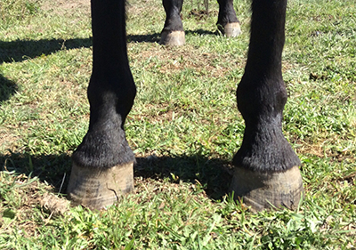
  • Some of these horses have a problem with standing square in front, and will always keep one foot further forward. This can persist despite all attempts to improve the horse’s body and to train the horse to halt squarely.
  • Horses with the more serious malformations will often stand base-wide. Such horses can become very unbalanced on uneven ground, and sometimes in work. They easily become unbalanced when a hoof practitioner works on a forefoot. 


A bilateral C6 – unilateral C7 horse showing a toe-out stance and hoof distortion. The ventral part of the transverse process was transposed onto the left side of C7.


  • With such asymmetry in the skeletal structure, these horses have serious lateral flexion issues that can’t be overcome. When required to elevate the forehand, many will experience difficulties, due to the absence of correctly inserted musculature and incorrect articulation through the joints of the lower neck. 
The horse may have one very prominent, widely positioned scapula.
  • A high level of asymmetry may be seen in the shoulders, with one scapula sometimes positioned very wide, with no improvement after chiro, osteopathy or bodywork. This is particularly so with the C6-C7 problem and associated first sternal rib abnormalities.
The ventral aspect of the neck may show some scoliosis.


  • There may be scoliosis along the entire spine.
  • There may be an obvious scoliosis to the underside of the neck.
  • The problem may lead to heavily asymmetric loading of the forefeet, so may be accompanied by a severe high foot/low foot issue (this is not in itself a sign of the C6-C7 problem).

If you suspect your horse has the C6-C7 issue

First, note that many horses do just fine with a C6 problem. It is those with the bilateral C6 and unilateral/bilateral C7 issue that tend to show the more worrying problems.

If your horse is showing ongoing signs of instability, it’s important to seek veterinary advice, so that neurological issues can be ruled out. (As this a recently recognised problem, it may be worth printing out the abstracts from the journal articles listed at the end of this page and handing them over.)

If the more severe malformations are identified by radiograph, it is important to remember that in some cases this can cause discomfort and pain to the horse, and it is not going to improve over time.

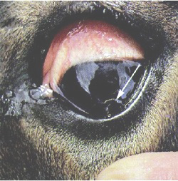
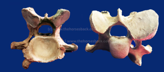
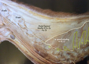
On the contrary, the cervical vertebrae of some older horses with the C6 and C7 malformations often display advanced osteoarthritis of the articular processes, as shown in the header image of a 19-year-old Thoroughbred’s malformed C7.


Where does this knowledge take us?

At the moment, that question is wide open. The findings published by Sharon May-Davis have triggered ongoing research on an international level. There are certainly ramifications for breeders in more than one equine sporting industry.

See Sharon May-Davis’s December 2020 presentation on ECVM here (article continues below):

Connections have been made with a number of falls on the racetrack that have caused injury, and worse, to both horse and jockey, as well as other runners. Similar things can be said for the sport of eventing, where unforced errors can have equally catastrophic effects.

It is entirely possible that at higher levels, pre-purchase examination radiographs will come to include a check on C6. While it’s not possible to radiograph the deeply positioned C7, we do at least know that this will only be present if the C6 anomaly exists.

Vets in some countries are proving faster at picking this up than others. While papers are being published, it clearly takes some time for information to filter down.

And until more is known, this problem is being unknowingly propagated every breeding season.

Of course, many horses harbouring the milder manifestations of this problem at C6 level are functioning very well. All horse owners can do is be aware that this issue exists, make use of this information if a problem arises, and await further research findings.

Literature on the malformation

In no particular order: the following peer reviewed journal articles address ECVM, as it is now known. Please remember when reading papers that no single paper proves one thing – a review of many papers and critical thinking is always essential. Apologies for not having standardized the formatting, but the links should be present.

  • The Occurrence of a Congenital Malformation in the Sixth and Seventh Cervical Vertebrae Predominantly Observed in Thoroughbred Horses, May-Davis, Sharon, Journal of Equine Veterinary Science , Volume 34 , Issue 11 , 1313 – 1317
  • Variations and implications of the gross morphology in the longus colli muscle in thoroughbred and thoroughbred derivative horses presenting with a congenital malformation of  the Sixth and Seventh Cervical Vertebrae May-Davis, S, Walker, C. Journal of Equine Veterinary Science 35 (7), 560-568
  • Characterization of the Caudal Ventral Tubercle in the Sixth Cervical Vertebra in Modern Equus ferus caballus. May-Davis, S., Dzingle, D, Saber, E., Blades Eckelbarger, P. Animals 13 (14), 2384
  • Morphology of the Ventral Process of the Sixth Cervical Vertebra in Extinct and Extant Equus: Functional Implications, May-Davis, S., Hunter, R, White, R. Animals 13 (10), 1672
  • Variations and Implications of the Gross Morphology in the Longus colli Muscle in Thoroughbred and Thoroughbred Derivative Horses Presenting With a Congenital Malformation of the Sixth and Seventh Cervical Vertebrae, May-Davis, Sharon et al., Journal of Equine Veterinary Science , Volume 35 , Issue 7 , 560 – 568
  • Congenital Malformations of the First Sternal Rib, May-Davis, Sharon, Journal of Equine Veterinary Science , Volume 49 , 92 – 100
  • Ex Vivo Computated Tomographic Evaluation of Morphology Variations in Equine Cervical Vertebrae,  Veraa, S. et al,  Veterinary Radiology & Ultrasound, Vol. 57, Issue 5
  • Prevalence of Anatomical Variation of the Sixth Cervical Vertebra and Association with Vertebral Canal Stenosis and Articular Process Arthritis in the Horse, Spriet, M. and M Aleman, Veterinary Radiology & Ultrasound, Vol. 57, Issue 3
  • Anatomical Variation of the Spinous and Transverse Processes in the Caudal Cervical Vertebrae and the First Thoracic Vertebra in Horses, Santinelli, I. et al, Equine Veterinary Journal, Vol. 48, 45–49
  • Congenital variants of the ventral laminae of the sixth and seventh cervical vertebrae are not associated with clinical signs or other radiological abnormalities of the cervicothoracic region in Warmblood horses. Dyson S, Phillips K, Zheng S, Aleman M. Equine Veterinary Journal, 2025 Vol.2 :419-430
  • Neck pain but not neurologic disease occurs more frequently in horses with transposition of the ventral lamina from C6 to C7. Henderson CS, Story MR, Nout-Lomas YS. J Am Vet Med Assoc. 2024 May 29;262(9):1215-1221


Filed Under: Bodywork, Sharon May-Davis Tagged With: C6, C7, ECVM, equine dissection, GA, homepage, Sharon May-Davis, skeletal malformation, slider, Thoroughbred anatomy

Meet Spinalis, the Forgotten Muscle in Saddle Fitting

December 20, 2016 by Jane @ THB 43 Comments

Spinalis Header

It’s barely mentioned in saddle fit or anatomy books, yet the muscle Spinalis cervicis can hugely impact on the spinal health and movement of the horse, particularly with poor tack fit.

Meet muscle Spinalis cervicis et thoracis, a far more important muscle than is generally realized. As a deep muscle, it’s influential in mobilizing and stabilizing that hidden area of the spine at the base of the neck, the cervico-thoracic junction, deep between the scapulae.


Where to Find this Muscle

As part of the deeper musculature, Spinalis is as hidden in books as it is in life. Usually, it’s a single entry in the index.

Spinalis Standard
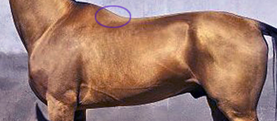
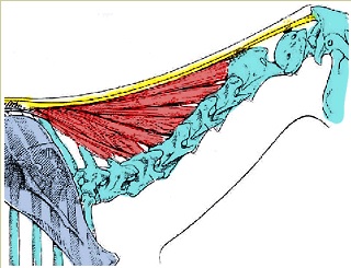
At best, it has no more than a bit part in anatomical illustrations,  usually as a small triangular area at the base of the withers. This is also where we can palpate it.

The reality is quite a bit more interesting. It’s actually a muscle of three parts – dorsalis, thoracis and cervicis. These names denote its many insertions, for it links the spinous processes of the lumbar, thoracic and cervical vertebrae.

  • Bradley_2.1
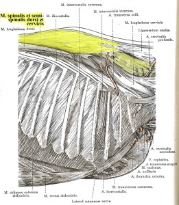
Further back along the spine, it lies medially to Longissimus dorsi, and in fact integrates with this larger, better known muscle, attaching to the processes of the lumbar and thoracic vertebrae.
  • When it reaches the withers, it becomes more independent, attaching to the processes of the first half dozen thoracic vertebrae (T1-T6). Here, the cervical and thoracic portions overlap and integrate to share a common attachment. (The part we palpate, at the base of the withers, is the thoracic section.)
  • Heading into the neck, as Spinalis cervicis, it attches to the last 4 or 5 cervical vertebrae (C3/C4-C7). Only the lamellar portion of the nuchal ligament runs deeper than this muscle.

Dissection 2
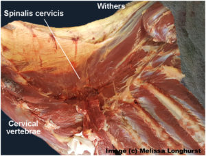
Its integration with other muscles is complex, and its close relationship with Longissimus dorsi partially explains why it doesn’t get much consideration as a muscle in its own right.

It is the more independent section, Spinalis cervicis, between withers and neck, that we are interested in, although its influence is present along the entire spine.

© All text copyright of the author, Jane Clothier, https://thehorsesback.com. 

What Does Spinalis Do? 

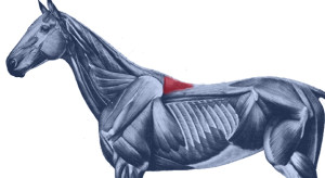
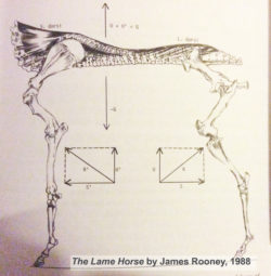
In his 1980s’ Guide to Lameness videos, Dr. James Rooney, first director of the Gluck Equine Research Center, University of Kentucky, referred to Spinalis as part of the suspension bridge of muscles supporting the spine (Longissimus dorsi achoring from the lumbosacral vertebrae, Spinalis thoracis et dorsalis from the upper thoracics). He also refers to this extensively in The Lame Horse (1988).

In fact, the suspension bridge analogy only really makes sense if Spinalis dorsi is considered.

Spinalis cervicis is usually credited with a role in turning the head to left to right, and raising the head.

Bradley Spinalis-1Older texts, such as Bradley’s 1922 veterinary dissection guide, Topographical Anatomy of the Horse, mention its role in stabilizing the spine.

This creates a point of interest. Given that the nuchal ligament (lamellar portion) doesn’t attach to C6 and frequently only weakly with C5 (see the findings of anatomist Sharon May-Davis, in this earlier article ), Spinalis cervicis suddenly appears pretty important in stabilizing and lifting the base of the neck, particularly as it does so at the point of greatest lateral bending.


Spinalis and Poor Saddle Fit

Anyone who has been involved in close examination of the horse’s back will recognize Spinalis thoracis where it surfaces close to the skin, on either side of the withers.

When a horse has been ridden in an overly tight saddle, this small area of muscle can become pretty hypertrophic – raised and hardened. Typically, the neighbouring muscles are atrophied. When Spinalis is palpated, the horse often gives an intense pain response, flinching down and raising the head.

GerdHeuschman
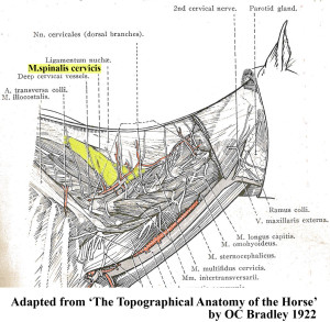
What often happens is this. An overtight saddle fits over the base of the withers like a clothes peg, pinching Trapezius thoracis and  Longissimus dorsi. However, it frequently misses Spinalis thoracis where it surfaces, wholly or partially within the gullet space. Often, the muscle is partially affected.

It’s as if the neighbouring muscles are under lockdown. Free movement of the shoulder is restricted and the horse’s ability to bear weight efficiently while moving is impeded. In response to the surrounding restriction and its own limitation, this muscle starts to overwork.

Result? The horse, which was probably already moving with an incorrect posture, hollows its back even further, shortening the neck and raising its head.  As this becomes even more of a biomechanical necessity, all the muscles work even harder to maintain this ability to move, despite the compromised biomechanics.

Working harder and compensating for its neighbours, Spinalis becomes hypertrophic. It is doing what it was designed to do, but it’s now overdoing it and failing to release. We now have a rather nasty vicious circle.


Spinalis photo

Here, Spinalis thoracis stands out due to atrophy of the surrounding musculature. In this TB, a clearly audible adjustment occurred in the C4-C5 area after the muscle was addressed. 


Vicious CircleThe Inverted Posture and Asymmetry

Of course, saddle fit is not the only cause of an inverted posture. However, any horse that holds its head and neck high for natural or unnatural reasons is more vulnerable to saddle fit issues, thus starting a cascade effect of problems.

Are there further effects of this hypertrophy? Consider the connections.

  • When saddles are too tight, they’re often tighter on one side than the other. This can be due to existing asymmetry in the horse, such as uneven shoulders, uneven hindquarters, scoliosis, etc.
  • On the side with greater restriction, the muscle becomes more more hypertrophic.
  • With its attachment to the spinous processes of the lower cervical vertebrae, there is an unequal muscular tension affecting the spine.
  • Without inherent stability, the neck and head are constantly being pulled more to one side than the other, with the lower curve of the spine also affected.
  • Base of neck asymmetry affects the rest of the spine in both directions and compromises the horses ability to work with straightness or elevation.
  • There is also asymmetric loading into the forefeet.
  • We haven’t even started looking at neurological effects…

This isn’t speculation. I have seen this pattern in horses I’ve worked on, many times over.


So, How Do We Help?

In working with saddle fit problems, the saddle refit may be enough to help the horse, if the riding is appropriate to restoring correct carriage and movement. Obviously, the horse’s musculoskeletal system is complex and no muscle can be considered in isolation. As other muscles are addressed through therapeutic training approaches, with correct lateral and vertical flexion achieved, M. spinalis will be lengthened along with the surrounding musculature.

I hold with a restorative approach:

  1. Refit the saddle, preferably with the help of a trained professional,
  2. Remedial bodywork, to support recovery from the physical damage,
  3. Rest the horse, to enable healing of damaged tissue and lowering of inflammation, and
  4. Rehabilitate the horse, through the appropriate correct training that elevates the upper thoracics while improving lateral mobility.

This is particularly important where saddle fit has been a major contributor to the problem. I have frequently found that in these cases,correction will take longer to achieve, as the debilitating effects of poor saddle fit (especially long-standing issues) can long outlast the change to a new, better-fitting saddle. In bodywork terms, the hypertrophic M. spinalis cervicis is often the last affected muscle to let go.

It’s as if Spinalis cervicis is the emergency worker who will not leave until everyone else is safe.


Bodywork Notes

I am fortunate, in that my modalities enable the gentle release of joints through a non-invasive, neuromuscular approach.  The responses I’ve had from horses when M. spinalis cervicis et thoracis has been addressed in isolation have been hugely informative.


** Questions, thoughts or comments? Join us at The Horse’s Back Facebook Group. 


Appendix: Spinalis in the Textbooks

I’m going to add Spinalis references to this post on a regular basis, as I come across them. It’s interesting to see how much, or how little, the muscle is referenced in various textbooks.


Equine Back Pathology

This image, from Equine Back Pathology, ed. F Henson 2009, shows acute atrophy of Longissimus dorsi due to neurological damage. It’s still possible to see the raised attachment/origin of Spinalis cervicis et thoracis – the highlighting is mine. Spinalis does not appear in the book’s index. (added 23 Dec 2016)


nuchal and spinousI have also altered this image, in order to show M. spinalis cervicis more clearly. This is Fig 2.16 from Colour Atlas of Veterinary Anatomy Vol 2, The Horse, R Ashdown and S Done. Spinalis cervicis is within the bounded area and it’s possible to see how it overlies the lamellar part of the nuchal ligament, lamellar portion. (added 23 Dec 2016)


S&G
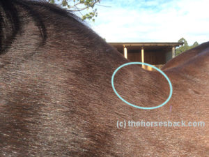
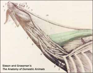
The muscle is tinted green in this image from Sisson and Grossman’s The Anatomy of Domestic Animals, Volume 1, fifth edition 1975.  Here, it is labelled Spinalis et semi-spinalis cervicis. This anatomical figure is credited to an earlier text, Ellenberger and Baum, 1908. (added 23 Dec 2016)


James Roony dedicates two pages to the ‘suspension bridge’ theory of the vertebral column in The Lame Horse (1988). His interest is in Spinalis dorsii section of the muscle and its effect behind the withers, in conjunction with  Longissimus dorsii. (added 4 Jan 2017)


Schleese diagram
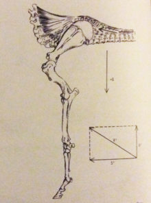
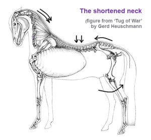
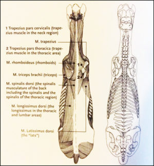
Master Saddler Jochen Schleese refers to Spinalis dorsi and its function in stabilizing the withers in Suffering in Silence, his passionate book about saddle fitting from 2014. “This muscle area is especially prone to significant development – especially with jumpers – because it is continually contracted to accommodate the shock of landing”. The surface area of the muscle is indicated in the anatomical figure, reproduced here. (added 23 Dec 2016)


In his seminal text addressing issues of modern dressage training, Tug of War, 2007, Gerd Heuschmann includes Spinalis cervicis in the triangle formed by the rear of the rear of the cervical spine, the withers, and the shoulder blades, “… an extensive connection between the head-neck axis and the truck… it explains how the position and length of the horse’s neck directly affects the biomechanics of the back.” (added 31 Dec 2016)


Filed Under: Bodywork, Saddle Fit Tagged With: Anatomy, equine bodywork, forgotton muscle, GA, homepage, saddle fit, saddle fitting, slider, spinalis, spinalis cervicis, spinalis dorsalis, spinalis thoracis

Debunked: The Lie That’s Told About Adjustable Gullet Saddles

May 19, 2014 by Jane @ THB 63 Comments

adjustable-header-2

One of the best innovations in the world of saddle making has been the interchangeable gullet plate in the synthetic saddle tree. I mean, there’s no getting away from it, it’s brilliant. With the removal of a few screws, every horse owner can adjust their own saddle in minutes. Easy.

saddleWhy so good? Well, they can be fitted to a lot of horses. They can accommodate the changing shape of a growing young horse, as well as seasonal weight gain and loss, or the development of back muscle through training.

So what’s my problem with them?

Unfortunately, such saddles are often accompanied by extra inventions,
this time originating in the marketing department.

Before I go on, I must declare an interest here. I fit saddles. What’s more, I fit saddles with interchangeable gullet plates. I’m not going to say which brand, because that’s not what this post is about. I say this simply to demonstrate that I’m not against adjustable saddles.

My problem is very much with the misleading statements that are made in order to sell them, and in particular the notion that these saddles can be adjusted to fit any horse. Not just a single weight-changing or shape-changing horse, or a few horses in the same yard, but any horse.

They can’t. It’s not true. They simply can’t.


Back to the horse’s back

XchangeLet there be no doubt that many horses experience a lot of pain from ill-fitting saddles that are too tight, or too wide, at the front of the tree. Most people are familiar with the sight of horses with white hair behind the shoulder blade, and areas of mild to profound muscle wastage.

The so-called wither profile is incredibly important for this reason. Gaining a correct fit across the gullet (and I mean gullet in the Australian sense – referring to the front of the saddle tree only, rather than the entire channel) is a highly important aspect of saddle fitting.

Yet it isn’t the only aspect. Astonishing as it may seem, horses are 3-dimensional organic structures. Yes! And they have many profiles in that area where the saddle sits.

S/W Ver: 96.66.76RThink about horses’ backs. The gullet plate matches the profile across the withers. But what about the profile along the withers, as well? Withers have different heights and lengths…

There are other profiles, too. There’s along the spine. There’s across the back at the rear of the saddle area, close to the last ribs. All of these profiles have both lengths and angles.

This is one of the reasons why many experts in the world of saddle making and fitting refer to the 9 points of saddle fitting. Several of these points involve the length and angle of the profiles I’ve just mentioned.

genesisGoing back a few years, the common view was that there are 5 points. Times have moved on, anatomy and biomechanics are better understood, and saddle design has evolved dramatically to reflect more recent ideas about how a saddle should interact with the horse’s body and movement, as well as the rider’s. And yet…

Fitting saddles isn’t like buying a pair of socks

Going by a single measurement might be OK for some things, but it isn’t for saddles. There’s more than one measurement involved, and I’m not just talking about the rider’s seat size. Think again about horses’ backs.

  • We have high withers, middling withers and rangy tabletops. High withers can extend way back into the area of the saddle.
  • Looking along the spine, we can see dippy backs, straight backs and bumpy backs.
  • Looking across the spine, we can spot angular A-frame backs and smooth, flat and pudgy backs.
  • It’s easy to spot uphill and downhill backs.
  • Not to mention short backs and long backs (or, to be more accurate with saddle fitting, rib cages).
  • And spines may have wide spinal processes or narrow ones.
  • And how about round rib cages that spring out nearer the spine, or narrow, flat-sided rib cages that drop sharply away, and everything in between?
  • This is before we even look at damaged backs, uneven shoulders, laterally curved spines, and all manner of physical issues affecting the horse, rider and the saddle in between.

Horses have a combination of these features. Many horses have one or two that can make saddle fitting a bit tricky.  Some have combinations that make saddle fitting an utter nightmare.

The saddle’s tree must reflect all those variations. It’s what makes saddle fitting such an interesting challenge, and occasionally a very hard one.


More on this Topic

Exclusive posts on Saddle Fit are available on The Horse’s Back Patreon, including The Deep, Deep Pain Caused by a Poor Saddle Fit and The Princess and the Pea… or, the Saddle and the Ding in the Back. Join today and have access to 25 archive posts straight away. Membership from US$ 7.50 per month.

But what about adjusting the flocking?

images6

Well, what about it? Adjusting flocking is the saddle fit version of fine-tuning. It is not changing the overall fit of the saddle.

Adjusting the flocking when the tree is the wrong shape is like (ahem) whistling in the wind.

It’s like adding an extra hole to your belt in an attempt to make a pair of jeans fit, despite the fact that the waist is a size too narrow and the legs 6  inches too short.

Adjusting the flocking only works when the tree is already a fundamentally good fit. The same goes for any flocking substitute, such as risers or wedges inserted into the panels. It is not enough to make a saddle fit the horse, when the tree is the wrong shape.

The message is being massaged

screwdriver

Adjustable gullet plates are now free of the original designer’s patent restrictions and a number of companies are now using them.

As already said, that’s great, providing the saddles are fitted well.

And who determines that? It can be hard to be sure when certain departments continue to make this ongoing, inaccurate claim about their brand of saddles being adjustable to all horses.

It’s marketing at its worst. It’s not just misleading, it’s plain untrue. Worse, it’s willful mis-education that leads horse owners into the mistaken belief that because they have the right gullet plate, then their saddle fits and their horse can’t possibly be in any pain. 

It bugs me that people are being misled. It bugs me far more that horses end up being the silent incumbents of a problem with so much potential to lead to back pain. (And I have worked with the results first-hand.)

As I said earlier, when the saddle fits, FANTASTIC. In fact, FANTASTIC with bells on.

And when it doesn’t, it’s the horse who suffers, no matter how many professionals are saying that black is in fact white, and that with the right ‘system’, an adjustable saddle can be made to fit any horse.

It can’t.

June 2019 update: 5 years after publishing this article, I received the following message from the Ruiz Diaz company in Argentina:

“I am part of the team producing Pessoa, PDS and Anky saddles. I just want to let you know that we have been working on our saddles for better fitting on the rest of the body and not only the withers. Thanks for your article once again, it was really nice to read your words and realize that with a wrong concept we have been saying that the x-change system was fitting every horse but there is much more apart the horse withers.”


Questions, thoughts or comments? Join us at The Horse’s Back Facebook group. 


Filed Under: Saddle Fit Tagged With: anky saddles, bates saddles, buying saddles, GA, genesis saddles, homepage, pessoa saddles, saddle fit, saddle fitting, thorowgood saddles, wintec saddles

The Disturbing Truth About Neck Threadworms and Your Itchy Horse

June 12, 2013 by Jane @ THB 284 Comments

disturbing-truth-feature

Look on any ivermectin or moxidectin-based wormer packet and you’ll see a long list of parasites. Tucked in neatly at the end – it’s nearly always at the end – you’ll see the words Onchocerca cervicalis, otherwise known as neck threadworms.

Also known as neck threadworms, these critters vary in length from 6cm to 30cm (think the length of a regular ruler). Astonishingly, they live in the horse’s nuchal ligament.

Yes, the nuchal ligament. It runs the full length of the neck, from poll to withers, with a flat ligament part connecting with the cervical vertebrae.

Apparently, most horses have Onchocerca. For many they’re not a problem, but some horses develop a reaction to their microscopic larvae (the microfilariae). This is known as Onchocerciasis. The horses become itchy, mostly around the head, neck, chest, shoulders and underside of the belly. That’s why owners often make the understandable assumption that their horse has Queensland itch or sweet itch.


A quick introduction to neck threadworms

Original article by Jane Clothier, posted on https://thehorsesback.com, June 2013. All text and photographs (c) Jane Clothier. No reproduction without permission. Links to this page are fine.

Onchocerca is what’s known as a parasitic filarial worm (nematode). One reason these worms get relatively little attention is that they never live in the intestines. The microscopic larval form live in the horse’s skin, mostly around the head, neck, shoulders, chest and underside of the belly. It is the adult worm that later makes its home in the nuchal ligament.

The problem is global and horses in most countries have been found to have this parasite. Unfortunately for those of us who keep horses in warmer, humid climates, it’s more frequent here. The biting insect that serves as a carrier is the Culicoides fly, which is also connected to Queensland Itch (aka Sweet Itch, Summer Itch, etc.).

It’s an unfortunate coincidence of environment that leads to many cases of neck threadworms being missed, because they’re assumed to be Itch.


Does your horse have “the itch” – or neck threadworms?

It’s a humdinger of a thought. If your horse is itchy, something different could be happening to what you think is happening.

  •  Your horse has the ‘regular’ itch (ie, Queensland, sweet, whatever it’s called in your region) and are reacting to midge spit – and nothing else. (The point of this article certainly isn’t to try and say that all itch cases are due to neck threadworms. Just some.)
  •  Your horse has neck threadworms and its inflammatory reaction to them has increased its sensitivity, so it’s now reacting to fly bites everywhere – in other words, Queensland/sweet itch has been triggered as a secondary response.
  • Your horse only has neck threadworms, in which case they’re probably rubbing along the mane and particularly the base of mane, around the neck and face, under the chest and down the ventral line (under the belly), but not on the tail head – or at least, relatively little.

Are you by any chance now thinking other horses you know? If so, they might be suffering from Onchocerciasis. There’s a lot of it about.


So how do we identify neck threadworms?

Neck threadworms have a distinctive life cycle, but as is so often the case, the problem presents in  different ways, depending on the individual.

In my brumby Colo, it started with him scratching the underside of his neck on posts. That was about 3 months before I had an inkling it might be neck threadworms. How I wish I’d known  what it was at that point, so that I could have nipped the problem in the bud…

A pony with the Itch and neck threadworms. It's Autumn and she's stopped rubbing out her entire mane, but is still itching that tell-tale area in front of the withers. Her coat has raised in a temporary histamine reaction to the ivermectin wormer.
A pony with the Itch and neck threadworms. It’s Autumn and she’s stopped rubbing out her entire mane – it has grown back – but is still itching that tell-tale area in front of the withers. Her coat is raised in a temporary histamine reaction to the ivermectin wormer.

I’ve also seen it manifest as a new, previously unseen itchy and scurfy patch on the lower part of the neck of a horse who’d never been itchy. And I’ve heard of a local horse who suddenly started furiously itching his face, bang in the middle of the forehead, to the point that it bled. He had never been itchy before.

These are the classic early signs, usually recognised by the owner only through miserable hindsight. Other signs include small lumps forming along the underside of the horse and on its neck and face, weeping spots, and a scaly crest to an area of the mane through rubbing.

The base of the mane, just in front of the withers, seems to be party central where neck threadworms are concerned.


The real nastiness of neck threadworms

The microscopic larvae can travel to the eye, although this is rare.

It just gets better: the larvae can travel to the horse’s eyes, where they can cause untold damage. This cheering sentence from Scott and Miller’s Equine Dermatology sums it up: “O. cervicalis microfilariae may also invade ocular tissues, where they may be associated with keratitis, uveitis, peripapillary choroidal sclerosis, and vitiligo of the bulbar conjunctiva of the lateral limbus.”

Oh heck. Nobody’s sure how common this is. All I know is that I don’t want to find out the hard way.

Consider this: in humans, a slightly different strain of Onchocerca infestation is known as River Blindness.

Please remember this detail when you’re deciding whether to worm for neck threadworms or not.


The very strange lifecycle of the neck threadworm

These worms have a complicated existence. They’re among the shapeshifters of the parasitic worm world, developing through several larval stages before reaching adulthood.

The first stage microfilariae live in the horse, close to the skin. Their numbers are highest in the spring and decrease to their lowest point in mid-winter. They live in clusters, which is why you may first notice patches of scurfy skin where the horse has started itching. This is a reaction to the dead or dying larvae.

Itching down the midline. Mine have itched neck and shoulders only - so far. (Photo courtesy of blog, Baba Yaga's Mirror)
Itching down the midline. Mine have itched neck and shoulders only – so far. (Photo courtesy of blog, Baba Yaga’s Mirror)

At this point, our good friends the culicoid flies make a contribution, by biting the horse and ingesting a good number of microfilariae along with blood. Within the fly, the larvae then develop through a further stage (or two). They are then deposited back into a horse when the flies bite. The flies can do this for an impressive 20 to 25 days after first hoovering up the larvae.

Back in a host horse, the larvae then make their way via the bloodstream to the connective tissue of the nuchal ligament, which runs along the crest of the neck. Here they moult and develop into adult worms. The adults live for around 10 years and in this time, the females release thousands of microfilariae (larvae) very year.

Original article by Jane Clothier, posted on www.thehorsesback.com, June 2013. All text and photographs (c) Jane Clothier. No reproduction without permission, sorry. Links to this page are fine.

No matter where the adult worms settle, the itchiness is caused by the microfilariae that aren’t lucky enough to be consumed by a fly and are instead left to die off.

The next part’s really not fair. The more the horse itches and breaks the skin, the more the flies will bite exactly where the microfilariae are located, before transporting them to the same or another horse, to start all over again.

Unsurprisingly, horses with most lesions have higher microfilariae counts – it’s a perfect ascending spiral of parasite-induced discomfort.

The Onchecerca life cycle lasts for 4 to 5 months.


Can we test for neck threadworms?

The microfilariae can be identified in the living horse through a biopsy of the nuchal ligament. Published veterinary research shows you won’t get any indication within 34 days of worming, so the timing is critical.

Worming with ivermectin can lead to weeping spots in the mane. This was after they'd cleared.
Worming with ivermectin can lead to weeping spots in the mane. These can be painful. This photo was taken after they’d cleared, leaving bald areas. Sometimes the hair grows back white.

A dose of ivermectin-based wormer is the quickest way to tell if your horse has them. If the microfilariae are present, the horse usually responds with intense itching – and I mean, manically intense, demented itching – around 48 to 72 hours after worming.

It may develop weeping, gunky spots at the base of the mane. (If you live in a paralysis tick area, it’s similar to the localised reaction you see in response to the ticks.) These are very specific spots around 1cm in diameter, with hair loss after they’ve erupted.

My brumby responded this way, rolling furiously and rubbing vigorously against posts. Unsurprisingly, he was also hard to handle for a few days. He was definitely sore at the base of the neck, where the weeping eruptions came out, and didn’t want to be touched there. I have to say that the scale of his reaction came as a shock to me, so take heed and be prepared with some soothing salves.


What can we do about adult neck threadworms?

Here’s the depressing answer: not much. But we can manage them.

The nuchal ligament runs from poll to wither and links with the vertebrae. Yellow = funicular part, home to neck threadworms.
The nuchal ligament runs from poll to wither and links with the vertebrae. Yellow = funicular part, home to neck threadworms. (Image copyright Sustainable Dressage.)

The adults live for 10-12 years and happily inhabit the nuchal ligament. What often happens is that the horse’s body throws down calcification around the adult worms in an attempt to isolate the foreign body. In some horses, you can feel a collection of  pea-like bumps in the nuchal ligament. In the ones that I’ve checked, this was just in front of the withers.

The slightly better news it that the worms are so fine and the lumps so small that it doesn’t seem to affect the function of the ligament, which is tough and fundamentally taut anyway. However, I’ve not yet knowingly seen a horse with a long history of neck threadworms – I’d be interested in doing so.

Heavier calcification is usually most prevalent in horses in their late teens. It figures, as the adult wormers are older, and longer. Apparently they intertwine and live in small clumps. Mid-aged horses have mainly shown inflamed tissue around live parasites.

In horses less than 5yo, the parasites can be present but there’s relatively little immunological response. So if your horse has suddenly developed itchiness at the age of 5 or 6, you could be looking at the presence of this parasite.

Original article by Jane Clothier, posted on www.thehorsesback.com, June 2013. All text and photographs (c) Jane Clothier. No reproduction without permission, sorry. Links to this page are fine.


Managing the initial outbreak

Do you worm your horses? Do you want to reduce the itching at the cost of having to worm more? I know I do, but I realise that some people can’t abide the thought of chemical wormers, or their increased use. But here’s what you can do if you want to reduce that dreadful itching and virtually eliminate the possibility of eye damage.

Unfortunately, there’s no single recommended protocol for worming against neck threadworms, so you’re in fairly uncharted territory.

  • wormerTo address the initial outbreak, the advice ‘out in the field’ is to use a regular dosage of an ivermectin-based wormer, multiple times until symptoms subside. The recommended interval I’ve seen is a week, but do check with your *equine* vet first.
  • I’ve also read forum posts by US horse owners stating that a double dosage at fortnightly intervals is the most effective treatment. It’s usually around three doses, or until symptoms subside. One reason is that lower doses do not kill off enough larvae, allowing resistance to develop amongst those that remain. Wormers are certainly tested as safe at higher dosages, but again, horses are individuals, so always check with your *equine* vet first.
  • I’ve read that an injection of ivermectin can be more effective, with off-label use of a product such as Dectomax being recommended as the heavy artillery when all else has failed. Again, do check with your *equine* vet.

Some say that an ivermectin and praziquantel wormer is more effective. One small comfort is that these wormers are available in the lower price ranges. It’s a consideration, because if you’re worming multiple horses, this won’t be a cheap time. It may even be worth looking at the large bottles of liquid wormer used by studs for greater economy.

Published research has shown that moxidectin-based wormers are equally as effective in addressing the microfilariae (but don’t double-dose with this one – only with ivermectin).

Whichever option you follow, it’s worth following this worming protocol with prebiotics, probiotics and ‘buffers’ such as aloe vera to support a healthy gut lining.

Reducing the larval population

After the initial worming, it’s a matter of management. What you’re trying to do is keep the numbers of microfilaraie low, so that the horse’s itching is reduced. Remember, most horses show little reaction, although the parasites are present. The aim has to be to bring them down to levels the horses’ systems can deal with, while taking other measures to boost the horses’ immune system.

  • Some vets say a single dose every 6-8 weeks during the fly season.
  • Others say every 3 months, timed in accordance with the larval lifecycle, which is 4 to 5 months.
  • In humid sub-tropical zones, where all parasite burdens are dramatically higher, I’ve heard of people doing it as frequently as once a month.

Beyond that, you’re back to the barrier treatments – fly rugs, lotions and potions to deflect the flies and to insulate the skin, lotions to soften the skin and heal the lesions, fly screens on shelters during the day, etc. And don’t forget about boosting your horse’s immune system generally through sound nutritional approaches.


And if we do nothing?

If we don’t address the problem one way or another, we have very itchy horses, for their entire lives.

Researchers say that the calcification in the ligaments has no effect, but you’ve got to wonder. There’s no guarantee that those scientists had a highly developed understanding of equine biomechanics. Maybe they did, but… who knows. A lot of the small amount of research available is over 20 years old and the knowledge base has since grown.

There’s a small but serious risk of damage to the eyes.

On the plus side, Onchocerciasis hasn’t been found to have any association with fistulous withers.

How to put together a program of treatment for your horse with neck threadworms (and maybe the Itch) – How to Fight the Big Fight against Neck Threadworms

To recap…

Onchocerciasis is so often masked by the itch that awareness, even in the regions where it’s rife, is low.

And in those same regions, there are so many highly prevalent and deadly parasites – the worms that cause colic, that drag down the horse’s condition, that can kill through spontaneous mass emergence from encysted larval stages – that the neck threadworm larvae simply doesn’t get much of a look-in.

To repeat, I’m not saying that all cases of itch are neck threadworms. Just that these parasites may be involved and can be a contributory factor in a heightened immunological response that leads to Queensland itch (or sweet itch, or whatever you know it as).

However, some horses definitely have neck threadworms. The earlier we can identify and manage it, the better.

We can’t eliminate the neck threadworms, but we can certainly manage the effects and make our horses’ lives more comfortable.

(c) Jane Clothier – no reproduction without permission – jane@thehorsesback.com


Here are some of the links I’ve been accessing for this article.


Equine Onchocerciasis: Lesions in the Nuchal Ligament of Midwestern US Horses. G. M. Schmidt, J. D. Krehbiel, S. C. Coley and R. W. Leid. Vet Pathol 1982 19: 16.

Efficacy of ivermectin against Onchocerca cervicalis microfilarial dermatitis in horses. Herd RP, Donham JC, Am J Vet Res. 1983 Jun;44(6):1102-5. http://www.ncbi.nlm.nih.gov/pubmed/6688162

Onchocerciasis, Submitted by EquiMed Staff. http://equimed.com/diseases-and-conditions/reference/onchocerciasis

Onchocerciasis, the Neck Thread Worms and Midline Dermatitis in Horses.

The Hypersensitivity of Horses to Culicoides Bites in British Columbia. Gail S. Anderson, Peter Belton and Nicholas Kleider. http://www.ncbi.nlm.nih.gov/pmc/articles/PMC1680856/pdf/canvetj00574-0044.pdf

Onchocerca in Horses from Western Canada and the Northwestern United States: An Abattoir Survey of the Prevalence of Infection, L Polley. http://www.ncbi.nlm.nih.gov/pmc/articles/PMC1790494/

Research of skin microfilariae on 160 horses from Poland, France and Spain. M.T. FRANCK et al. Am J Vet Res. 1983 Jun;44(6):1102-5. http://www.revmedvet.com/2006/RMV157_323_325.pdf

Efficacy of ivermectin against Onchocerca cervicalis microfilarial dermatitis in horses. Herd RP, Donham JC. http://www.ncbi.nlm.nih.gov/pubmed/6688162

Equine Dermatology. By Danny W. Scott, William H. Miller Jr. via Google Books [link now unavailable]

Baba Yaga’s Mirror. Personal blog. [link now unavailable]


Filed Under: Neck Threadworms, Parasites Tagged With: GA, homepage, horses itch, neck threadworms, onchecerciasis, onchercerca, queensland itch, slider, summer itch, sweet itch

Footer

Contact Me

Send your questions this way…

Get in Touch

  • Store Support
  • Shipping & Returns
  • Contact

Copyright © 2025 · Parallax Pro on Genesis Framework · WordPress · Log in

MAY Red Light Products Now In Stock! Dismiss