• Skip to main content
  • Skip to footer

The Horse's Back

Body Talk for Thinking Owners

  • Home
  • Articles
  • BEMER
  • Store
    • Store
    • Shipping & Returns
    • Support
  • Equine Healthworks
    • Bodywork for Horses
    • Reviews
    • Leave a Review
  • About & Contact
    • About
    • Contact

GA

Not Quite Classical… Better Lungeing for the Rest of Us

July 14, 2019 by Jane @ THB 8 Comments

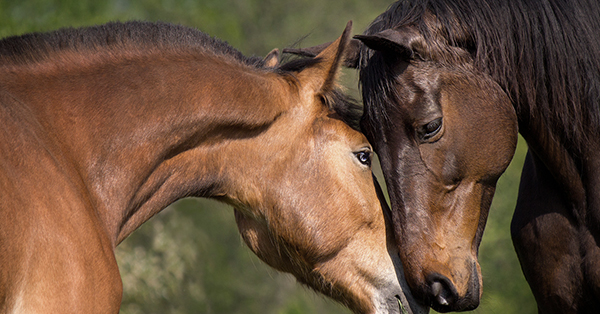
Thinking as a bodyworker, if there were one thing I could change in the training of ridden horses, it would be the way that many people lunge their animals.

I’d be so happy if the standard practice were a simple, gentle and biomechanically correct approach that brings profound improvements to the horse’s back health and readiness for riding.

It’s not a science and it needn’t be. For most people, it’s easy to learn, easy to do and easy to continue with. At even a basic level, it conditions the horse to carry weight and to not only balance itself, but to move effectively while carrying the weight of the rider.

Every rider and horse can benefit from this, no matter the ridden goals or discipline. Horses may just happen to have a back we can sit on, but their bodies are not designed to function in the way that we ask.

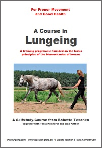
Here is a simple and affordable resource that will help you to do it better: A Course in Lungeing. It’s something I’ve recommended to bodywork clients over the years, with positive results. (It does involve ditching the gadgets and moving with your horse, but you don’t mind that, do you?)

Let’s look at why I believe it’s so good.


© All text copyright of the author, Jane Clothier, https://thehorsesback.com. No reproduction of partial or entire text without permission. Sharing the link back to this page is fine. Please contact me for more information. Thank you!

** Questions, thoughts or comments? Join us at The Horse’s Back Facebook Group. 

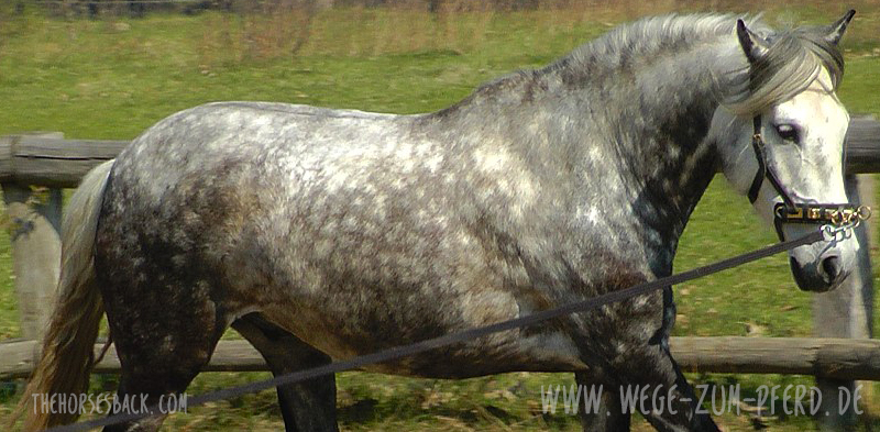
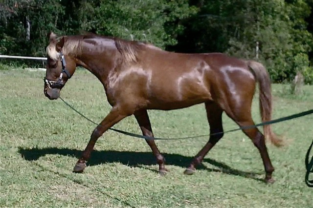
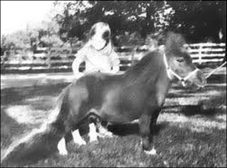
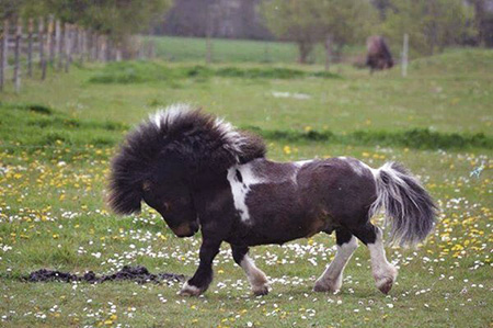
What often passes for lungeing

Oddly enough, many people think you don’t have to learn anything in order to lunge a horse.

It does involve ditching the gadgets…

It’s assumed that you just need to buy the right equipment. In all too many cases, it’s then a matter of sending the horse in a circle, achieving as much trotting in as short a time as possible.

There are plenty of goals, including many that don’t have much to do with preparation for biomechanically correct movement while being ridden.

Lungeing to warm up the horse. Lungeing to build topline. To get the horse ‘into an outline’. To make the horse listen. To make the horse understand. To ‘get the buck out’. To get weight off the horse.

It’s also what many people do when they haven’t got time to ride. It’s not good and it’s not the best option for any horse.


It takes time to prepare the horse’s back

Lungeing shouldn’t be part of a rush. It isn’t a ‘hack’, ie. some kind of shortcut or saving of time and effort, adopted to fill the gap between feeding and going to work on days when there aren’t enough hours for riding.

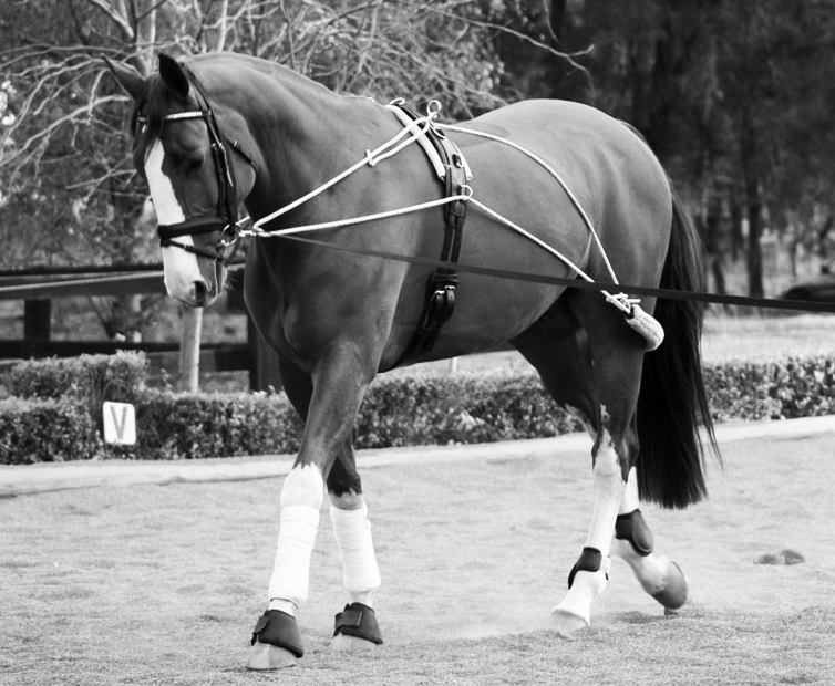
In the last few years, a renewed interest in a more classical, biomechanically correct approach to training has brought simple, in-hand training techniques to wider audiences. Older methods that aimed to prepare horses for ridden careers, which have been overlooked in the great rush to do everything faster, have come back into focus.

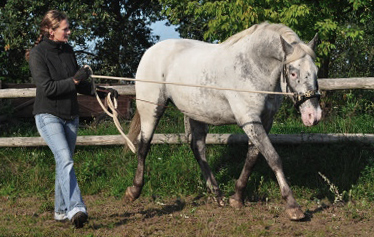
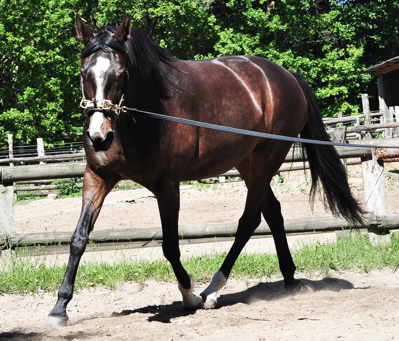
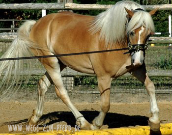
This doesn’t involve sending horses in circles around a largely stationary human. It involves walking with the horse, working with it and shaping it. It’s about educating the horse to move and to carry itself in a way that is then taken forward into ridden work.

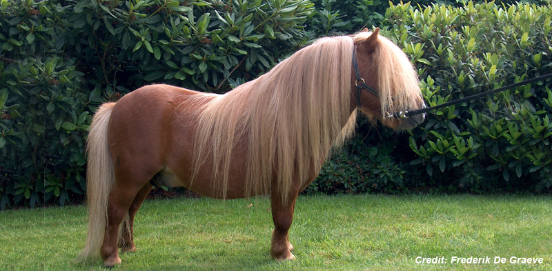
Lungeing for ‘everyhorse’

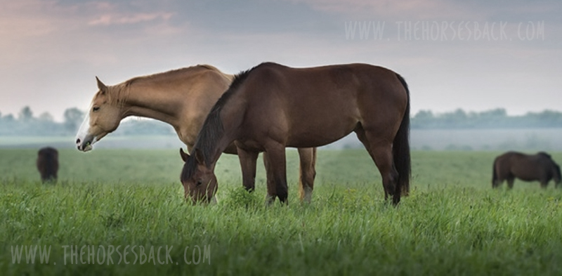
While horses differ enormously in terms of conformation, their needs are basically the same for carrying a rider. This is true no matter what discipline their athletic capabilities and fate destine them for. It is equally true for the trail riding horse.

Pien, Anglo Arabian

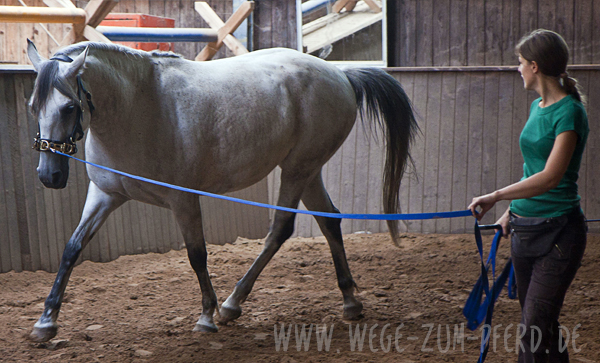
Biomechanically correct lungeing prepares the horse to do this. Put very, very simply, this means lungeing so that on a bend, the horse weights inside and outside legs evenly and doesn’t lean in.

Instead, this even weighting means it is able to freely lift up through the base of neck and withers, between the shoulders (i.e. the thoracic sling). Meanwhile, the hindquarters are active and load bearing. The horse moves lightly with elevation and is able to do so freely and at all paces.

Biomechanically correct lungeing helps to develop the back and teach self-carriage, which makes it humane as well as effective.

Why would anyone wish for less for their horse?  The wonderful thing is that it’s not hard to do.


Therapeutic benefits of correct lungeing

As a bodyworker, I have repeatedly witnessed the  improvements in horses that were helped to work correctly in this way.

Clients and associates have had success rehabilitating horses, including (and especially) ex-racehorses with serious sacroiliac dysfunction.

Fanta, 12-year-old Westphalian

The strength in their bodies, the suppleness, and the ability to work softly in a magnificent shape without any kind of restriction or force is wonderful to see.

I’ve also noticed how much horses seem to enjoy working in this way, as their body and movements develop and their spirits lift. Pride becomes visible in their movement and attitude.

These horses go on to flow under the saddle. Again: who wouldn’t want that for their horse?

The thing is, once this approach is learned, the improvements come quickly. Dramatic changes can happen through 2-3 short sessions a week. It’s not time consuming at all – and you don’t need to be into dressage to do it.

Which brings us to A Course in Lungeing.


So how do we relearn lungeing?

A Course in Lungeing makes these time-tested approaches available to the owners of any horse, no matter what breed, age or conformation, or ridden discipline.

It’s perfect for people who don’t want to study for qualifications or who can’t afford or manage to attend clinics with classical trainers (you can do this in your paddock).

The course was developed by German horsewoman, Babette Teschen, based on her many years of teaching correct lungeing in workshops in Europe.

Lunging training stood out as being really helpful for my work,” writes Babette Teschen. “In as much as you learn to lunge your horse according to biomechanical principles, you are doing what is best for the health and spirit of your animal partner: you give it the means to fulfill what you want of it in a healthy way.”

What I love is their strong focus on musculoskeletal health, so much so that they’ve included contributions from an equine osteopath and an equine acupuncturist.

More info and to purchase ‘A Course in Lungeing’

What you receive with ‘A Course in Lungeing’

With this training, you can help your horse to learn to bend in a circle instead of falling in, to raise its inner shoulder, and to move with the hindquarters tracking the forelegs (think of a train on the tracks rather than a motorbike on a bend).

Anthony, 5 year old Haflinger

When your horse can do this, it will be able to free up the neck and ‘let go’ from the withers. It can develop rhythm and freedom of pace, with good ground coverage. The horse’s back muscles are freed up to do their work effectively, with its hindquarters taking up the load.


What you’ll receive: 

  • View the first 30 pages of the book for free.
  • 250 pages of instruction material plus videos of various exercises.
  • Extensive basic information of anatomy and biomechanics.
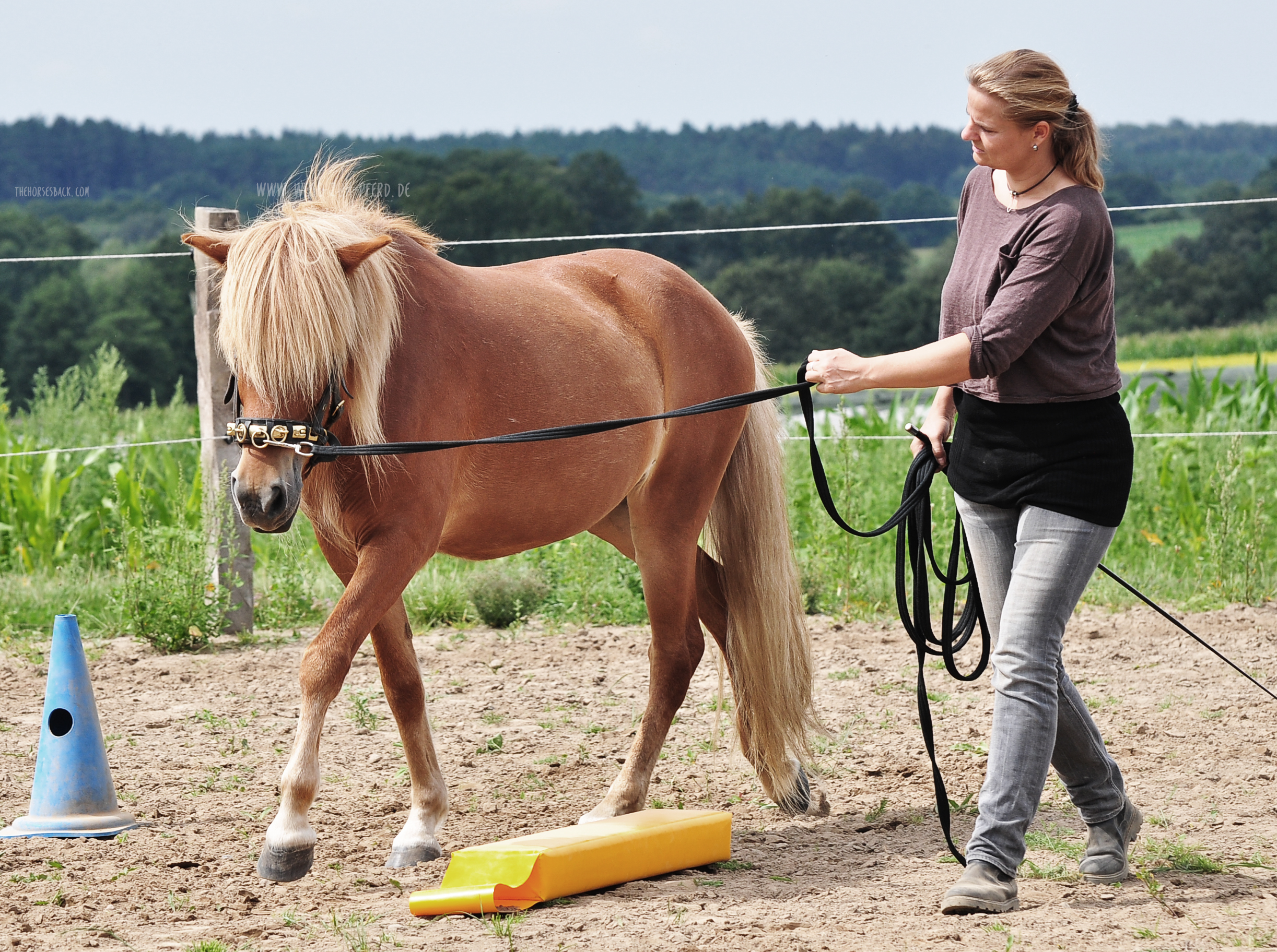
  • Clearly presented exercises that will help you and your horse to master the problem of moving in a circle with ease and enjoyment such as the Cone Slalom, the Stepped Pathway, and many more.
  • Photos and diagrams with explanations and illustrations.
  • A Media Library with videos, and a substantial .pdf by osteopath Maike Knifka on the theme of Physiotherapy and the Lungeing Course, with supporting videos.
  • Acupressure tips from acupuncturist Patrizia Harneit on video.
  • A special on working with horses with extra paces, such as Icelandic horses.
  • Tips for working with older horses and those with health problems.


The book is available as a PDF in English.

More information and to purchase ‘A Course in Lungeing’

Note: this post was published in July 2019, simply because I believe in the approach.

This has resulted in many purchases of the course. I’ve been happy to facilitate that, as it helps so many horses.


Bondi, an Australian Stock Horse whose owner, the late Eva Thaler, introduced me to this course many years ago. 


Filed Under: Bodywork, Viewpoint Tagged With: brachial plexus, classical dressage, classical training, GA, Lungeing, lunging, thoracic sling

The Very Big Problem of Dwarfism in Horses

February 22, 2019 by Jane @ THB 9 Comments

In this guest post, Dr. Sonja Dominik describes equine dwarfism and what the latest tests mean for horse breeders. Sonja is a Research Scientist in Quantitative Genetics at CSIRO, Australia’s national science agency.

She is currently a Superstar of STEM, appointed by Science & Technology Australia to increase the public visibility of women in science. To top that off, she was also a competitor in the 2018 series of Australian Ninja Warrior.

Sonja writes :


There’s small and then there’s very small.

There’s cute – and then there’s dwarfism, which isn’t cute at all.

Equine dwarfism is a very complex genetic condition. However, a few facts apply to all dwarf types in horses.

First, dwarfism in horses is caused by a disruption of the structural processes in bone and / or ligament development. It can potentially occur in any horse breed, but is most prevalent in Miniature horses, Shetland ponies and Friesians, but has also been described in Mustangs and donkeys.

All characterized dwarf types in horses are disproportional, meaning that only some parts of their bodies – eg.  their limbs – are reduced in size.

Dwarfs can suffer secondary conditions, usually due to their skeletal deformations, and multiple health problems, such as metabolic, digestive or respiratory disorders.

In genetic terms, dwarfism is a recessive condition (more on that below). Not all dwarf types are genetically characterized, but genetics tests are available for some.

Bond Tiny Tim (b.1970) was an American dwarf miniature horse breeding stallion, whose name appears throughout miniature horse pedigrees. Image copyright unknown.


Horses with dwarfism aren’t just tiny

Four types of equine dwarfism have been defined, based on their physical characteristics. However, there are a lot of overlaps and there are dwarf horses that are difficult to fit into any of the described types.

1. Short legs, long bodies.

Achondroplasia is the most common form of dwarfism. Affected horses have short-limbs with a normal trunk, although often with an elongated back.

The term achondroplasia actually means ‘without cartilage formation’, although that is not quite correct…. these dwarfs have cartilage, but the problem is that cartilage is not turned into bone while they are gestating.

However, this type of dwarf can lead a relatively normal life.

Foal with achondroplasia. These miniature horses can lead relatively normal lives. Image copyright unknown.


2. Large heads, distorted features.

Brachycephalic dwarfs have a bulging forehead, with a short and flat nasal bridge, overly large eyes, and nostrils that are higher than what is considered normal.

They also have a short neck and limbs, and often have spine deformations. This type of dwarf often has a shortened life span.

Thumbelina (b. 2001), a brachycephalic dwarf miniature, was celebrated as the smallest horse in the world at 43cm tall. Image: Brad Barket, Getty Images


3. Multiple deformities.

Diastrophic dwarfs can have twisted limbs and / or multiple limb deformities and other characteristics such as a domed head and roached back and a pot belly.

Due to the severely deformed limbs, affected animals would require splints or surgery to move properly.

Diastrophic dwarfs are prone to colic due to the small size of the abdominal cavity and pressure on the intestines. Image copyright unknown.

4. No bone.

Hypochondrogenesis is the most severe condition of dwarfism where the bones are not ossified at all. Affected fetuses are normally aborted before birth.


Here’s what we know about dwarfism genetics

Recently, a genetic test for dwarfism in miniature horses was developed at the Gluck Equine Research Center, University of Kentucky.

This tests for achondroplasia-like dwarfism, which is caused by mutations in the so-called ACAN gene.

Skull of a dwarf miniature horse compared with a full size horse’s skull. Dwarfs experience more dental problems due to the large size of their teeth in relation to their skulls. Miniature skull courtesy of S. May-Davis. Image (c) J. Clothier

Every horse carries the ACAN gene, which encodes the protein Aggrecan, an integral part of the extracellular matrix in cartilaginous tissue. Four variants of the gene have been identified that cause the above types of dwarfism – there might be even more.


What does a genetic test tell us?

Every horse inherits two copies of the genetic code, one from each parent. This is really important in our understanding of dwarfism and genetic test results, because all known dwarfism types are recessive conditions.

What this means is that only horses carrying two copies of affected genes, ie. one from each parent, will actually be dwarfs. Horses with one affected and one unaffected gene will be a carriers.

Thumbelina as a foal. Image: Youtube video via www.sunnyskyz.com blog.

Carriers are normal in appearance, but there is a 50% chance that they will pass the affected copy of the gene on to their offspring. This means that if two carriers are bred, there is a 25% chance that the offspring is affected by dwarfism.


And there’s a second gene too

As well as ACAN, another gene has been identified that causes dwarfism if it is mutated. This gene is B4GALT7, which, if mutated, leads to disrupted bone and cartilage formation. (In humans is associated with  Ehlers-Danlos Syndrome, a group of connective tissue disorders.)

This mutation is prevalent in the Friesian breed – affected horses have also short limbs, deformation of the rib cage, and hyperextension of the fetlock joint5.

Dwarf Friesian foal with hindlimb hyperextension. Image copyright unknown.

Dwarfism in the Friesian horse is also recessive and again, only horses with two copies of affected genes are dwarfs.


Why is dwarfism only present in certain breeds?

There are a number of possibilities.

  • Selective breeding may have played its part: horses with favorable features have sometimes been carriers of a defective version of a gene.
  • A single copy of the defective gene might have had a favorable effect on small size, so animals with a single copy might have been preferentially selected.
  • However, as size in horses is influenced by a large number of genes, the defective version of the genes might be linked to versions of other genes that cause small size.  It could have “hitchhiked” during the selection for small stature.

In this way, selection for smaller stature, obviously without genetic testing, has led to a higher frequency of the defect gene in the Miniature horse population.1

It is less clear which favorable feature might have been selected for in the Friesian horse, although we do know that the stallions Paulus 121 (b.1913) and Us Heit 126 (b.1917) each sired 7 dwarf foals.

Image copyright unknown.


What is skeletal atavism?

Skeletal atavism has similar physical features to dwarfism, with horses being deformed and with shortened limbs. It also occurs in Miniature horse and Shetland ponies.

The characteristics include abnormal bone growth, which is evident in the upper limb bones, including the ulna in the forelimb and the fibula in the hindlimb.1

In atavistic horses, the ulna extends from the humeroradial to the carpal joint (ie. elbow to knee) and the fibula from the femerotibial to the tarsal joint (ie. stifle to hock).6

This can cause splayed legs and create movement difficulties.6

Atavistic ulna and radius from a miniature horse (L) and normal ulna and radius (R). Miniature bone sample courtesy of S. May-Davis, TB bone sample J. Clothier (Image (c) J. Clothier

Atavistic characteristics have been observed in fossil records of earlier ancestors of a species, and have then reappeared quite recently.4  The earliest report in recent times was in Shetland ponies in 1958.7

Genetically speaking, these are not new mutations, but are ones that became dormant, only recently being re-expressed.2

Like dwarfism, atavism is a recessive condition, which means that affected horses need to have inherited an affected copy of the gene defect from each parent.


How can we avoid dwarfism?

The occurrence of dwarf horses is a matter of chance, but the fewer horses in a population that carry the affected genes, the lower the chances that two carrier horses will be bred and produce a dwarf foal.

To avoid dwarfism and reduce the frequency of affected genes in a population, carriers of affected genes should not be bred.

Koda, adopted permanent resident at Yarrambat Veterinary Hospital in  Australia, has experienced “a barrage of health problems and major surgical procedures” Image (c) thevetsurgery.com.au

How do we know that a horse is a carrier?

There are only two ways to know if a horse is a carrier:

1. If the horse ever had a dwarf foal, it is clearly a carrier.

2. Genetic testing.

In North America, you can test for dwarfism in Friesians here and for dwarfism in miniature horses here.

“Cuteness overload”… or a health and welfare issue? Image (c) SWNS.com


What tests are available?

Genetic tests to identify carriers of the variants that cause dwarf appearance are now available for dwarfism in Miniature horses and Friesian horses, and for skeletal atavism in Shetland ponies.

Genetic testing is straight forward. All that is required is a sample of hairs, including hair bulbs, that is sent to a testing laboratory and the test will establish if the sample originates from a carrier of defect genes that cause dwarfism.

Hair follicles are all that is needed for a low cost, genetic test. Image (c) www.imagenesmi.com


If a test is positive, it can be devastating. However, if your horse is a carrier and is only bred with horses that are also tested and shown to be unaffected non-carriers, all offspring will be unaffected.

However, the defect gene will still be passed on to the offspring, with a 50% chance to create a new carrier and dwarfism gene remains in the population.

That means you’re depending on future owners to do the right thing – and they very well may not do so.


Finally, here’s how different versions of the ACAN gene affect the horse (adapted from Eberth et al. 2018)

Examples of some mutations. Image (c) Genetic Testing at Gluck – https://gluck.ca.uky.edu/disease-mutation – click on image for link.


If you love horses and love your breed, then the truly responsible option is to test every horse you breed from. If you then breed and sell a mare or entire horse colt that’s a carrier, it remains that someone less responsible than yourself may then breed it to an untested stallion. 


If you’ve enjoyed Sonja’s writing, do take a look at another of her public posts on The Conversation’s blog, about the giant cow Knickers – yes, THAT giant cow.  Read her article: Yes, Knickers the steer is really, really big. But he’s far short of true genetic freak status


References:

  1. I.J.M. Boegheim, P.A.J. Leegwater, H.A. van Lith, W.Back (2017) Current insights into the molecular genetic basis of dwarfism in livestock. The Veterinary Journal 224: 64.
  2. J.M.Cantu, C. Ruiz (1985) On atavisms and atavistic genes. Ann Genet 28: 141.
  3. J.E. Eberth, K.T. Graves, J.N. McLeod (2018) Multiple alleles of ACAN associated with chondrodysplastic dwarfism in Miniature horses. Animal Genetics 49: 413.
  4. B.K. Hall (1995) Atavisms and atavistic mutations. Nature Genetics 10: 126.
  5. P.A. Leegwater, M. Vos-Loohuis, B.J. Ducro, I.J. Boegheim, F.G. van Steenbeek, I.J. Nijman, G.R. Monroe, J.W.M. Bastiaansen, B.W. Dibbits, L.H. van de Goor, I. Hellinga, W. Back, A. Schurink (2016) Dwarfism with joint laxity in Friesian horses is associated with a splice site mutation in B4GALT7. BMC Genomics 17:839
  6. Rafati, L.S. Andersson, S. Mikko, C. Feng, T. Raudsepp, J. Pettersson, J. Janecka, O. Wattle, A. Ameur, G. Thyreen, J. Eberth, J. Huddleston, M. Malig, E. Bailey, E.E. Eichler, G. Dalin, B. Chowdary, L. Andersson, G. Lindgren, C.-J. Rubin (2016) Large Deletions at the SHOX Locus in the Pseudoautosomal Region Are Associated with Skeletal Atavism in Shetland Ponies. G3 Genes Genomes Genetics 6: 2213.
  7. Tyson, J.P. Graham, P.T. Colahan, C.R.Berry (2004) Skeletal Atavism in a Miniature Horse. Veterinary Radiology and Ultrasound 45 (4): 315

Links:

https://thehorse.com/18222/12-miniature-horse-health-risks 

https://www.ofhorse.com/view-post/Equine-Dwarfism-Not-a-Desireable-Trait,

https://www.facebook.com/aryathedwarfpony/photos/facts-about-equine-dwarfism-1-equine-dwarfism-is-most-prevalent-in-miniature-bre/800155847006325/

http://www.littlemagicshoes.com/page5.html

https://www.vgl.ucdavis.edu/services/horse/Friesian.php

https://gluck.ca.uky.edu/sites/gluck.ca.uky.edu/files/2022-01/dwarfism_miniatures_submission_form_april_2017.pdf

http://www.friesianhorses.com.au/healthdwarfism.htm

Filed Under: Guest Posts, Uncategorized Tagged With: Dwarfism, friesian horses, GA, genetic testing, horse breeding, miniatures

All You Need to Know About the New Biological War on Worms

July 7, 2018 by Jane @ THB 48 Comments

Australia has declared a new war on worms – and this time it’s biological.

You may already have read about this wonder from Down Under, the “world’s first biological control for grazing animals”, on the equine news sites.

Instead of dosing our horses continually with chemical wormers, we now have the option of fighting parasites by feeding our horses an innovative new product: a supplement containing a fungus that passes through the horse to kill the larvae in manure.

Called BioWorma®, this new supplement can, through continued use as part of a worming strategy, dramatically reduce roundworm larvae in pasture by an average of 84% (across livestock species – the lowest effect recorded has been 50%, the highest over 90%).

Perhaps, like me, you have quite a few questions about exactly what this means for horse health.

Recently, I caught up with Chris Lawlor, CEO of International Animal Health Products, the commercial producer of BioWorma®, at the Australasian Equine Science Symposium 2018. (The other partners included scientists at CSIRO, Australia’s national science agency.)

Between his presentation, our conversation and other reading, I’ve taken the opportunity to get my head around this remarkable new product. 


© All text copyright of the author, Jane Clothier, https://thehorsesback.com. 


What is this biological wormer and how does it work?

Duddingtonia flagrans is a nematophagous fungus that is found in pasture and manure around the world. The term nematophagous is Greek and means ‘worm-eating’.

Good news for horse owners is that once the fungus is resident in a manure pile, it creates a microscopic net that traps, paralyses and consumes the larvae of parasitic worms – and in particular, nematodes.

This means it basically consumes the larvae of large and small roundworms, the gastrointestinal parasites that are amongst the dangerous for our horses.


How do we use this biological wormer?

Duddingtonia flagrans has been added to a protein meal that we can feed as a supplement to horses (vets can obtain it ‘neat’).

It is best used strategically within a rotational paddock system. In practice, this means that you take the following recommended steps:

  1. Deworm your horse or herd with conventional chemical wormers. All horses in the same paddock must be treated, or this process becomes pointless.
  2. Conduct a fecal egg count to see how much your horse is shedding.
  3. Once the egg count is low, move your horse into a fresh paddock just after deworming (yes, I know this isn’t always possible, but please read on – there’s another option).
  4. Give Bioworma in the horses’ feed, so that fresh manure in the new paddock is hosting worm-eating Duddingtonia. Repeat once or twice a year, until roundworm egg counts are negligible.
  5. Continue to use chemical dewormers for tapeworms, bots, neck threadworms, etc.

If you’re not in a position to rotate paddocks but are able to clear manure from your paddocks, then you can:

  1. Deworm your horse or herd with conventional chemical wormers.
  2. Conduct a fecal egg count to see how much your horse is shedding.
  3. Once the egg count is low, give Bioworma in the horses’ feed, just after deworming.
  4. Continue clearing manure from the paddock.
  5. Give Bioworma in the horses’ feed, once or twice a year, until roundworm egg counts are negligible.
  6. Continue to use chemical dewormers for tapeworms, bots, neck threadworms, etc.

Here’s the current research paper that covers the equine trials, in the July 2018 issue of Veterinary Parasitology. It’s Open Access so everyone can read it.


Exactly which parasites are targeted?

Duddingtonia flagrans loves consuming the larvae of the following worms:

  • Large strongyles (large red worms), including Strongylus spp., Triodontophorus spp. and Oesophagodontus spp
  • Small strongyles (small red worms or cyathostomes), including Cyathostomum spp., Cylicocyclus spp. and Cylicostephanus spp
  • Stomach Hair Worm (Trichostrongylus axei)
  • Ascarids (Parascaris equorum)
  • Threadworms (Strongyloides westeri)
  • Pinworms (Oxyuris equi)

It doesn’t have any effect on:

  • Tapeworms, which have an intermediate host – the forage mite, which isn’t on the Duddingtonia menu.
  • The larvae of the botfly (Gasterophilus), which lays its eggs on the horse’s legs or coat.
  • Onchocerca cervicalis (neck threadworms), which are not gastointestinal and are transmited by culicoides flies.

So, you’ll still need to deworm for tapeworms with a praziquantel or pyrantel product, and for bot larvae and neck threadworms with an ivermectin or moxidectin product.

Even so, that’s a lot of worms taken care of with this product, including the most dangerous of the lot.


And what’s the dosage?

As mentioned earlier, vets can buy straight Bioworma®, while horse owners can buy Livamol® with Bioworma®.

I have to be honest and say that I’m a bit puzzled by this – Livamol® is an existing nutritional supplement from International Animal Health Products, which is marketed as a coat conditioner.

This means you’re actually going to be paying for two products, one of which you may not really want or need. It doesn’t appear to be necessary to the effectiveness of Bioworma®, so this appears to be a purely commercial decision. I understand that a carrier is needed for this to be administered in feeds – but why not simply a protein meal, which would presumably be cheaper?

That said, the 20 years’ development period is a long time for a company to hang in there, and return on investment has been a very long time coming.

Moving on… Dosage per 100kg bodyweight is 100g per horse per day. This doesn’t sound much if you only have one horse. If you have more, it does add up.

It needs to be fed during seasons when larvae are thriving, so definitely the warmer months of the year.

Another point to bear in mind is this. If you live in a warmer, more humid zone where parasites thrive (eg, north and north eastern coastal areas of Australia), you’ll need to use it more often.

None of this means it isn’t a great development! Just that there’s more to consider – it’s not simply a case of feed this product and say goodbye to using chemical wormers.


Is it safe – and when can we buy it?

Yes, it’s safe for domestic animals, wildlife and the environment. It’s been tested in 19 trials and three different safety studies for everything from environmental effects to toxicology and residues before being licensed by the Australian Pesticides and Veterinary Medicines Authority (APVMA) in May 2018. (You can see the APVMA’s licensing sheet here.)

BioWorma® will be available in Australia and New Zealand from July 2018, and in the US soon after. Europe will come on board within the next year or two.

I’ll add price information once this is available.

Questions, thoughts or comments? Join us at The Horse’s Back Facebook Group. 


Filed Under: Parasites Tagged With: biological wormer, bioworma, deworming, GA, neck threadworms, roundworms, strongyles, worming, worms

An Unwelcome Side Effect: Transitional Vertebrae in Horses

May 1, 2018 by Jane @ THB 24 Comments


They can lead to scoliosis, spinal arthritis, flexion and straightness problems, saddle fit issues, secondary lameness, hoof problems and soft tissue trauma. So, what on earth are transitional vertebrae, and why haven’t we heard more about them?

To answer the first part of that question, transitional vertebrae are hybrids that appear where one group of vertebrae changes to another. They show mixed features of each group.

They can be found along the spine, where:

  • the cervical (neck) meet the thoracic vertebrae,
  • the thoracic meet the lumbar vertebrae,
  • the lumbar meet the sacral vertebrae (sacrum),
  • where the sacrum meets the caudal vertebrae (tail bones).

As for why we’ve not heard much about them, the answer is probably that they’re rarely identified while a horse is alive.

However, they can lead to some very real problems in the living horse due to the asymmetry they cause along the spine – and they’re far more common than you might think.

A transitional vertebra at L1. (c) J. Clothier

 The affected process or rib can hurt when the horse bends into it, as the abnormal rib/process is literally ‘stabbing’ into soft tissue.”


© All text copyright of the author, Jane Clothier, https://thehorsesback.com.


Thoracic and lumbar transitional vertebrae

Here are the three main types of variation, as shown in this diagram from one of the few research papers to mention this issue.

Here, we’re going to look at the first two – labeled A and B – which are the most common manifestations.

The three kinds of thoracolumbar transitional vertebrae. (c) American Journal of Veterinary Research. (Annotated in green by J. Clothier) Haussler, K.K., Stover, S.M., Willits, N.H. Developmental variation in lumbosacropelvic anatomy of Thoroughbred racehorses (1997); American Journal of Veterinary Research, 58 (10), pp. 1083-1091

A ‘process-like rib’ at T18

Labeled ‘A’ in the above diagram, this is a transitional vertebra at T18 (the last thoracic vertebrae) – a rib that thinks it might be a transverse process, lacking an articulation or joint with the vertebral body.

A normal facet on one side, a non-articulated process-like rib on the other (c) J. Clothier

Instead, the process-like rib is solidly attached, meaning there is no independent movement whatsoever. At its end point, it’s joined by costal cartilage to the preceding rib, partially restricting that rib’s movement, too.

This is a problem, as the caudal ribs are not directly attached to the sternum because they need to move more.

The abnormality can be on one or both sides of the vertebra, although single side is most common.

A ‘rib-like process’ at L1

Labeled ‘B’ in the above diagram, this is a transitional vertebra at L1 (the first lumbar vertebrae). Again, it’s usually one-sided, although two sides also occur.

Here, we’re looking at a transverse process that rather than being fairly short, wide and flat, instead extends outwards like a misshapen rib. There’s no articulation with the vertebral body.

The first lumbar vertebrae (L1) of this Quarter Horse mare is a transitional vertebra. (c) Melissa Longhurst, www.equinebodybalance.com.au 

The above image shows an abnormal L1 found in a Quarter Horse mare. This mare was asymmetric throughout her body, and had a history of unsoundness both fore and rear throughout her lifetime.

Effect on the horse

Scoliosis is the major effect of transitional vertebrae. It’s an asymmetry that in these cases can be lifelong and permanent.

I’ve seen it a few times now in skeletons and on horses that have subsequently been euthanized for unrelated reasons – the spine curves in the affected direction, ie. the horse’s ‘short side’ is the same as the abnormal rib/process that is causing restriction.

The above bones were from a TB gelding who was in his late teens. Over his lifetime, the additional pressure on the side of the abnormal L1 had caused greater bone development in the vertebra further forward. In this photo, T18, the last thoracic vertebra, has been cut to show this impact.

Cases are highly individual and the degree of impact depends on how abnormal the vertebra is, plus other factors affecting the horse’s musculoskeletal balance – including tack and riders. However, we can consider the following points.

There can be an obvious localized effect:

  • The affected process or rib can hurt when the horse bends into it, as it is literally ‘stabbing’ into soft tissue.
  • The attachments of the deep, short muscles involved in segmental stabilization at L1 and T18 are affected, also affecting proprioception and posture.
  • The abdominal muscles involved in breathing and flexion during locomotion are restricted over an affected T18.
  • The diaphragm inserts onto T18, meaning its function is also affected.
L1 transitional vertebra on the left side causing scoliosis along the spine, including the sacrum. (c) Melissa Longhurst, www.equinebodybalance.com.au


This can affect overall spinal health and biomechanics:

  • Scoliosis means that bending to the affected side can be uncomfortable, while bending to the opposite side can be highly limited.
  • Achieving straightness may be impossible. Scoliosis can extend through the withers and into the neck.
  • Impinging transverse processes and vertebral arthrosis at other vertebral joints further limit movement.
  • These restrictions make lifting the back problematic. 

And then there can be a host of secondary effects:

  • In the heavily pregnant mare, existing discomfort due to a T18 may worsen.
  • Achieving saddle fit is difficult on an asymmetric horse with scoliosis.
  • Abnormal loading can lead to recurrent lameness and persistent hoof issues.
  • Unrelated pathologies can scale up uncontrollably, as the horse cannot compensate effectively.


More on this Topic

Take a closer look at the vertebrae featured in this article (Equine Healthworks is my practice page in NSW, Australia – also on Facebook.)


Questions, thoughts or comments? Join us at The Horse’s Back Facebook Group. 


Can we spot transitional vertebrae in the living horse?

Yes, sometimes.

Unilateral transitional vertebra at T18. (c) J. Clothier

As this TB mare (above and below) was unable to maintain weight due to the physical stresses she was experiencing, her rib outline was fairly clear.

In her case, the last rib felt wider and flatter than the other ribs. The space between the rib and the point of hip was also noticeably narrower on the affected side (although this would be true of any horse with scoliosis, it’s a matter of putting the picture together, sign by sign).

The problem is visible here. This mare’s body condition and tension reflects the stresses caused by the T18 transitional vertebra, which was later confirmed at necropsy. (c) J. Clothier

There were other reasons for suspicion. Even when all the surrounding tissue was relaxed, there was no ‘spring’ when the rib was palpated with a flat hand. That’s not definitive, but it’s a cause for concern.

Do something that most people never do – stand on a fence or mounting block and take a photo down the horse’s spine, when it’s standing square…”

This veteran grey Arabian, below, is one I’d also consider a suspect. Again, we can see a very obvious protruding last rib on the offside and a lack of straightness. Even with musculoskeletal bodywork and spinal mobilization, the rib remained just as pronounced.

Arabian mare with a suspect rib. Photo: J. Clothier

Incidentally, I’ve also worked on this horse’s offspring, and the younger horse has the same profile to the ribs, on the same side, accompanied by a history of inexplicable back pain – and lack of straightness. 

Note: It’s important to eliminate other causes first, as horses will often have this appearance at the last rib, without it being caused by a transitional vertebra. What’s happening is that the rib is protruding because the vertebra is immobilised in a rotated position. When chiropractic, osteopathy or bodywork restores mobility to the spine, the rib returns to its normal position. 


Ongoing hoof issues

In the bay TB mare, spinal asymmetry (scoliosis, with bend to the right) had led to excessive loading of the near fore. This was no doubt compounded by constantly training and racing in a clockwise direction, plus the classic long toe/low heel frequently found in ex-racehorses.

As a result, her near fore had constant hoof wall separation, bacterial infection (seedy toe / white line disease) and a deep P3 problem that would never come right.

Here’s the hoof capsule and P3. Yes, the poor girl suffered, despite extensive efforts to reconstruct that hoof.

P3 and hoof capsule, near fore, TB mare. Photo: J. Clothier


Patreon members can view videos of this mare and further photos. Go to: www.patreon.com/thehorsesback for more details.


The TB gelding mentioned earlier also had chronic issues in the opposing fore hoof, with wall separation, damage to P3 and evidence of earlier laminitis.


How many horses are affected?

Who knows? The study mentioned earlier (Haussler et al, 1997) found that 22% of Thoroughbreds examined at necropsy, having died or been euthanized at the racetrack, had thoracolumbar transitional vertebrae.

Transitional vertebra at T18 (above ground skeleton, damaged by scavengers)    (c) J. Clothier


To date, I’ve come across 3 in above-ground skeletons (2 x T18, 1 x L1), plus one in a horse later euthanized (1 x T18). These were TBs and Australian Stock Horses.

And as mentioned, I’ve suspected the T18 issue here and there amongst clients’ horses.

Although found mostly in Thoroughbreds, transitional vertebrae are seen across a range of breeds. And certainly, with equine dissection having taken off in quite a big way in the equine care industry, more and more of these anomalies are being observed.


Questions, thoughts or comments? Join us at The Horse’s Back Facebook Group. 


Should we be concerned?

The answer is, inevitably, both yes and no.

On the positive side, if the numbers harbouring this problem are as high as it seems, we have to assume that many horses are coping just fine. 

For as with any musculoskeletal anomaly, horses can compensate very well.

However, when another problem is added to the mix, things can head south very quickly indeed.

And it can all happen without us ever knowing that a skeletal anomaly is an underlying factor. When this happens, owners often have a lot of unanswered questions about their horses – and often large vets bills.

Transitional vertebrae at T18. (c) J. Clothier

It’s the TB or TB-derived breed horse that is most likely to present this (although not exclusively). If you’re buying one and you view a horse with an obvious T18 that really stands out, you might want to get that checked.

At the very least, do something that most people never do – stand on a fence or mounting block and take a photo down the horse’s spine, when it’s standing square or close to square.

If there’s a clear scoliosis along the spine, be cautious (this is a good rule of thumb anyway, no matter what the cause is). If you see an overly pronounced rib on the concave side, be doubly cautious.

And if you believe your horse may have one, the answer is the same as always: be aware, take a 360 degree approach in ensuring that hooves, tack, training and riding are as good as they can be, and your horse will have the best possible chance of functioning well without cause for concern.

(c) Melissa Longhurst, www.equinebodybalance.com.au


Filed Under: Bodywork Tagged With: equine anatomy, equine bodywork, equine vertebrae, GA, Thoroughbred, transitional vertebrae

All You Need to Know About the Hidden C6-C7 Malformation That’s Bringing Horses Down

July 16, 2017 by Jane @ THB 71 Comments


What if you were to learn that your horse is living with a hidden malformation? A skeletal abnormality that could be affecting it every day, changing the way it moves, creating a string of other physical problems, and possibly underlying the hard-to-pinpoint problems you’ve been noticing for months or even years ?

And that might even be causing a level of inherent instability that could be putting the rider in danger?

Sadly, this isn’t a hypothetical question. Instead it’s a reality that is only now being slowly uncovered.

And like the proverbial stone rolling down a mountain, the issue is gathering momentum as the equine industry, owners, breeders and researchers learn about it.

  • It’s a skeletal malformation and it can’t be corrected.
  • It’s congenital, ie inherited, so is present from birth.
  • It has been in some lines of TBs for hundreds of years.
  • It creates biomechanical issues due to asymmetry and lack of anchor points for key muscles.
  • At its worst, it can contribute to neurological issues such as Wobbler syndrome.
  • Some horses are so unstable, they are more prone to falling (not good news for jockeys).
  • It can cause constant pain and associated behavioural changes.
  • It’s primarily found in Thoroughbreds, Thoroughbred crosses and Warmbloods, but has also been identified in European breeds, Quarter Horses, Arabs and Australian Stock Horses.


Note: in the years since this post was written with direct support from Dr Sharon May-Davis, research has moved on. The body of literature now includes papers from Sharon and also from other researchers who do not reach similar conclusions about the issues caused by this malformation. I have included a list of journal papers and webinars on the topic at the end of the post, for reference if you wish to deepen your knowledge. You can also go view the full list here (opens in a fresh tab).


The problem behind this is ECVM, a congenital malformation of the C6 and C7 cervical vertebrae (ie, base of neck) – and it’s pretty nasty.

I’ve written about the work of Sharon May-Davis on this blog before and here I’m going to do so again. Through her many dissections per year, gross anatomist Sharon has become the first person to comprehensively document and quantify this problem.

In doing so, and publishing her findings in peer-reviewed journals, she has triggered a minor research avalanche as others take up the subject.

Update: since this article was published in July 2017, this malformation has been labelled Equine Complex Vertebral Malformation (ECVM). Some amendments have been made to include this term. To preserve existing links to the page, I’ve not updated the title and URL.

© All text copyright of the author, Jane Clothier, https://thehorsesback.com. No reproduction of partial or entire text without permission. Sharing the link back to this page is fine. Please contact me for more information. Thank you!


A hidden problem: this OTTB had the C6-C7 malformation but presented few outward signs.


Twenty years + of research into ECVM

Sharon May-Davis’s path with this research began some 20 years ago. In February 1996, a Thoroughbred called Presley came down unimpeded in a race in Grafton, NSW, fracturing his pelvis, a hock bone, and right front fetlock.

Three years later, Sharon examined his bones, and saw something strange in his last two cervical vertebrae and his first ribs.

Fast forward to 2014, when Sharon published the first of her four peer-reviewed papers in the Journal of Equine Veterinary Science, concerning a congenital malformation in the sixth and seventh cervical (neck) vertebrae.

Although the problem had been mentioned briefly in papers, this was the first time that a researcher had accurately described and quantified the problem in its various forms.

Sharon’s unique perspective, gained as an anatomist who dissects between 15 and 20 horses per year, had certainly placed her in a position to do so.


The horse’s seven cervical vertebrae – made simple

Horses have seven vertebrae in their necks, labelled C1 to C7. Of these, four have unique shapes. Most horse people are familiar with C1, the first vertebrae known as the atlas, as it can be both seen and felt by hand with its distinctive ‘wing’ at the top of the neck.

Cervical vertebrae from C1 (top) to C7 (bottom), view from above (L) and below (R).

Almost as well-known is C2, the second vertebrae, known as the axis.

Both atlas and axis have unique shapes for a special reason: they support the heavy skull and anchor the muscles that control the head’s movement.

Heading down the neck, C3, C4 and C5 are broadly similar in shape, with each being a bit shorter and blockier than the one above.

However, C6 and C7 are both slightly different on the ventral (lower) side, for here they provide insertion points for muscles arising from the chest.

  • C6 has transverse processes (the protrusions extending outwards) that are different to those of neighbouring bones, with two distinctive ridges running the vertebrae’s length. C6 also has two large transverse foramen, the openings that the arteries pass through.
  • C7 is the shortest and squattest cervical vertebrae of all. Its transverse processes are shorter, while there are also two facets that articulate with the first ribs. C7 has no transverse foramen.

At least, that’s how the vertebrae should be in a normal horse.


So, what is wrong with the malformed C6 and C7 vertebrae in ECVM?

In certain horses, these last two vertebrae are rather different, being malformed.

Sharon has identified the manifestations of this problem as a congenital (inherited) malformation affecting some Thoroughbred horses, and horses with Thoroughbred blood in their ancestry.

In C6, there is a problem with the two ridges of the transverse processes, as one or both can be partially absent.

When both are partially missing, it is common for one or two ridges (ie, parts of the transverse processes) to appear on C7 instead.

Also, the articular processes (the oval surfaces on the upper side, where each vertebrae links to its neighbours) can be radically different sizes. There can also be an additional arterial foramen or two.

The level of asymmetry can be radical.

The secondary problems the ECVM malformation causes

Being at the base of the neck, the asymmetry of C6 and C7 can cause alignment problems all the way up the vertebral column, leading to osteoarthritis of the articular facets.

It can also contribute to Wobbler Syndrome (Cervical Vertebral Stenotic Myelopathy), due to narrowing and/or malalignment of the vertebral foramen/canal, the opening through which the spinal cord passes. Not all Wobbler cases have this particular malformation, though.

A further problem is that the lower part of the longus colli muscle, which is involved in flexing the neck, would normally insert on the transverse processes of C6 and C7. When these processes are malformed, the normal insertions are not possible.

This means there is a serious symmetry problem in the junction of the thorax and neck, which can have a deeper effect on the horse’s neurology and proprioception, as well as respiration.

Asymmetry and narrowed vertebral foramen (canal) contribute to DJD and Wobbler Syndrome.


In a few cases, horses with both the C6 and C7 problem also have malformations of the first sternal rib, on one or both sides. This can cause problems beneath the scapular and further issues with muscular attachments.

Associated stability problems can have far-reaching consequences for the horse, not to mention some serious safety issues for the rider. The safety issue can’t be stated often enough.


Why isn’t ECVM – the C6-C7 problem – more widely known ?

Why hasn’t this problem been noticed in regular veterinary interventions?

The answer is quite simple. While neurological issues may have been diagnosed, the exact cause has often remained hidden.

Both Thoroughbred horses and Warmbloods are known to have higher incidences of Wobbler Syndrome than other breeds, and while this is certainly not always due to C6-C7 malformation, the malformation has been found in some when dissected.

For example, the following dissection image appears in a veterinary account of large animal spinal cord diseases. It clearly shows a malformed C7 vertebrae, very similar to the one in the above image, but without giving any further categorisation.

The difficulty lies in the deep location of the lower cervical vertebrae. While normal radiographs can show all or some of C6, they are unable to penetrate the deeper tissues beneath the shoulder to image C7.

Photo from another online article: the familiar asymmetry of a malformed C7 is clearly visible in a dissected set of vertebrae.  (c) veteriankey.com (click image to access full article.)


Nevertheless, the malformation can be identified in radiographs of C6, once you know where to look.

Since Sharon’s first paper appeared, the School of Veterinary Medicine, University of California, Davis, has reviewed its history of radiographs from horses with Wobbler Syndrome.

Researchers found that 24 cases out of 100 (close to 25%) showed malformation of one or both C6 transverse processes. This study also clarified how to identify the problem on standard radiographs of C6.

In another study, the Faculty of Veterinary Medicine, Utrecht University, completed CT scans on horses’ necks and found the various forms of the malformation in 26 horses out of 78 (33%). Unlike radiographs, the CT scans enabled identification of the C7 and first rib issues, although of course this imaging was conducted post mortem.

Is this rare, or are many horses affected?

While the problem has been identified primarily in TBs, it affects most breeds with TB blood in the ancestry to some degree.

Sharon May-Davis reports that to date, published, peer-reviewed journal papers have tallied 136 out of 471 horses as exhibiting congenital malformation of C6.

These have been in a range of breeds including Thoroughbreds (39%), Thoroughbred crosses (27%), Warmbloods and European breeds (30%), Quarter Horses (11%), and Arabs (11%). Standardbreds have also shown the problem, although the numbers included in studies are very small.

A common question is whether it’s known which TB lines predominantly carry this problem. The answer is: Yes. However, it is now so disseminated amongst the modern equine population beyond TBs, that it is of little help to identify them.

It must be remembered that these horses are those already brought to veterinary attention and/or euthanized for a related or unrelated reason, so the percentages may be higher than those for the general horse population. At the same time, the malformation might have played a major part in the horses’ decline, due to the many locomotory and postural problems it can lead to.


How do we identify these horses in life?

It’s all very well looking at these bones post mortem, you might say. Yet how can I tell if my horse has this problem? Or a horse that I might want to buy?

Some answers are forthcoming. As Sharon has frequently assessed horses before dissecting them – usually from video – she has been able to observe that many of these horses lack stability. (Indeed, in many cases, it is this very instability has directly led to the horse being euthanized, and ending up on the dissection table.)

As her research has progressed, she has also been able to identify many biomechanical and locomotion traits that make these horses ‘suspicious’ or at least ‘of interest’. Unsurprisingly, these problems have been particularly noticeable in horses with both a malformed C6 and C7.

For owners and equine professionals, here are some signs that can raise initial suspicions. All can also be caused by other problems, so a group of signs is more common than an individual indication.

  • Some of these horses have a problem with standing square in front, and will always keep one foot further forward. This can persist despite all attempts to improve the horse’s body and to train the horse to halt squarely.
  • Horses with the more serious malformations will often stand base-wide. Such horses can become very unbalanced on uneven ground, and sometimes in work. They easily become unbalanced when a hoof practitioner works on a forefoot. 


A bilateral C6 – unilateral C7 horse showing a toe-out stance and hoof distortion. The ventral part of the transverse process was transposed onto the left side of C7.


  • With such asymmetry in the skeletal structure, these horses have serious lateral flexion issues that can’t be overcome. When required to elevate the forehand, many will experience difficulties, due to the absence of correctly inserted musculature and incorrect articulation through the joints of the lower neck. 
The horse may have one very prominent, widely positioned scapula.
  • A high level of asymmetry may be seen in the shoulders, with one scapula sometimes positioned very wide, with no improvement after chiro, osteopathy or bodywork. This is particularly so with the C6-C7 problem and associated first sternal rib abnormalities.
The ventral aspect of the neck may show some scoliosis.


  • There may be scoliosis along the entire spine.
  • There may be an obvious scoliosis to the underside of the neck.
  • The problem may lead to heavily asymmetric loading of the forefeet, so may be accompanied by a severe high foot/low foot issue (this is not in itself a sign of the C6-C7 problem).

If you suspect your horse has the C6-C7 issue

First, note that many horses do just fine with a C6 problem. It is those with the bilateral C6 and unilateral/bilateral C7 issue that tend to show the more worrying problems.

If your horse is showing ongoing signs of instability, it’s important to seek veterinary advice, so that neurological issues can be ruled out. (As this a recently recognised problem, it may be worth printing out the abstracts from the journal articles listed at the end of this page and handing them over.)

If the more severe malformations are identified by radiograph, it is important to remember that in some cases this can cause discomfort and pain to the horse, and it is not going to improve over time.

On the contrary, the cervical vertebrae of some older horses with the C6 and C7 malformations often display advanced osteoarthritis of the articular processes, as shown in the header image of a 19-year-old Thoroughbred’s malformed C7.


Where does this knowledge take us?

At the moment, that question is wide open. The findings published by Sharon May-Davis have triggered ongoing research on an international level. There are certainly ramifications for breeders in more than one equine sporting industry.

See Sharon May-Davis’s December 2020 presentation on ECVM here (article continues below):

Connections have been made with a number of falls on the racetrack that have caused injury, and worse, to both horse and jockey, as well as other runners. Similar things can be said for the sport of eventing, where unforced errors can have equally catastrophic effects.

It is entirely possible that at higher levels, pre-purchase examination radiographs will come to include a check on C6. While it’s not possible to radiograph the deeply positioned C7, we do at least know that this will only be present if the C6 anomaly exists.

Vets in some countries are proving faster at picking this up than others. While papers are being published, it clearly takes some time for information to filter down.

And until more is known, this problem is being unknowingly propagated every breeding season.

Of course, many horses harbouring the milder manifestations of this problem at C6 level are functioning very well. All horse owners can do is be aware that this issue exists, make use of this information if a problem arises, and await further research findings.

Literature on the malformation

In no particular order: the following peer reviewed journal articles address ECVM, as it is now known. Please remember when reading papers that no single paper proves one thing – a review of many papers and critical thinking is always essential. Apologies for not having standardized the formatting, but the links should be present.

  • The Occurrence of a Congenital Malformation in the Sixth and Seventh Cervical Vertebrae Predominantly Observed in Thoroughbred Horses, May-Davis, Sharon, Journal of Equine Veterinary Science , Volume 34 , Issue 11 , 1313 – 1317
  • Variations and implications of the gross morphology in the longus colli muscle in thoroughbred and thoroughbred derivative horses presenting with a congenital malformation of  the Sixth and Seventh Cervical Vertebrae May-Davis, S, Walker, C. Journal of Equine Veterinary Science 35 (7), 560-568
  • Characterization of the Caudal Ventral Tubercle in the Sixth Cervical Vertebra in Modern Equus ferus caballus. May-Davis, S., Dzingle, D, Saber, E., Blades Eckelbarger, P. Animals 13 (14), 2384
  • Morphology of the Ventral Process of the Sixth Cervical Vertebra in Extinct and Extant Equus: Functional Implications, May-Davis, S., Hunter, R, White, R. Animals 13 (10), 1672
  • Variations and Implications of the Gross Morphology in the Longus colli Muscle in Thoroughbred and Thoroughbred Derivative Horses Presenting With a Congenital Malformation of the Sixth and Seventh Cervical Vertebrae, May-Davis, Sharon et al., Journal of Equine Veterinary Science , Volume 35 , Issue 7 , 560 – 568
  • Congenital Malformations of the First Sternal Rib, May-Davis, Sharon, Journal of Equine Veterinary Science , Volume 49 , 92 – 100
  • Ex Vivo Computated Tomographic Evaluation of Morphology Variations in Equine Cervical Vertebrae,  Veraa, S. et al,  Veterinary Radiology & Ultrasound, Vol. 57, Issue 5
  • Prevalence of Anatomical Variation of the Sixth Cervical Vertebra and Association with Vertebral Canal Stenosis and Articular Process Arthritis in the Horse, Spriet, M. and M Aleman, Veterinary Radiology & Ultrasound, Vol. 57, Issue 3
  • Anatomical Variation of the Spinous and Transverse Processes in the Caudal Cervical Vertebrae and the First Thoracic Vertebra in Horses, Santinelli, I. et al, Equine Veterinary Journal, Vol. 48, 45–49
  • Congenital variants of the ventral laminae of the sixth and seventh cervical vertebrae are not associated with clinical signs or other radiological abnormalities of the cervicothoracic region in Warmblood horses. Dyson S, Phillips K, Zheng S, Aleman M. Equine Veterinary Journal, 2025 Vol.2 :419-430
  • Neck pain but not neurologic disease occurs more frequently in horses with transposition of the ventral lamina from C6 to C7. Henderson CS, Story MR, Nout-Lomas YS. J Am Vet Med Assoc. 2024 May 29;262(9):1215-1221


Filed Under: Bodywork, Sharon May-Davis Tagged With: C6, C7, ECVM, equine dissection, GA, homepage, Sharon May-Davis, skeletal malformation, slider, Thoroughbred anatomy

  • « Go to Previous Page
  • Page 1
  • Interim pages omitted …
  • Page 3
  • Page 4
  • Page 5
  • Page 6
  • Page 7
  • Interim pages omitted …
  • Page 9
  • Go to Next Page »

Footer

Contact Me

Send your questions this way…

Get in Touch

  • Store Support
  • Shipping & Returns
  • Contact

Copyright © 2025 · Parallax Pro on Genesis Framework · WordPress · Log in

Red light / near infrared devices now in store! Dismiss