• Skip to main content
  • Skip to footer

The Horse's Back

Body Talk for Thinking Owners

  • Home
  • Articles
  • BEMER
  • Store
    • Store
    • Shipping & Returns
    • Support
  • Equine Healthworks
    • Bodywork for Horses
    • Reviews
    • Leave a Review
  • About & Contact
    • About
    • Contact

GA

Learn Equine Osteopathy to Restore Function and Health to Horses

October 20, 2022 by Jane @ THB 3 Comments

This Diploma is open to experienced equine therapists, physiotherapists and chiropractors and animal care professionals such as trainers, vet techs, breeders, trainers, and farriers.

The past decade has seen an explosion in hands-on equine therapies, with an unprecedented number of courses in musculoskeletal therapies appearing.

There are surely more equine bodyworkers than ever before. It’s a competitive world out there for therapists!

Perhaps, like many, you’ve found there’s a ceiling on the level of training you can access.

You may be adept at working with the soft tissue structures of the horse’s body, yet when it comes to addressing deeper structural concerns, you’re sometimes at the limits of your  skills.

Thankfully, there’s now an opportunity to dramatically raise your knowledge and skills, while gaining the professional edge in your mission to help horses.


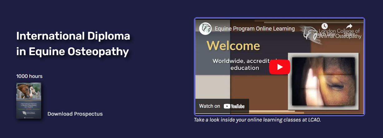
Introducing The International Diploma in Equine Osteopathy

Receive 45-50 % OFF by using the links on this page and clicking the Horse’s Back when you apply!

International leader in animal osteopathic education, the London College of Animal Osteopathy (LCAO), has recently launched one of the highest levels of training currently available outside universities.

The International Diploma in Equine Osteopathy (Int’l DipEO) is led by LCAO founder and renowned osteopath Professor Stuart McGregror, D.O., who shares the knowledge and techniques he has taught internationally since 1998.


OFFER: 40% Off All Programs 

Equine program regular fee: US $3,800  With 40% off: US $2,280

Download Prospectus

Application Form

Unlock another 5-10% discount by mentioning The Horse’s Back when you apply.

The Osteopathic Knowledge You’ll Gain 

It’s the perfect balance of theory and practice

Drawing on 30 years’ experience of teaching equine osteopathy, Stuart will instruct in the following areas, building your capacity as a therapist.

  • Understand the systemic and functional anatomy, physiology, and biomechanics of the horse.
  • Learn how to perform performing a physical examination using observation, palpation skills, and clinical reasoning skills.
  • Be able to deliver an effective treatment using appropriate osteopathic techniques.
  • Recognize indications and contraindications to osteopathic treatment.
  • Understand your role as an osteopath within today’s animal health care model.
  • Understand the osteopathic philosophy and principles as applied to horses.
  • Understand the osteopathic approach to health and disease based on structure-function models.

OFFER: 40% Off All Programs

Download Prospectus

Application Form

Unlock another 5-10% discount by mentioning The Horse’s Back when you apply.


Helping Horses with Equine Osteopathy

How you can become a more effective practitioner

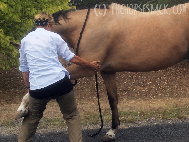
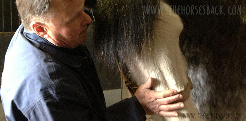
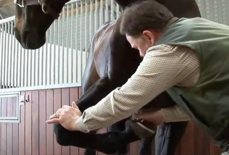
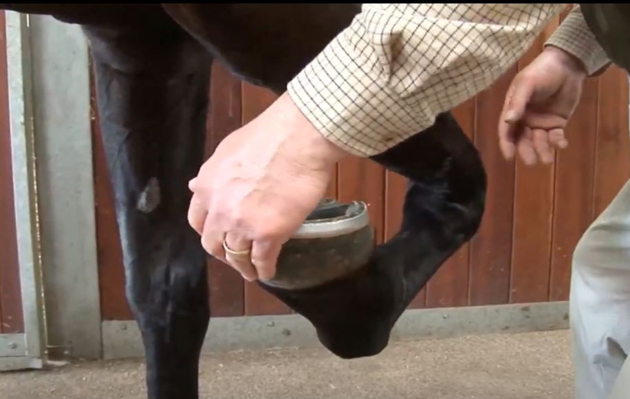
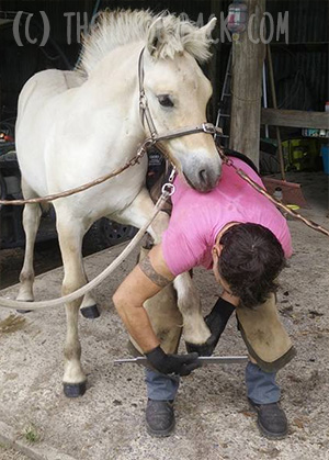
Stuart will teach you a general osteopathic treatment with its roots deep in classical osteopathy. Called Osteopathic Articular Balancing (OAB), it improves the horse’s function by relieving pain, maximizing movement, and increasing performance.

As a gentle manipulation of the whole body, OAB removes tensions and restrictions, restoring health through the assessment and treatment of muscles, joints, tendons, and ligaments.

“One of the main principles in osteopathic medicine is that treatment should restore health to the local tissues. This involves the restoration of blood supply, nerve supply, and lymphatic drainage. Where any of these are absent, the tissues can only be in poor health.” – Stuart McGregor

Download Prospectus

Application Form

Unlock another 5-10% discount by mentioning The Horse’s Back when you apply.


Learn Better Assessment and Treatment of Horses

Treat with more effective skills

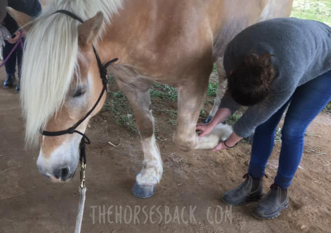
Putting OAB into practice with the horse, you’ll examine the functional anatomy of each joint and its accompanying structures.

You’ll also consider the direct and indirect relationships that extend through the skeletal system, taking in posture, gait assessment, and palpation.

Next, you’ll mobilize associated bones using the ‘functional’ osteopathic techniques taught by Stuart. These are gentle, slow and small controlled movements, as opposed to high velocity, short lever thrusts.

OFFER: 40% Off All Programs

Download Prospectus

Application Form

Unlock another 5-10% discount by mentioning The Horse’s Back when you apply.


Learn a Truly Holistic Approach…

Explore the many factors affecting horses’ bodies

Besides treating the horse’s whole body with equine osteopathy, you’ll learn how to bring other considerations and assessments into your treatment approach. You’ll be learning about:

  • The effect of gravity and rider weight on the vertebral column and pelvis.
  • Pathologies associated with specific sports.
  • The differences in the functions of hindlimbs and forelimbs.
  • A range of congenital and developmental conditions.
  • Various health issues.
  • Incorrect riding.
  • Poorly fitting tack.
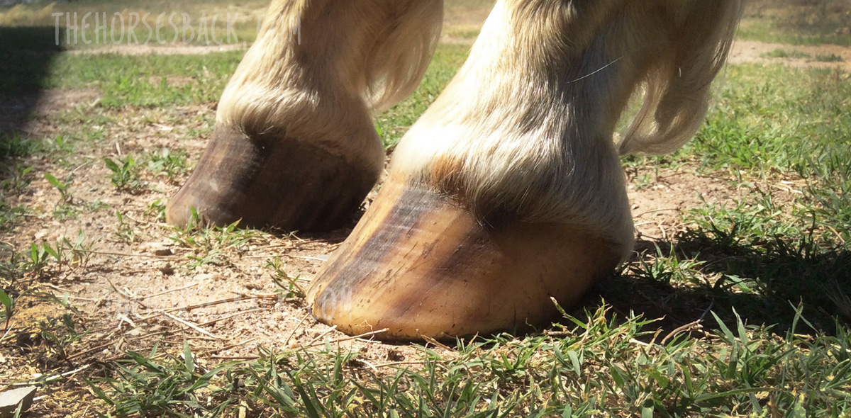
  • Inferior hoof balance.
  • Dental issues.
  • Behavior.


My Personal Experience

I only write posts on this blog about subjects I believe in. I have personal experience of Prof McGregor’s equine osteopathy training, having organised his first course for equine therapists in Australia – this program includes techniques that I use with every horse I see.

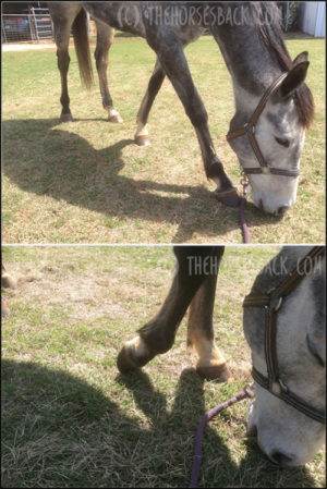
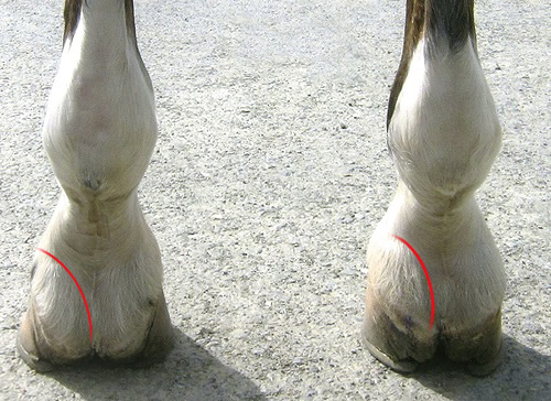
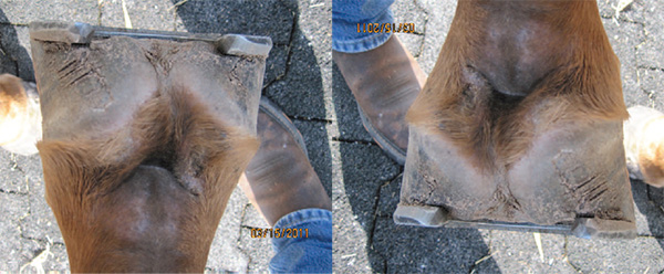
In fact, I used these techniques when first assessing the lateral cartilages explored in this article: Beyond Sidebone: Pastern Pain and the Lateral Cartilage.

Prof McGregor is also very supportive of this blog. For these reasons, we’re offering an extremely generous discount to anyone who books via this page.

These links will save you up to 50% (seriously!), while this blog receives a benefit that helps to cover my considerable writing time and publication costs.


Are You Ready for 1,000 Hours of Equine Osteopathy Training?

It’s fascinating and deeply rewarding!

This training will help you to develop as an effective practitioner with the knowledge and skills to truly help horses.

To qualify, you’ll need to complete at least 1,000 hours of core knowledge and clinical learning. You’ll submit written and video asssessments, and a thesis, and take an elective class in advanced clinical skills – available in Europe, the US or Australia.

As an equine professional, you’ll expand your practice and develop treatment styles relevant to your current practice, through:

  • Pre-clinical studies to build your core knowledge (500 hours).
  • Clinical studies, guiding you through osteopathic research (500 hours).
  • Advanced masterclass, with hands-on clinical training in advanced modalities such as cranial osteopathy, short lever and functional techniques.
  • Individual tutoring sessions.
  • Open group discussions.

OFFER: 40% Off All Programs

Download Prospectus

Application Form

Unlock another 5-10% discount by mentioning The Horse’s Back when you apply.


Education That Suits Your Lifestyle

Benefit from this flexible program

You can study for the International Diploma in Equine Osteopathy in a way that suits you.

Most of the content is delivered through recorded lectures, video tutorials, downloadable presentations, digital textbooks, and additional readings.

This means you can work at your own pace, at times of the day that work for you.

But you’re never alone with your study, for you can constantly connect with your course instructor and student forum.

This means that you can complete in your own time and, best of all, benefit from lifetime access to the online resources.

The LCAO uses a best practice model derived from the academic programs of higher education institutes, with an eLearning platform powered by Moodle software.

Download Prospectus

Application Form


Who Can Apply?

The International Diploma of Equine Osteopathy is open only to approved therapists and animal care professionals

As not everyone can take this qualification, you need to apply to be admitted. The LCAO will consider applications from:

  • DVMs, VMDs, animal physiotherapists and chiropractors
  • Osteopaths and osteoapthic manual therapists (OMTs)
  • Registered MSK practitioners, ie massage therapists, osteopathic manual therapists
  • Veterinary technicians, nurses and assistants
  • Equine bodywork therapists with 7+ years of experience
  • Equine professionals with 7+ years of experience, eg trainers, breeders, farriers, equine massage therapists and bodyworkers, yard managers
  • Graduates and students of animal science degree programs

Download the prospectus to learn more, or apply straightaway.

Download Prospectus

Application Form


More About Prof Stuart McGregor, D.O.

Stuart McGregor has taught hundreds of animal osteopaths over a period of some 30 years.

He graduated from the UK’s European School of Osteopathy in 1984, having completed his dissertation on The Principles of Osteopathy Applied to the Horse. This was the first known work about osteopathy for horses.

He soon began treating horses and dogs, and it was not long before he set up the Osteopathy Centre for Animals in Oxfordshire, England.

Other osteopaths and veterinarians soon came across his work and were keen to learn more, so in 1998 he began to teach. Since that time he has refined his methods and instruction, leading to the training that he practices and teaches today.

“In our treatment, there is something we call ‘intent’. This is where we apply the techniques intending to enable healing. We imagine ourselves inside the tissues being treated and then bring about positive change.” – Stuart McGregor


OFFER: 40% Off All Programs

Equine program regular fee: US $3,800  With 40% off: US $2,280

Download Prospectus

Application Form

Unlock another 5-10% discount by mentioning The Horse’s Back when you apply.


Treat Horses and Dogs with the LCAO

If you’re keen to expand your practice to include both horses and dogs, you may be interested in the Animal Osteopathy qualifications, which address both species.

  • International Diploma in Equine Osteopathy (Int’l DipEO)
  • International Diploma in Animal Osteopathy (Int’l DipAO)
  • International Diploma in Canine Osteopathy (Int’l DipCO)

For licensed veterinarians (DVM/VMD) and final year students of veterinary programs, LCAO also offers the following postgrad programs:

  • Postgraduate Diploma in Equine Osteopathy (PGDipEO)
  • Postgraduate Diploma in Animal Osteopathy (PGDipAO)
  • Postgraduate Diploma in Canine Osteopathy (PGDipCO)


Filed Under: Bodywork Tagged With: diploma in equine osteopathy, equine osteopathy, GA, international diploma in equine osteopathy, london college of animal osteopathy

Beyond Sidebone: Pastern Pain and the Lateral Cartilage

May 31, 2022 by Jane @ THB 4 Comments


It’s a problem of a pressing nature. In some horses with upright, contracted and sheared heels, I’ve seen first hand how the lateral cartilage can painfully impinge the back of the pastern. Sometimes this leads to a gait anomaly and in some horses, this has looked an awful lot like lameness.

Full disclosure here: I’m a bodyworker, not a hoof care practitioner. But everything I’m going to mention I gleaned from the horses’ reactions to focused palpation. My take is that I’m working above the hairline, so I’m OK to offer my observations…

But first, a point about language. Different countries favor different anatomical terms and depending on your part of the world, the lateral cartilages may variously be called collateral, ungual or ungular cartilages.

Anatomy’s just like that. In this article, I’ll be sticking with lateral.

My thanks to go Paige Poss, Anatomy of the Equine, who wrote this interesting article in The Horse about the heel structures of the foot, and to Megan Matters of Hoofmatters for their help discussing this post with me.

Ed. OK, it seems I need to say this: I am not saying this is the only source of caudal hoof pain. There are obviously many and hoofcare is a huge topic. What I’m saying is that there’s a specific location for pain that bodyworkers, and anyone for that matter, can palpate for, and it’s often very useful to know about it, so you can start to do something about it.


What and where are the lateral cartilages?

Palpable edge of lateral cartilage at back of foot (c) Jane Clothier

The lateral cartilages of the hoof are interesting in that they are attached to the pedal bone (third phalanx), extending up and above the hoof capsule.

Here’s an image showing where they can be easily palpated in the living horse. The red line shows the palpable edges, where they rise above the heel bulbs at the back of the foot. Each cartilage then extends about two thirds of the way forwards on either side, still above the coronary band.

Now, here’s a wonderful illustration showing where they’re located in relation the skeletal structures. This shows the short pastern bone, the navicular and the pedal bone.

What’s their function? This I’ll leave to the experts. Here’s a quote from a paper by Sue Dyson and Annamaria Nagy, who in turn reference Prof Robert Bowker in describing how many other structures connect to the cartilages:

The lateral cartilages (c) imaios.com

“The cartilages of the foot are connected to surrounding structures, such as the digital cushion, proximal, middle and distal phalanges and navicular bone by small ligaments… The cartilages of the foot are thought to reduce concussion to structures within the foot (Bowker et al. 1998) and to assist blood flow by compression of the venous plexuses of the foot during loading”. [1]


How high heels can affect the lateral cartilages

Now, I’m coming at this from the direction of one particular issue. To sum it up: when there’s a high heel or mediolateral imbalance in the lower limb and hoof, the lateral cartilage can be pressed against the short pastern bone.

Sometimes, this can be painful. When we press the cartilage, the horse pulls the foot away.

(c) http://www.drkerryridgway.com/

Let’s take a quick look at how the hoof balance and in particular high heels can create these pressure points.

In this next photo, the foot on the right is clearly higher and the heels are more contracted.

As you can see, this has an effect on the position of the lateral cartilages, bringing them higher and closer into the pastern.

Collapsed heels (c) Paige M Poss, Anatomy of the Equine

Now for a dissection image from Paige M Poss showing these structures in a flat hoof, both before and after the skin and hoof capsule are removed.

The cartilages are well away from the bone, because with an undeveloped digital cushion, the heels in this horse had collapsed. As you can see, the cartilages are wide of the pastern here.

Upright, contracted heels (c) Paige M Poss, Anatomy of the Equine

Next, a companion image showing the same structures in a more contracted hoof. The heels are upright and the cartilages are pushed inwards.

There’s also a mediolateral imbalance, with one side being higher and more contracted than the other. As a result, the left cartilage is flatter and compressed inwards towards the bone.

Dissected hoof (c) Solounias, N. Royal Society Open Science. 2018

Now here’s an image of the transverse section of a hoof (ie. sliced vertically, left to right)  from a ppaer by Nikos Solounias.

Here we can see that on the right side, which is the more compressed side, the space between the cartilages – the thick, white upright structures – and the short pastern bone is narrower.

Where this leads us now is towards sheared heels, when there’s a dramatic difference in heel height. When this happens, not only is the heel higher, but the mediolateral imbalance causes narrowing of the pastern joints on that side as well. This tilt increases compression further, while the low side heel may become crushed.

Sheared heel (c) Morrison, S. AAEP Proceedings, 2013

Finally, here’s an image of badly sheared heels in a racehorse by Scott E Morrison, DVM, of Rood and Riddle Equine Hospital. I’ve taken the liberty of rotating it too, so you can see how high that heel is. It’s not a pretty foot.

That’s plenty of description for now. What we need to think about next is what the horse might be feeling.

What are the signs of pain?

In bodywork sessions when addressing the forelimb, I often start at the foot and work up. I rotate the pasterns and mobilise the joints, but before that, I check the lateral cartilages.

Palpation (c) Jane Clothier

As part of this, I raise the foot and press inwards on the peaks of the cartilages, shown as red spots on this image. At this point, some horses will attempt to pull the foot sharply away. That’s what happened with the right side of the foot shown in this image.

I also gently lift the cartilages a couple of millimeters outwards, with my thumbs placed on the palpable edge, shown with the white arrows. Again, some horses will snatch the foot away in response to this. They usually do this in relation to one cartilage only.

It’s a clear sign of pain, particularly if the horse does it a second time when either of these actions is repeated.


What’s actually causing this pain?

There are a few candidates for what’s actually hurting when the upper edge of the cartilage is pressed towards the pastern.

Sidebone (c) Onar, V. 2013

1.  Could it be the start of sidebone, which is the condition when the cartilage begins to ossify? There is some limited evidence that sidebone can be painful, linked primarily to fracturing ossified cartilage or pedal bone. So generally, this is unlikely.

2. Pain could also be caused by an injury to one of the many ligaments attached to the cartilage. It’s possible that tweaking the lateral cartilages outwards might cause a pain response here.

Ligaments attaching to the lateral cartilage (c) https://mink-studios.blogspot.com

3. There’s that palmar digital artery. If the cartilage is pressing against it, then there could be restriction. Might that be uncomfortable or painful? 

4. The region between the lateral cartilage and the pastern bone is also filled with stabilizing ligament. Constant pressure could potentially create lesions here.

5.  There might also be an indirect pressure on the deep digital flexor tendon that runs centrally down the back of the pastern. Might be, but seems less likely in the absence of an obvious lameness suggesting a lesion.

Digital vein, artery and nerve (c) Lancaster & Bowker, 2012 [larger labels added for clarity]
6. Then there’s nerve pain. The areas compressed between the lateral cartilages and bone is where the medial palmar digital nerve and lateral palmar digital nerve course down the inside and outside of the pastern, respectively. These are the major nerves anaesthetized during nerve blocking (abaxial sesamoid nerve block). Then there’s the myriad of branch nerves extending through the foot, sending sensory information back to the central nervous system.

The fact is that we could be looking at one or a combination of these factors when pain is present when we press the upper edge of the cartilage.

This pressure might be creating pressure on the nerve or artery or both.

With potential causes that are so small and focused, it’s impossible to tell without veterinary imaging and diagnosis. As so often, we answer a question with more questions.


Thermal imaging

I recently took this infrared thermal image of the rear of an upright foot.

Infra red thermal image of upright hoof (c) Jane Clothier

Now, there’s a lot to explain about thermal images, for there are many variables that affect the results. But I’m keeping this simple for now by showing just the one photo.

This hoof was already showing a higher thermal signature than the lower hoof (not shown – like I said, keeping this simple).

Note that it’s normal for the concave area at the back of the pastern and hoof to be warmer (although it shouldn’t be warm so far down the hoof).

What I’m wanting to show you is the spot marked with an oval. We have an obviously warmer spot just above the lateral cartilage here.

Granted, this is in an area where there’s vascular activity, as the palmar digital artery and vein are nearby.

(c) Jane Clothier

However, it’s not just that, for the thermal reading is 2 °C (35.6 °F) warmer on this upright hoof than in the same spot on the lower hoof. And yes, I compared them within the same image.

This doesn’t provide evidence of the reason for the increased heat. Inflammation within the hoof capsule is always a possibility, although I’d expect both veins to be affected.

I happen to know that this horse didn’t have an abscess.

However, the horse palpated positive for pain over the left lateral cartilage, which was tight against the pastern.

Given that his reluctance to load the hoof disappeared with the tiniest of micro-trims on that wall, it appears that pressure was a likely cause.


So what do we do?

All we can know is that hoofcare needs to be optimized, to ease pressure in every respect.

While I’m unable to say exactly what hurts, the effect of additional caudal hoof pain is clear.

If compression in this area is painful, descending hills and turning on that hoof is also going to be problematic at times. It could be behind some of the hesitations that can’t be otherwise explained.

Meanwhile, any trim that fails to adequately address heel height, sheared heels or mediolateral imbalance  is going to potentially contribute to this existing painful situation.

Why? Because it feeds into the ‘unloading spiral’.

When pain or discomfort is present, the horse is less inclined to load that foot, particularly at rest. The less loaded foot will then tend towards remaining upright, or may even become more upright, leading to an intensification of the problem.

As I’ve said, I’m not a hoofcare expert. But manifestations like this help me to advise clients when they should be talking to their farriery expert, and if that doesn’t work, maybe switching to another one.

References
    1. Dyson, S. and A. Nagy, Injuries associated with the cartilages of the foot, Equine Veterinary Education, 2011, 23, 581-593. doi.10.1111/J.2042-3292.2011.00260
    2. Solounias Nikos et al. 2018. The evolution and anatomy of the horse manus with an emphasis on digit reduction R. Soc. open sci. 5171782171782 
    3. Scott E Morrison, DVM, AAEP proceedings, vol. 59, 2013
    4. Onar V et al. Byzantine Horse Skeletons of Theodosius Harbour: 1.Paleopathology, Revue de Médecine Vétérinaire, 2012, 163(3):139-146
    5. Lancaster LS, Bowker RM. Acupuncture Points of the Horse’s Distal Thoracic Limb: A Neuroanatomic Approach to the Transposition of Traditional Points. Animals (Basel). 2012 Sep 17;2(3):455-71. doi: 10.3390/ani2030455


Filed Under: Bodywork Tagged With: bare hooves, barefoot horses, caudal hoof, collateral cartilage, GA, lateral cartilage, natural hoofcare, pastern pain, sidebone, ungual cartilage, ungular cartilage

Here’s a Round Up of My Premature and Dysmature Foal Research

May 26, 2022 by Jane @ THB Leave a Comment

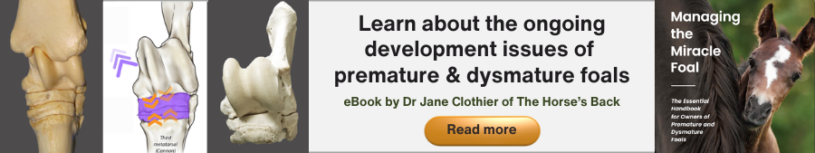
Here are abstracts, downloads and links for my research into the ongoing effects or premature or dysmature birth in horses.

These are the publicly available details of my thesis (full download) and published, peer-reviewed journal articles. The articles aren’t open access, but if you really want to read something, please contact me.

As always, huge thanks are due to the breeder owners who so very kindly allowed me to study their horses, and who provided such valuable images. Together, you’ve helped me to learn a lot and reach initial findings that I now hope to pass on.


Beyond the Miracle Foal: A Study into the Persistent Effects of Gestational Immaturity in Horses 

PhD Thesis, University of New England and CSIRO

Abstract

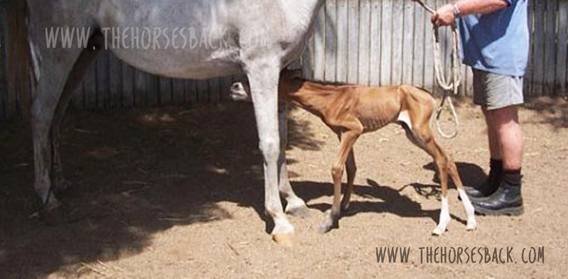
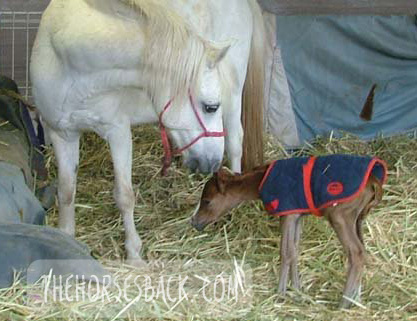
Breeding horses can be a financially and emotionally expensive undertaking, particularly when a foal is born prematurely, or full term but dysmature, showing signs normally associated with prematurity. In humans, a syndrome of gestational immaturity is now emerging, with associated long-term sequelae, including metabolic syndrome, growth abnormalities and behavioural problems.

If a similar syndrome exists in the equine and can be characterised, opportunities for early identification of at-risk individuals emerge, and early intervention strategies can be developed. This thesis explores the persistent effects of gestational immaturity manifest as adrenocortical, orthopaedic and behavioural adaptation in the horse.

Basal diurnal cortisol levels do not differ from healthy, term controls, but when subjected to a low dose ACTH challenge, gestationally immature horses presented a depressed or elevated salivary cortisol response, suggesting bilateral adaptation of the adrenocortical response. This may be reflected in behavioural reactivity, but the outcomes from a startle test were inconclusive.

A survey of horse owners indicated that gestationally immature horses tended to be more aggressive and active than controls, aggression being displayed mostly in families of Arabian horses. Case horses also tended to be more active, intolerant, and untrusting.

Gestationally immature horses have restricted growth distal to the carpal and tarsal joints, and this results in a more ‘rectangular’ conformation in adulthood compared to controls. They also often present with angular limb deformities that adversely affect lying behaviour and recumbent rest. This, however, can be mitigated using analgesic therapy, suggesting chronic discomfort.

Based on these findings, it is reasonable to postulate that a syndrome of gestational immaturity may persist, both clinically and sub-clinically, in affected adult horses. Further work is required to fully characterise this syndrome and validate the outcomes in larger populations, thereby providing a foundation for interventions applicable in the equine breeding industry.

Here is the downloadable doctoral thesis. This is a 236-page PDF.

Clothier, Jane  (author); Brown, Wendy  (supervisor); Small, Alison (supervisor); Hinch, Geoff  (supervisor)


Equine Gestational Length and Location: Is There More That The Research Could Be Telling Us?

Australian Veterinary Journal

Abstract

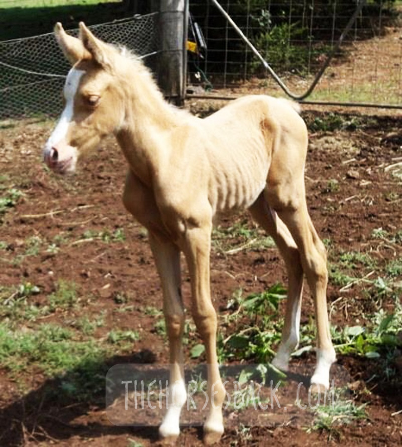
Clear definitions of ‘normal’ equine gestation length (GL) are elusive, with GL being subject to a considerable number of internal and external variables that have confounded interpretation and estimation of GL for over 50 years. Consequently, the mean GL of 340 days first established by Rossdale in 1967 for Thoroughbred horses in northern Europe continues to be the benchmark value referenced by veterinarians, breeders and researchers worldwide. Application of a 95% confidence limit to reported GL range values indicates a possible connection between geographic location and GL.

Improved knowledge of this variable may help in assessing the degree of the neonate’s prematurity and dysmaturity at or soon after birth, and identification of conditions such as incomplete ossification of the carpal and tarsal bones. Associated pathologies such as bone malformation and fracture, angular limb deformity and degenerative joint disease can cause chronic unsoundness, rendering horses unsuitable for athletic purpose and shortening ridden careers.

This review will examine both the factors contributing to GL variation and the published data to determine whether there is potential to refine our understanding of GL by establishing a more accurate and regionally relevant GL range based on a 95% confidence limit. This may benefit both equine industry economics and equine welfare by improving early identification of skeletally immature neonates, so that appropriate intervention may be considered.

The paper can be accessed here.

Clothier, J., Hinch, G., Brown, W. and Small, A. (2017), Equine gestational length and location: is there more that the research could be telling us?. Aust Vet J, 95: 454-461. https://doi.org/10.1111/avj.12653

Using Movement Sensors to Assess Lying Time in Horses With and Without Angular Limb Deformities 

Journal of Equine Veterinary Science

Abstract

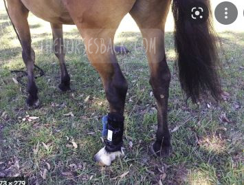
Chronic musculoskeletal pathologies are common in horses, however, identifying related effects can be challenging. This study tested the hypothesis that movement sensors and analgesics could be used in combination to confirm the presence of restrictive pathologies by assessing lying time. Four horses presenting a range of angular limb deformities (ALDs) and four non-affected controls were used.

The study comprised two trials at separate paddock locations. Trial A consisted of a 3-day baseline phase and 2 × 3-day treatment phases, during which two analgesics were administered to two ALD horses and two controls in a standard crossover design. Trial B replicated trial A, except that as no difference between analgesics had been evident in trial A, only one analgesic was tested. Movement sensors were used to measure the horses’ lying time and lying bouts.

In trial A, ALD horses’ basal mean lying time was significantly less than controls (means ± SD for ALD horses 213 ± 1.4 minutes and for controls 408 ± 46.7 minutes, P = .007); with analgesic administration, the difference became nonsignificant. In trial B, ALD horses’ basal mean lying time was also significantly less than controls (ALD horses 179 ± 110.3 minutes; controls 422.5 ± 40.3 minutes, P < .001), again becoming nonsignificant with analgesic administration. Given the increases in ALD horses’ lying time with analgesic administration, it is possible that their shorter basal lying time is associated with musculoskeletal discomfort. Despite the small sample size, movement sensors effectively measured this behavior change, indicating that they could be a useful tool to indirectly assess the impact of chronic musculoskeletal pathologies in horses.

The paper can be accessed here.

Clothier J, Small A, Hinch G, Barwick J, Brown WY. Using Movement Sensors to Assess Lying Time in Horses With and Without Angular Limb Deformities. J Equine Vet Sci. 2019; 75:5559. doi: 10.1016/j.jevs.2019.01.011


Prematurity and Dysmaturity Are Associated With Reduced Height and Shorter Distal Limb Length in Horses 

Journal of Equine Veterinary Science

Abstract

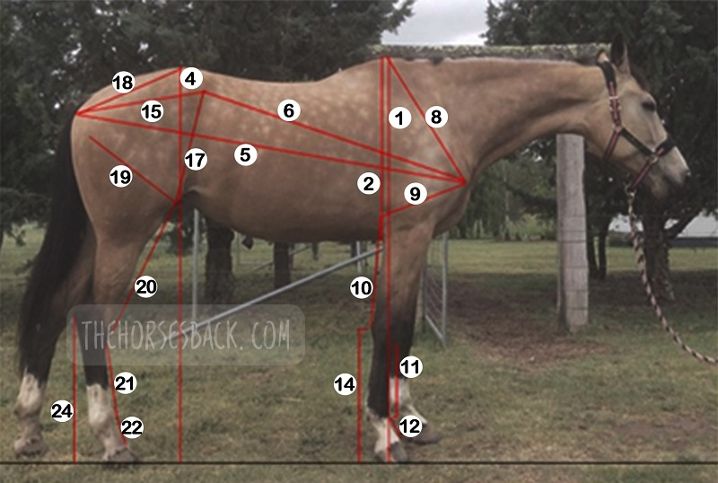
The long-term effects of gestational immaturity in the premature (defined as < 320 days gestation) and dysmature (normal term but showing some signs of prematurity) foal have not been thoroughly investigated. Studies have reported that a high percentage of gestationally immature foals with related orthopedic issues such as incomplete ossification may fail to fulfill their intended athletic purpose, particularly in Thoroughbred racing. In humans, premature birth is associated with shorter stature at maturity and variations in anatomical ratios, linked to alterations in metabolism and timing of physeal closure in the long bones.

We hypothesized that gestational immaturity in horses might similarly be associated with reduced height and different anatomical ratios at maturity. In this preliminary study, the skeletal ratios of horses with a history of gestational immaturity, identified through veterinary and breeder records, were compared with those of unaffected, closely related horses (i.e., sire, dam, sibling).

External measurements were taken from conformation photographs of cases (n = 19) and related horses (n = 28), and these were then combined into indices to evaluate and compare metric properties of conformation. A principal component analysis showed that the first two principal components account for 43.8% of the total conformational variation of the horses’ external features, separating horses with a rectangular conformation (body length > height at the withers), from those that are more square (body length = height at the withers). Varimax rotation of PC1 and analysis of different gestational groups showed a significant effect of gestational immaturity (P = .001), with the premature group being more affected than the dysmature group (P = .009, P = .012). Mean values for the four dominant indices showed that these groups have significantly lower distal limb to body length relationships than controls. The observed differences suggest that gestational immaturity may affect anatomical ratios at maturity, which, in combination with orthopedic issues arising from incomplete ossification, may have a further impact on long-term athletic potential.

The paper can be accessed here.

Clothier J, Small A, Hinch G, Brown WY. Prematurity and Dysmaturity Are Associated With Reduced Height and Shorter Distal Limb Length in Horses. J Equine Vet Sci. 2020 Aug;91:103129. doi: 10.1016/j.jevs.2020.103129. Epub 2020 May 22. PMID: 32684267.


Perinatal Stress in Immature Foals May Lead to Subclinical Adrenocortical Dysregulation in Adult Horses: Pilot Study 

Journal of Equine Veterinary Science

Abstract

The persistent endocrinological effects of perinatal stress due to gestational immaturity in horses are unknown, although effects have been reported in other livestock species. This pilot study tested the hypothesis that persistent adrenocortical dysregulation is present in horses that were gestationally immature at birth by assessing the salivary cortisol response to exogenous ACTH.Case horses (n = 10) were recruited with histories of gestation length < 315 d or dysmaturity observable through neonatal signs. Positive controls (n = 7) and negative controls (n = 5) were recruited where possible from related horses at the same locations.

Cases and positive controls received an intramuscular, low-dose (0.1 ug/kg) of synthetic ACTH (Tetracosactrin 250 mg/mL, Synacthen); negative controls received no ACTH. Saliva samples were collected from all horses at baseline T = 0 and at 30 min intervals post injection from T = 30 to T = 150. These were assayed for salivary cortisol concentration (SCC) using a commercially available ELISA kit (Salimetrics).All baseline values (T = 0) were within normal published ranges. Peak and AUC values (corrected for baseline) for case horses were significantly different (ANOVA P < .001) to positive controls, with either higher (H-cases) or lower (L-cases) SCC values, outside the 95% Confidence Interval of the reference population.

There was no significant effect of breed, age, sex, test month, or location on results. The results suggest that gestational immaturity may lead to subclinical adrenocortical dysregulation, with affected horses presenting an elevated or blunted response to a low-dose ACTH stimulation, despite normal basal levels.

The paper can be accessed here.

Clothier J, Small A, Hinch G, Brown WY. Perinatal Stress in Immature Foals May Lead to Subclinical Adrenocortical Dysregulation in Adult Horses: Pilot Study. J Equine Vet Sci. 2022 Apr;111:103869. doi: 10.1016/j.jevs.2022.103869. Epub 2022 Jan 21. PMID: 35074402.

Filed Under: Bodywork, Foals Tagged With: dysmature foals, equine anatomy, equine bodywork, equine dysmaturity, equine prematurity, GA, horse anatomy, immature foals, Premature foals

Yes, We Can Image for Transitional Vertebrae in Horses

September 23, 2021 by Jane @ THB 7 Comments

It’s been a question of mine for a while. Can diagnostic imaging show the presence of transitional vertebrae?

We’re seeing many bone samples from dissections, as shown in my previous article on transitional vertebrae.

But if we’re to help our horses that live with this issue, we need to identify it before they’re dead. (Yes, right?!)

Allow me to introduce a practicing vet and educator who is doing just that.


Imaging for Transitional Vertebrae 

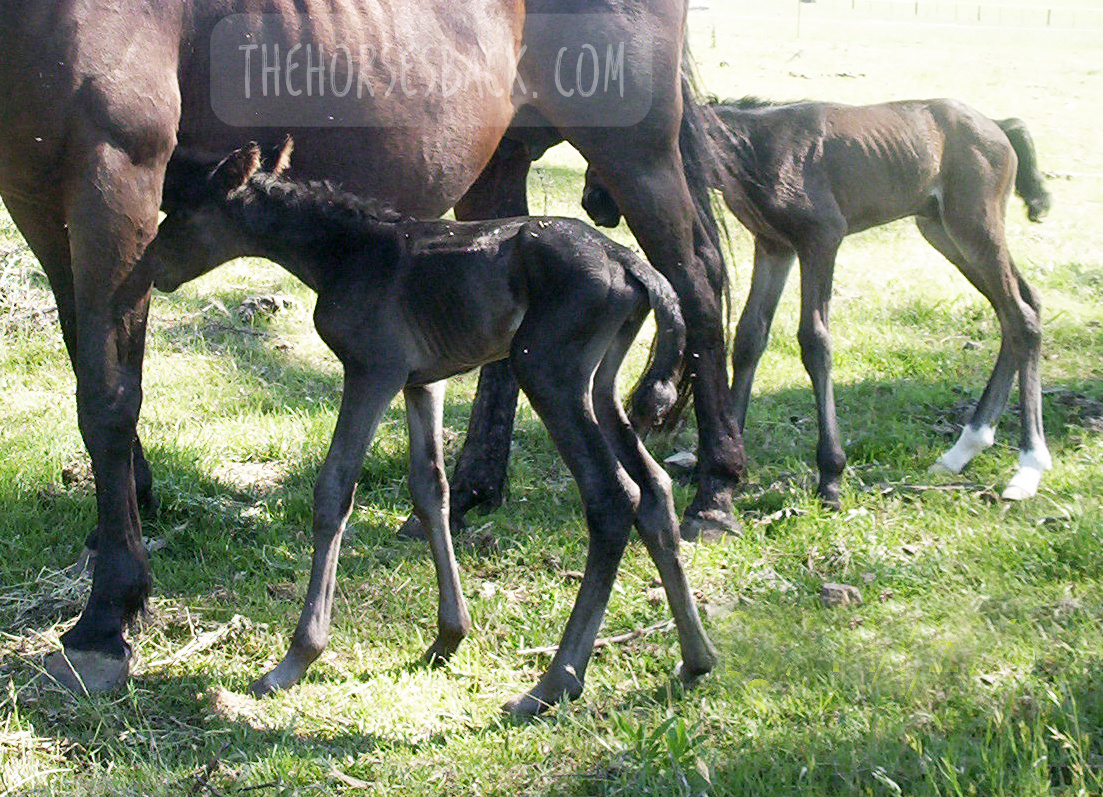
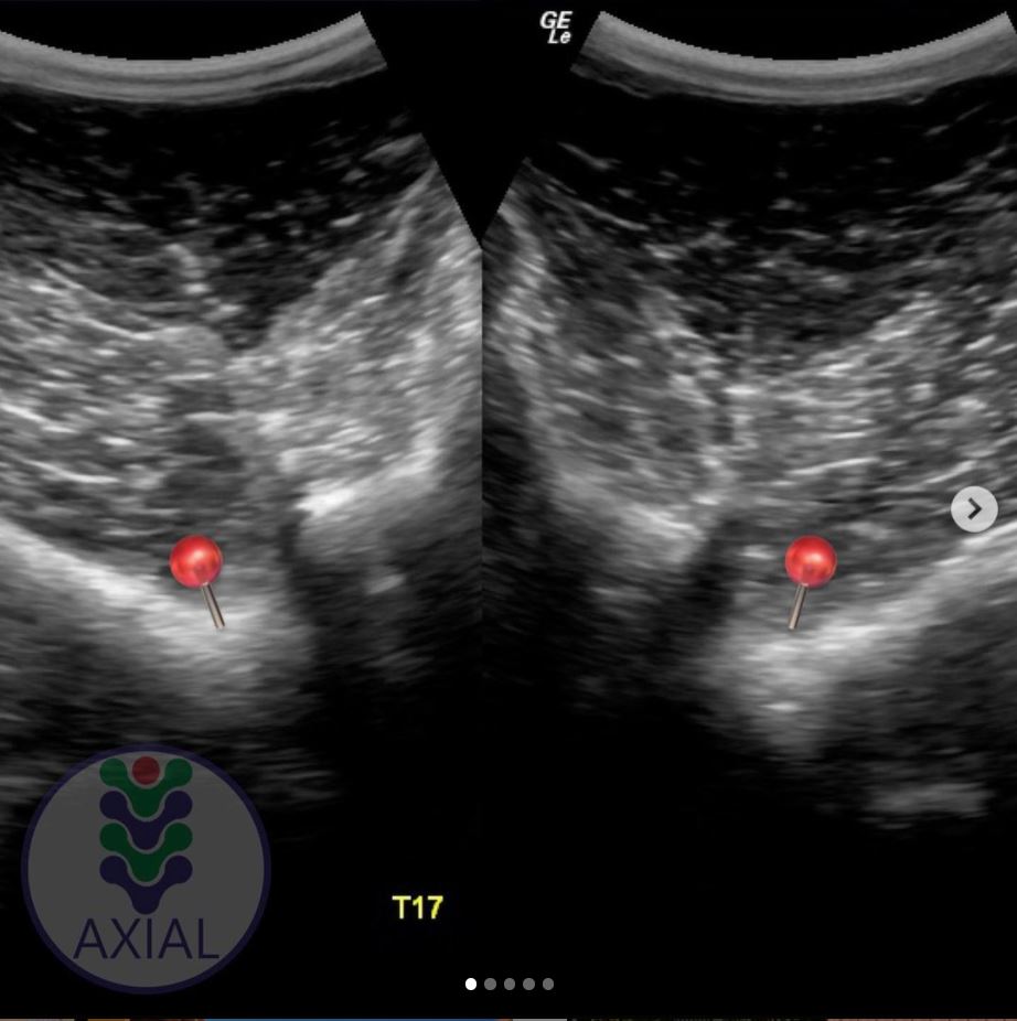
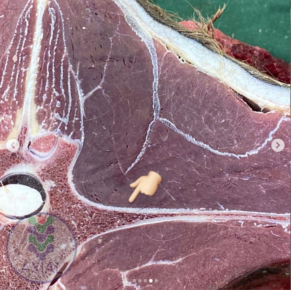
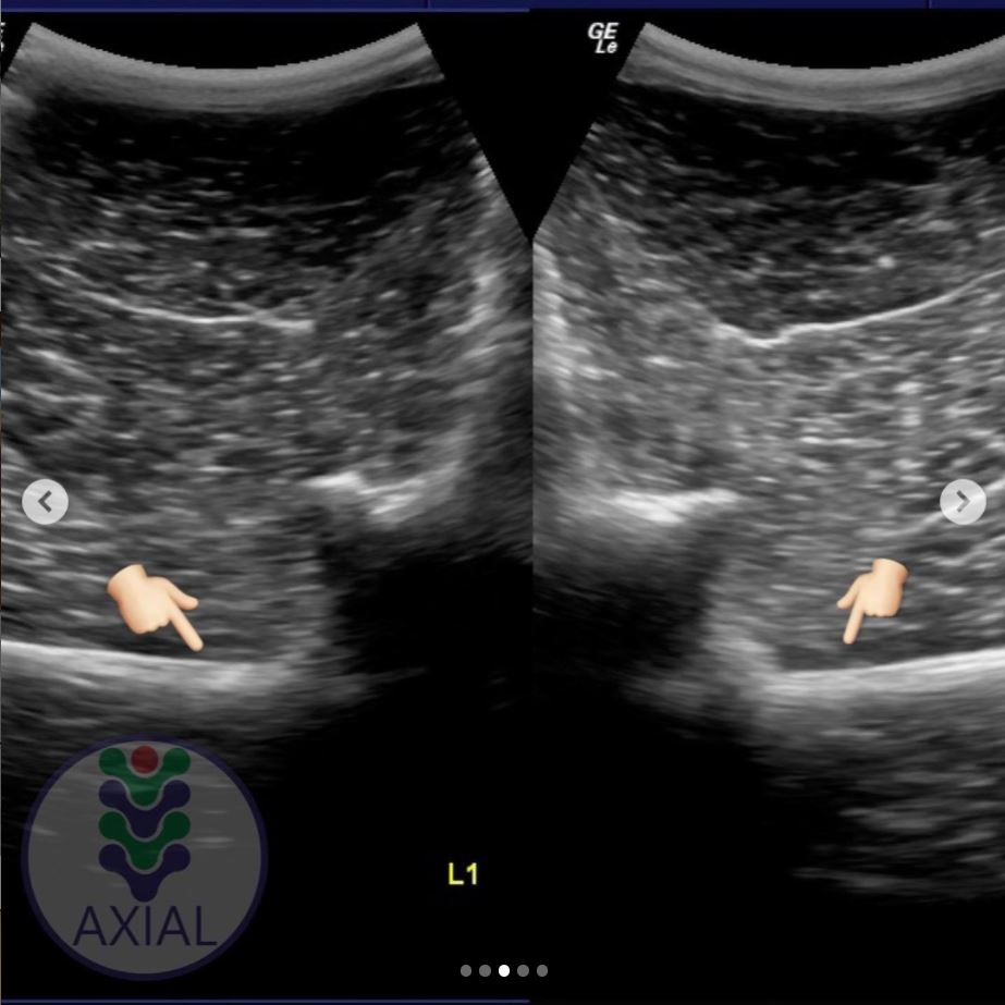
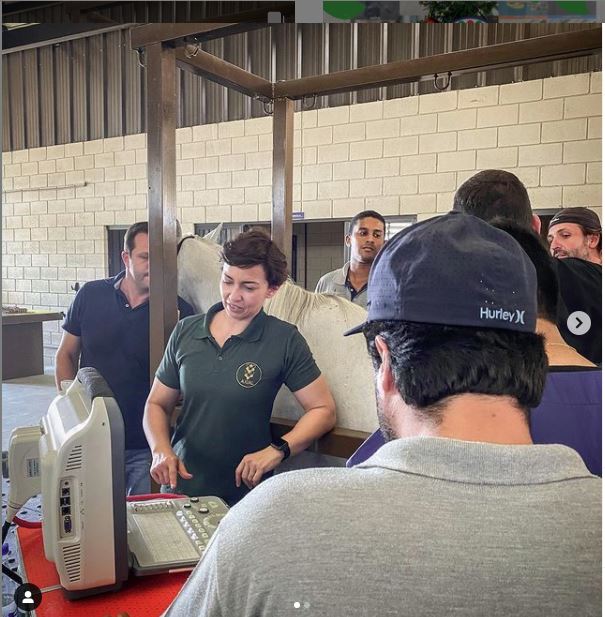
Meet Dr Brunna Fonseca, Associate Professor, educator and specialist in equine orthopedics, focusing on the spine and nervous system. She’s based in São Paulo, Brazil.

I’ve been following her Instagram for a while, because she posts brilliant videos and photos explaining what she does, and how, and why.

I was delighted to see a recent post on imaging for a transitional vertebra, which included fantastic visuals. Such a great communicator!

Dr Brunna has kindly given me permission to repost her images and descriptions here. So without further ado…

  • All images copyright of Axial Vet

Ultrasonograms

Ultrasonography for transitional vertebrae
Angle of transducer. Image: Equine Neck and Back Pathology: Diagnosis and Treatment, 2nd Edn. Ed. Frances M.D. Henson. © 2018 John Wiley & Sons, Ltd.

The following ultronographic images are each a composite of two images, one showing the left side and the other the right.

This textbook illustration helps to show the angle the image is taken at. This angle is usually used for imaging the articular facets of the vertebrae.

Additionally, the image at the top of this article shows a transitional vertebra at T18, like the mare being diagnosed by Dr Brunna.


1.  Can we recognise transitional vertebrae?

The first image shows two sides of a mare’s body. The hand icon gives us a strong hint of where to look… This appearance is very similar to that of the TB mare in my previous post.

Dr Brunna writes, “This mare has the T18 transitional vertebra, presenting a transverse process similar to the lumbar vertebrae on the right side, which causes the appearance of the horse to have the most visible rib on that side.

The occurrence of transactional vertebrae in the horse is not uncommon, especially in the thoracolumbar transition, which can occur in T18 or L1.”


2. Section of a thoracic vertrebra

This image is from a different horse showing a normal rib head and its joint with the vertebra.

Dr Brunna writes, “This is the image of a thoracic vertebra, showing the costotransverse joint.”


3. Image of a normal vertebra

Dr Brunna writes, “This is a T17 ultrasound image, where we can see the image of the normal costotransverse joints.”

This is the bay mare again.

As with the previous cross section, the red pins which show the facet joint between rib head and vertebra.


4. Section of a lumbar vertebra

This is cross section is of a normal lumbar vertebra from a different horse.

As you can see,  there is no joint between the  transverse process and the vertebral body.

The process is wide and flat, and integral to the vertebra.


5. Image of a lumbar vertebra

Here’s an ultrasound of the first lumbar vertebra (L1) in the bay mare.

As in the above cross section (picture 4), there is no joint between the transverse processes and the vertebral body.

We now have ultrasound images of the normal T17 and normal L1. As we will see, the transitional vertebra mixes elements from both.


6. Imaging transitional vertebrae

“This is an ultrasound image of T18, where we can see the image of the costotransverse joint on the left side (red pin) and image of the transverse process on the right side.”

So here’s the underlying skeletal issue in the bay mare.

The left side is a normal joint, being the same as the T17 thoracic vertebra (picture 3).

The right side is similar to the previous image of the lumbar vertebra (picture 5).

It is not identical, for while the process-like rib is joined to the vertebra, it is not the same shape and does not lie as flat as the lumbar process.


Want to Hear More From Dr Brunna Fonesca?

You can follow her Axial Vet Instagram page to see examples of her equine cases and their assessment, in images and videos.

An increasing number of captions are now translated into English.


Filed Under: Bodywork Tagged With: Anatomy, equine anatomy, equine bodywork, equine malformation, equine skeleton, GA, horse anatomy, transitional vertebra, transitional vertebrae

10 Rehab Tips for Horses with High-Low or Upright Hooves

January 1, 2021 by Jane @ THB 9 Comments


Sorting out that high-low hooves situation. It’s the farrier’s job, isn’t it?

Well yes, they’re clearly the primary professional. But there are plenty of things you can also do to help.

As their horse’s bodyworker, I’m often the first to tell an owner that unbalanced forefeet are causing problems right through the body.

Related issues can include ringbone, carpal arthritis, shoulder asymmetry, base of neck arthritis, atlas rotation, TMJ issues, spinal rotation, scoliosis and even sacroiliac dysfunction – all depending on the severity and duration of the hoof issue.

That’s not me being dramatic. In an older horse, that’s absolutely the type of problems I can find when there’s a long term high hoof.

And don’t forget, there are the saddle fit issues that go along with all of that.

Here, in plain speak, is the list of rehab tips that I offer my clients, so that they can help their farrier to help their horse.


© All text copyright of the author, Jane Clothier, https://thehorsesback.com. No reproduction of partial or entire text without permission. Sharing the link back to this page is fine. Please contact me for more information. Thank you! 


Before We Start: Choose Your Hoofcare Professional Well

It’s obvious, but this is essential.

Hoofcare professional
No problems here! Megan Matters trims 3.5 month-old Fjord, Lodur (c) Karen Groneng, Greenfield’s Stud

Engage a hoof professional who walks the walk as well as talking the talk.

This is someone who can tell you about hoof function and how the ‘normal’ hoof works during loading and movement.

This is someone who frequently updates their professional training.

Professionals who take continuing education are informed about current research into hoof function, as well as methods to address problems.

They know a lot more about current best practice and are more likely to demonstrate it, too.

This most definitely isn’t someone who relies solely on the apprenticeship they completed many years ago.

How it helps:

Skilled hoofcare professionals will aim to achieve a similar height in the two front hooves, even though the hoof angles may be different when viewed from the side.

They will trim each hoof according to its underlying structure, making corrections where needed.

They will NOT simply try to create similar angles and toe length, which creates stresses in a high hoof, and sets your horse up for numerous hoof and body problems.

With a skilled hoofcare practitioner on board, there is plenty more you can do to help your horse through this process of change. The more effort you put in, the more your horse will improve.


1. Use Variable Feeding Positions

Variable feeding positions were first described by Sharon May-Davis, who recognised their value as a form of passive physio.

Haynet at poll height
(c) Jane Clothier, equinehealthworks.com

This one is easy: hang a haynet at poll height to replicate eating from trees. This encourages the horse to stand square while eating.

You’ll gain the best results from small hole nets, as your horse will twist her head from left to right while eating. She’ll also drop down to eat hay from the ground.

Doing so activates the deep muscles beneath the neck vertebrae. It also activates the muscles beneath both shoulder blades as she shifts her weight one foot to the other.

If you feed more than one net a day, place the other at chest height.

How it helps:

Think thoracic sling and freeing up the restrictions that have arisen from stabilising the weight over different height limbs, as well as adopting a ‘scissor’ position to eat (which will have contributed to the problem in the first place).


2. Introduce Different Surfaces

The hoof balance is changing, but there may still be asymmetries in your horse’s body that are slow to shift.

Horses on varied footings
All horses seem to appreciate the stimulation of new surfaces during hoof rehab for a variety of issues. (c) Tanja Kraus Horsemanship

Standing your horse on different and unfamiliar surfaces can make the body’s self-adjustments happen quicker.

If you’re able to put some down, gravel provides wonderful under-hoof stimulation. Sand too. Your horse can tilt his hooves toe-down, heel-down or side-down, as he wishes.

Interlocking mats are also effective and can be used anywhere, as can commercially made physio mats and pads.

Watch your horse become curious, start relaxing and yawning, and you know that body adjustments are happening from within.

How it helps:

It can take the nervous system a while to wake up and catch up with what’s new.

The horse has sensory nerves in the feet, and new surfaces provide different proprioceptive.

It’s not just the hoof – changes in balance are registered from the many neuroreceptors of the lower leg.

As the parasympathetic nervous system (‘rest and repair’) is activated, musles are allowed to relax and reset.


3. Feed on a Slope

Again, think thoracic sling. The horse must open up the spine at the cervico-thoracic junction (base of neck).

Feeding on a slope
(c) Jane Clothier, equinehealthworks.com

It’s also next to impossible to adopt a scissor position (the foreleg grazing stance, see no. 7 below) when eating on a slope.

How it helps:

Your horse is encouraged to load into the front hooves equally, left and right.

Finally, the soft tissue structures at the back of the leg will be gently stretched on the limb with the upright hoof.


4. Rock the Withers

Standing alongside your horse when she is standing square, gently rock the withers from side to side.

Rocking the withers
(c) Jane Clothier, equinehealthworks.com

Hold one or two wither processes at a time (feel for the ‘buttons’) and swing gently from one side to another.

This makes the horse load into one forefoot, then back into the other.

Do this from both sides to ensure equal work, as most of us are either stronger pullers or stronger pushers.

How it helps:

The horse is being asked to mobilise between the shoulder blades. When there’s restriction, she’ll raise a hoof on the swing away from that leg.

Easing any restriction will help with relaxing the shoulder on the high side and allowing the joint angles to open out.

She may dislike the exercise at first if it’s uncomfortable, but it will improve over time. You are also gently mobilising the spine deep to the shoulders.


5. Massage the Neck Muscles

When there’s a high foot, the restriction and reduced movement on that side (the stride and therefore leg swing is often shorter) will transfer up through the shoulders to the neck.

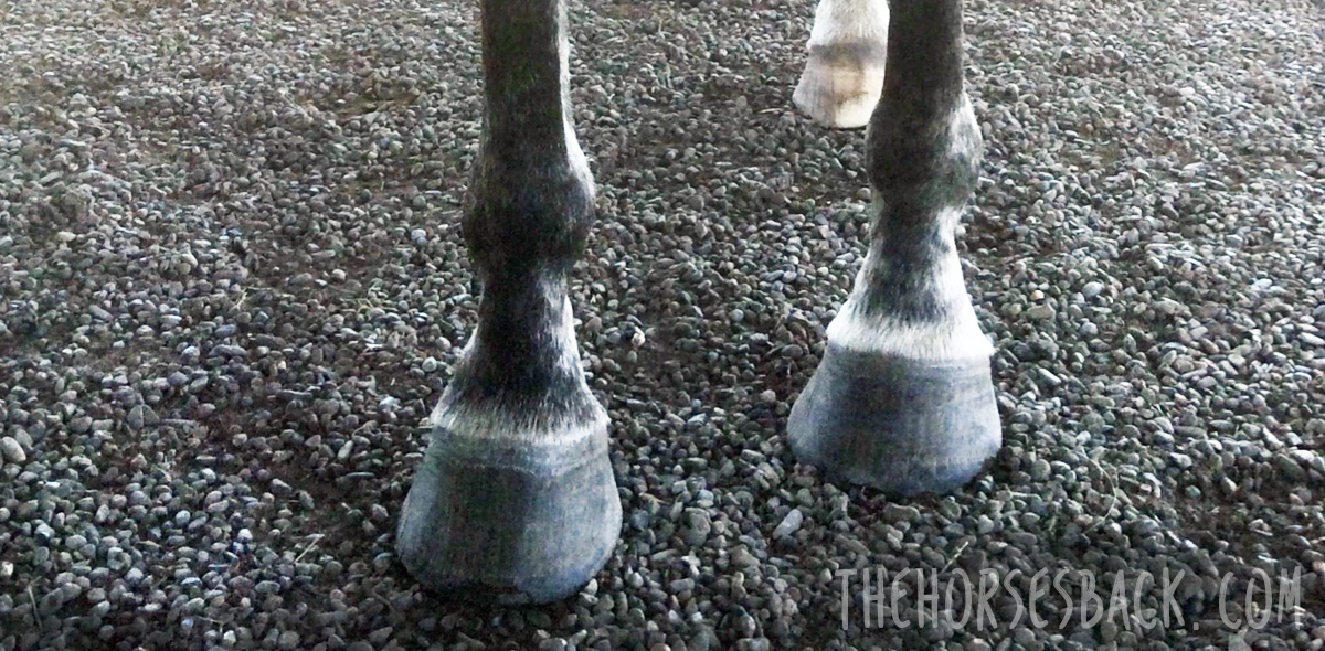
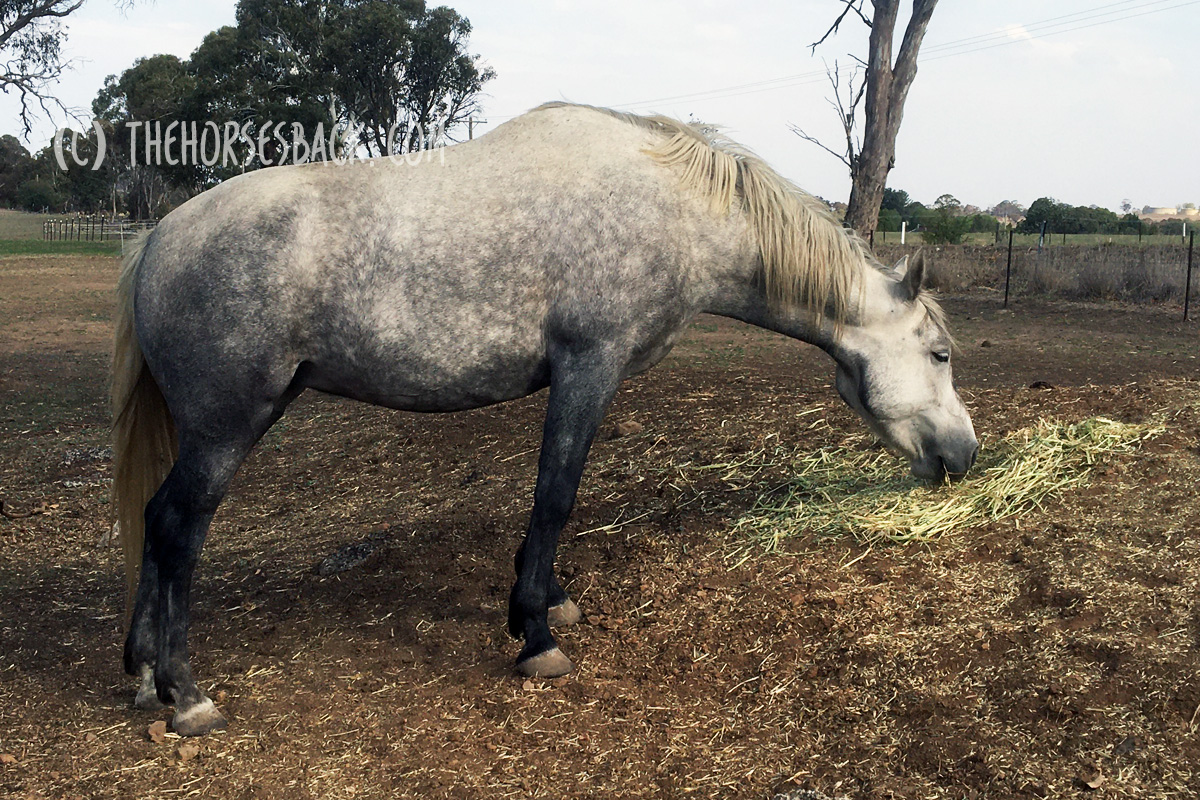
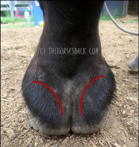
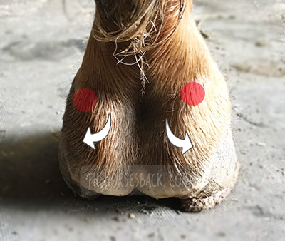
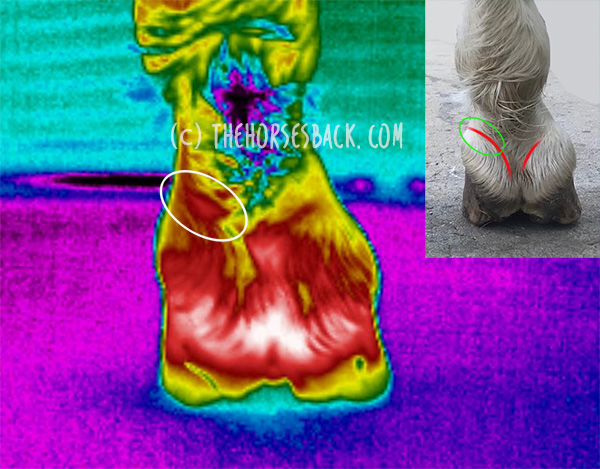
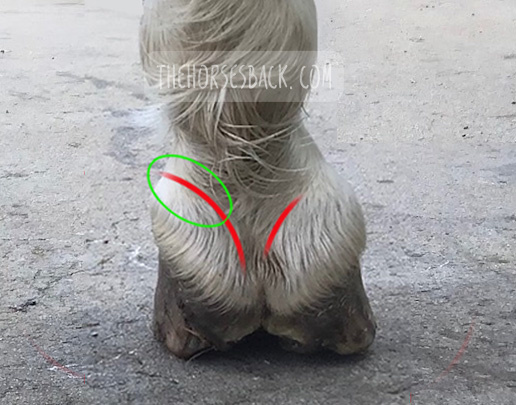
Tension also builds because the horse is trying – and failing – to balance their weight centrally.

This is a secondary effect, caused by the horse’s changed posture.

Pay particular attention to the fine muscles behind the poll, which will be imbalanced.

How it helps:

Releasing muscular tension will help the horse to move to a more symmetrical posture through training and postural improvement.


6. Good Old Carrot Stretches 

Use pieces of carrot to encourage stretches to either side.

This will help your horse to become more familiar with loading into both forehooves to a more equal degree.

Lateral carrot stretch
(c) Jane Clothier, equinehealthworks.com

There are many variations on these stretches and everyone has an opinion!

If you try to ensure that your horse is standing square in front, these stretches will help, whatever the exact approach.

How it helps:

The horse is stretching out the shoulder and neck muscles, including those beneath the shoulder blade, which are going to be more restricted on one side than the other.

This action will also stimulate and activate the deep spinal muscles, bring fresh ‘body awareness’ to the region (ie, proprioception).


7. In-Hand Grazing Tricks

Does your horse have an obvious ‘scissor’ grazing stance, as in the first photo (right)?

Grazing stance
Change is difficult at first! (c) Jane Clothier, equinehealthworks.com

If you horse’s high hoof is always the one at the back, then there is more you can do.

Try to spend up to 20 minutes a day feeding your horse in-hand.

Walk with her and let her graze, but ONLY allow her to do so when the upright foot is placed either level with the lower foot, or further ahead.

This means lots of stepping forward, stopping, stepping again, until she’s stood as you’d like.

This will be hard for your horse at first, as you can see here, but should become easier over time.

How it helps:

As grazing positions became long term, they also familiar and are adopted habitually. It’s like folding our arms or crossing our legs in one direction.

As the hooves become balanced, the horse will continue adopting the ‘old’ position.

This is a way of reprogramming your horse as the hooves start to become more balanced.


8. Daily Leg Stretches 

This is where little and often really helps. Perform gentle leg stretches forward.

Don’t haul the leg, but allow your horse to take up the last inch or so herself.

Foreleg stretch
(c) Jane Clothier, equinehealthworks.com

If there’s a lot of tension, go very gently and don’t stretch to the limit.

Let the cannon hang vertically from the knee, so you’re only extending the upper leg.

A hand behind the elbow will help and your encourage your horse to ‘pop’ the last part of the stretch.

Don’t force it. It’s not about physically lengthening muscles: you’re allowing signals to reach the brachial plexus (nerve centre behind the shoulder blade).

Watch out for your horse looking down and touching his own knee as you start doing this, almost as if he’s surprised to see it there.

How it helps:

This is far more than a muscle stretch! You are influencing the horse’s nervous system’s awareness of the forelimb’s joints and muscles, and the leg’s position in relation to the body and then the ground.

This is proprioception and it will benefit from some help in resetting itself at shoulder joint level.

This ‘resets’ the leg and improves awereness around various joints’ range of motion.


9. Joint Mobilization

The hoof height is changing and becoming more equalized, meaning there’ll be changes higher up the legs as well.

(c) Jane Clothier, equinehealthworks.com

The high hoof side will have to open out a bit, while the low hoof side will be slightly less extended than before.

This involves the soft tissue structures around joints, particularly those of the fetlock and pasterns.

Do you ever see horses shaking their hooves or rotating their lower limb? No.

Your horse is likely to love you for gently rotating and flexing the lower joints, allowing structures to move in different ways, where previously they have been limited.

Pasterns above an upright side are likely to feel stuck. Above an underrun hoof, they will feel loose.

How it helps:

Mobilizing the joints through gentle rotation helps to restore range of motion where it has been restricted.

Spaces will be opened up that allow fluid to circulate, lubricating the joint and bringing more freedom to tendon movement.

This all helps the horse to stand above the changed hoof, positioning the leg closer to where it needs to be.


10. Work Over Poles

Many horses with long toes have a tendency to trip. Once again, as the hoof balance changes, they need to adjust their sense of ‘where their feet are’.

Top: Ground poles (c) FEI.org Bottom: Extending the exercises to the trail (c) Tanja Kraus Horsemanship

Working over poles helps your horse to focus on individual hoof placement, in terms of both stride length and height of motion.

This can be done over ground and raised poles, or over logs on the trail.

For stronger input, back your horse over poles.

Lead him over with one fore foot, then back with that foot. Lead him over with both forefeet, then back with both. And so on, until the horse knows how to navigate during back-up with all four feet.

How it helps:

Once again, it’s about the proprioceptive input.

As well as ‘equalizing’ your horse’s awareness of its feet, you’re correcting for any lasting effects of pain.

Pain in one hoof and the joints above it has a negative effect on proprioception. Same if two hooves are affected by pain.

By doing this kind of work, you’re helping your horse’s nervous system to ‘square up’, along with the feet.


There’s more – there’s always more. For example, make sure that nutrition is adequately providing the building blocks for hoof growth, development and strength.

As you get going, you’ll find that these are not so much short term changes for rehabilitation, but part of a move towards a more proactiveway of managing your horse.

(c) Jane Clothier, thehorsesback.com


Filed Under: Bodywork Tagged With: bare hoofcare, bare hooves, barefoot horses, club foot, farrier, farriery, feeding positions, GA, hay high, high-low hoof, hoof trimming, natural hoofcare, upright hoof, variable feeding positions

  • « Go to Previous Page
  • Page 1
  • Page 2
  • Page 3
  • Page 4
  • Page 5
  • Interim pages omitted …
  • Page 9
  • Go to Next Page »

Footer

Contact Me

Send your questions this way…

Get in Touch

  • Store Support
  • Shipping & Returns
  • Contact

Copyright © 2025 · Parallax Pro on Genesis Framework · WordPress · Log in

Red light / near infrared devices now in store! Dismiss