• Skip to main content
  • Skip to footer

The Horses Back

Body Talk for Thinking Owners

  • Home
  • Articles
  • BEMER
  • Store
    • Store
    • Shipping
    • Returns
    • Support
  • Equine Healthworks
    • Bodywork for Horses
    • Reviews
    • Leave a Review
  • About & Contact
    • About
    • Contact

equine bodywork

10 Tips for the Risk-Free Use of Equine Massage Guns

February 10, 2023 by Jane @ THB Leave a Comment

Wait – you didn’t know there could be risks to your horse when using an equine massage gun? Then you’d best read on.

First, there are certainly benefits to  massage guns. They’re easy to use and time friendly when you’re time-poor.

They retail to suit every budget, and there’s no difference between ‘human’ and ‘horse’ devices.

They can help with pre-work warm-up and post-work relaxation, and may help to reduce post-work soreness.

But, but, but – you do need to switch on your mind at the same time as your gun.

And here’s why.

Note: this page includes some links. At no extra cost to you, if you make a purchase, these links may make a small contribution towards the costs of running this site, including my extensive writing time.

1.  Check the horse with your hands first.

This way, you can see if there are any adverse reactions to touching in certain areas. It’s better to know in advance, rather discover a problem with a device that isn’t giving you feedback.

Remember, these are designed primarily to be used by humans on themselves – humans know where their own spots are, or can tell you, but horses rely on you to be cautious.

2.  Vibration is fantastic for myofascia. 

Hydragun with 7 attachments, including 3 flat heads.

Soft tissue generally doesn’t need to be hammered.

Hint 1: using the side of the round massage head will achieve more vibration than percussion.

Hint 2: the less intrusive flat head used gently and flat to the surface also creates more vibration.

Please don’t use the hard pointy applicators, which are so much more likely to hit directly into a painful spot, or worse, penetrate an area of torn fascia.

3.  Go lightly and keep it moving. 

You’re not mashing potatoes here – this is a sensitive body. By minimizing pressure, you’re allowing more of a vibrational effect to happen.

You also don’t risk creating bruises (as can happen in humans, apparently). Your horse can’t tell you until it has already happened.

Don’t work closely over bone – and remember that older horses may have arthritic joints that don’t benefit from heavy percussion.

4.  Little and often is better. 

The Mini Atom is travel-sized with USB-C charging

Too much work starts to cancel out the good work that’s already been achieved through a light approach.

Massage guns are best used on small, targeted areas in short bursts.

And a practical conern is that you don’t want to overheat the device – many have an automatic shut-off after 10-15 minutes for this reason.

5.  Check your horse’s responses.

If your horse is showing any signs of tension, stop. The whole point here is relaxation. Tense muscles + percussion = high impact => pain.

If your horse starts leaning into it and groovin’, then fine, turn it up – but only if they say so.

It’s not a goal in itself (question why they’re so tense in that spot – maybe change something in training?). If there’s any flicker of discomfort in their eye, stop right now.

The Mini Atom Gun enables a focused, smooth and effective approach.

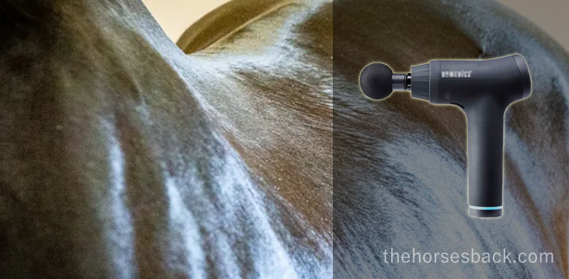
6.  Don’t go higher than the base of neck.

Think about it: you’ll be vibrating the skull. Try that on yourself if you’re not sure why it might be unpleasant!

Lots of horses have tension and pain around the poll and the temporomandibular joint (TMJ). It’s always better to do the upper neck with your hands and a whole lot of love.

7.  Ask why some muscles are always sore.

There may be muscles that are always sore, particularly as your horse steps up the work.

Maybe that spot would benefit from a gentle hand rub?

And maybe slow down the work pace down as well, if tension keeps developing?

Don’t be the Cozy Powell of massage gun users – think prevention and therapy, not heavy rock!

8.  Steer well clear of injuries.

If your horse is injured, don’t try to help with your massage gun. Doing so could likely disrupt the healing process.

Wait until the healing process is well under way and then start really lightly and only in adjacent areas. This is the best time to think about vibration rather than percussion.

9.  Take note of fresh pain responses.

If your horse is reactive in an area that was fine yesterday or last time you worked, stop. You may be right above an injury.

Worse, if you’ve been using too much pressure, you may have intensified an issue.

10.  Don’t forget nutrition.

If muscles are always super-tight in spite of careful conditioning and therapeutic work, there may be an imbalance in your horse’s nutritional and mineral intake that’s having a metabolic effect.


Interested in equine massage guns?

How do you know which massage guns for horses present really good options?

I don’t believe in buying on the basis of a label alone, but at the same time, demonstrated reliability is a good thing.

And don’t worry about whether it’s called an equine massage gun or not. They’re all the same technologies, whether marketed for human or horse use.

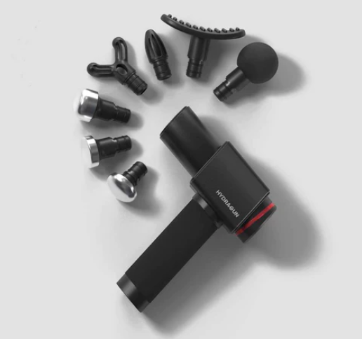
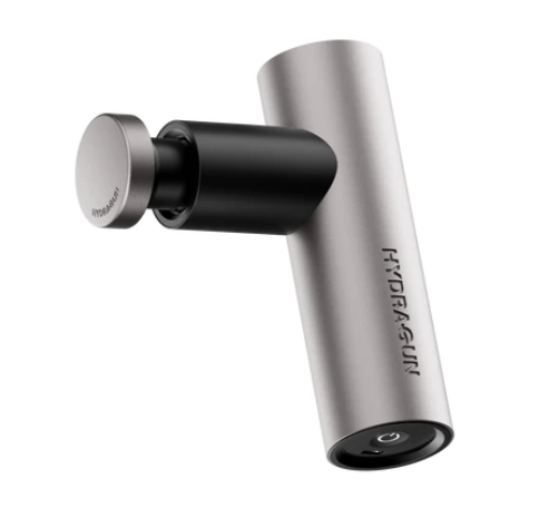
Learn more about the Hydragun’s massage gun and the Atom Mini Massager in The Horse’s Back Store. Renowned for being the quietest devices on the market, these excellent, durable devices are built with smoothly operating components within high-grade aluminimum casing. For the equine bodyworker, they offer a reliable solution that is so lightweight it won’t tax your body.


Filed Under: Bodywork Tagged With: best massage guns for horses, equine bodywork, equine massage, equine massage guns, equine sacroiliac, GA, homedics massage guns, massage guns for horses

Here’s a Round Up of My Premature and Dysmature Foal Research

May 26, 2022 by Jane @ THB Leave a Comment

Here are abstracts, downloads and links for my research into the ongoing effects or premature or dysmature birth in horses.

These are the publicly available details of my thesis (full download) and published, peer-reviewed journal articles. The articles aren’t open access, but if you really want to read something, please contact me.

As always, huge thanks are due to the breeder owners who so very kindly allowed me to study their horses, and who provided such valuable images. Together, you’ve helped me to learn a lot and reach initial findings that I now hope to pass on.

Beyond the Miracle Foal: A Study into the Persistent Effects of Gestational Immaturity in Horses 

PhD Thesis, University of New England and CSIRO

Abstract

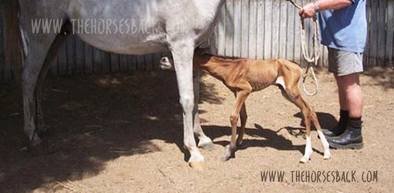
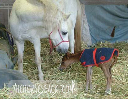
Breeding horses can be a financially and emotionally expensive undertaking, particularly when a foal is born prematurely, or full term but dysmature, showing signs normally associated with prematurity. In humans, a syndrome of gestational immaturity is now emerging, with associated long-term sequelae, including metabolic syndrome, growth abnormalities and behavioural problems.

If a similar syndrome exists in the equine and can be characterised, opportunities for early identification of at-risk individuals emerge, and early intervention strategies can be developed. This thesis explores the persistent effects of gestational immaturity manifest as adrenocortical, orthopaedic and behavioural adaptation in the horse.

Basal diurnal cortisol levels do not differ from healthy, term controls, but when subjected to a low dose ACTH challenge, gestationally immature horses presented a depressed or elevated salivary cortisol response, suggesting bilateral adaptation of the adrenocortical response. This may be reflected in behavioural reactivity, but the outcomes from a startle test were inconclusive.

A survey of horse owners indicated that gestationally immature horses tended to be more aggressive and active than controls, aggression being displayed mostly in families of Arabian horses. Case horses also tended to be more active, intolerant, and untrusting.

Gestationally immature horses have restricted growth distal to the carpal and tarsal joints, and this results in a more ‘rectangular’ conformation in adulthood compared to controls. They also often present with angular limb deformities that adversely affect lying behaviour and recumbent rest. This, however, can be mitigated using analgesic therapy, suggesting chronic discomfort.

Based on these findings, it is reasonable to postulate that a syndrome of gestational immaturity may persist, both clinically and sub-clinically, in affected adult horses. Further work is required to fully characterise this syndrome and validate the outcomes in larger populations, thereby providing a foundation for interventions applicable in the equine breeding industry.

Here is the downloadable doctoral thesis. This is a 236-page PDF.

Clothier, Jane  (author); Brown, Wendy  (supervisor); Small, Alison (supervisor); Hinch, Geoff  (supervisor)


Equine Gestational Length and Location: Is There More That The Research Could Be Telling Us?

Australian Veterinary Journal

Abstract

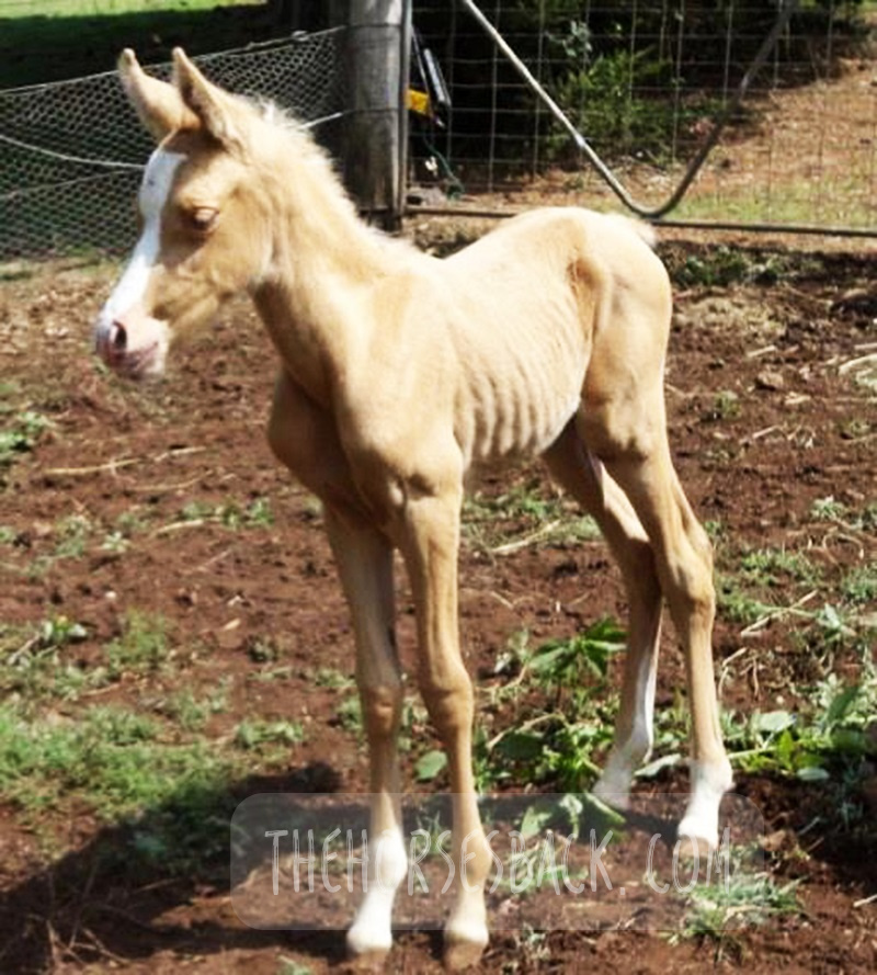
Clear definitions of ‘normal’ equine gestation length (GL) are elusive, with GL being subject to a considerable number of internal and external variables that have confounded interpretation and estimation of GL for over 50 years. Consequently, the mean GL of 340 days first established by Rossdale in 1967 for Thoroughbred horses in northern Europe continues to be the benchmark value referenced by veterinarians, breeders and researchers worldwide. Application of a 95% confidence limit to reported GL range values indicates a possible connection between geographic location and GL.

Improved knowledge of this variable may help in assessing the degree of the neonate’s prematurity and dysmaturity at or soon after birth, and identification of conditions such as incomplete ossification of the carpal and tarsal bones. Associated pathologies such as bone malformation and fracture, angular limb deformity and degenerative joint disease can cause chronic unsoundness, rendering horses unsuitable for athletic purpose and shortening ridden careers.

This review will examine both the factors contributing to GL variation and the published data to determine whether there is potential to refine our understanding of GL by establishing a more accurate and regionally relevant GL range based on a 95% confidence limit. This may benefit both equine industry economics and equine welfare by improving early identification of skeletally immature neonates, so that appropriate intervention may be considered.

The paper can be accessed here.

Clothier, J., Hinch, G., Brown, W. and Small, A. (2017), Equine gestational length and location: is there more that the research could be telling us?. Aust Vet J, 95: 454-461. https://doi.org/10.1111/avj.12653

Using Movement Sensors to Assess Lying Time in Horses With and Without Angular Limb Deformities 

Journal of Equine Veterinary Science

Abstract

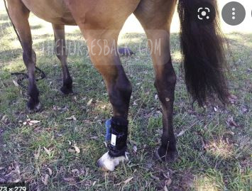
Chronic musculoskeletal pathologies are common in horses, however, identifying related effects can be challenging. This study tested the hypothesis that movement sensors and analgesics could be used in combination to confirm the presence of restrictive pathologies by assessing lying time. Four horses presenting a range of angular limb deformities (ALDs) and four non-affected controls were used.

The study comprised two trials at separate paddock locations. Trial A consisted of a 3-day baseline phase and 2 × 3-day treatment phases, during which two analgesics were administered to two ALD horses and two controls in a standard crossover design. Trial B replicated trial A, except that as no difference between analgesics had been evident in trial A, only one analgesic was tested. Movement sensors were used to measure the horses’ lying time and lying bouts.

In trial A, ALD horses’ basal mean lying time was significantly less than controls (means ± SD for ALD horses 213 ± 1.4 minutes and for controls 408 ± 46.7 minutes, P = .007); with analgesic administration, the difference became nonsignificant. In trial B, ALD horses’ basal mean lying time was also significantly less than controls (ALD horses 179 ± 110.3 minutes; controls 422.5 ± 40.3 minutes, P < .001), again becoming nonsignificant with analgesic administration. Given the increases in ALD horses’ lying time with analgesic administration, it is possible that their shorter basal lying time is associated with musculoskeletal discomfort. Despite the small sample size, movement sensors effectively measured this behavior change, indicating that they could be a useful tool to indirectly assess the impact of chronic musculoskeletal pathologies in horses.

The paper can be accessed here.

Clothier J, Small A, Hinch G, Barwick J, Brown WY. Using Movement Sensors to Assess Lying Time in Horses With and Without Angular Limb Deformities. J Equine Vet Sci. 2019; 75:5559. doi: 10.1016/j.jevs.2019.01.011


Prematurity and Dysmaturity Are Associated With Reduced Height and Shorter Distal Limb Length in Horses 

Journal of Equine Veterinary Science

Abstract

The long-term effects of gestational immaturity in the premature (defined as < 320 days gestation) and dysmature (normal term but showing some signs of prematurity) foal have not been thoroughly investigated. Studies have reported that a high percentage of gestationally immature foals with related orthopedic issues such as incomplete ossification may fail to fulfill their intended athletic purpose, particularly in Thoroughbred racing. In humans, premature birth is associated with shorter stature at maturity and variations in anatomical ratios, linked to alterations in metabolism and timing of physeal closure in the long bones.

We hypothesized that gestational immaturity in horses might similarly be associated with reduced height and different anatomical ratios at maturity. In this preliminary study, the skeletal ratios of horses with a history of gestational immaturity, identified through veterinary and breeder records, were compared with those of unaffected, closely related horses (i.e., sire, dam, sibling).

External measurements were taken from conformation photographs of cases (n = 19) and related horses (n = 28), and these were then combined into indices to evaluate and compare metric properties of conformation. A principal component analysis showed that the first two principal components account for 43.8% of the total conformational variation of the horses’ external features, separating horses with a rectangular conformation (body length > height at the withers), from those that are more square (body length = height at the withers). Varimax rotation of PC1 and analysis of different gestational groups showed a significant effect of gestational immaturity (P = .001), with the premature group being more affected than the dysmature group (P = .009, P = .012). Mean values for the four dominant indices showed that these groups have significantly lower distal limb to body length relationships than controls. The observed differences suggest that gestational immaturity may affect anatomical ratios at maturity, which, in combination with orthopedic issues arising from incomplete ossification, may have a further impact on long-term athletic potential.

The paper can be accessed here.

Clothier J, Small A, Hinch G, Brown WY. Prematurity and Dysmaturity Are Associated With Reduced Height and Shorter Distal Limb Length in Horses. J Equine Vet Sci. 2020 Aug;91:103129. doi: 10.1016/j.jevs.2020.103129. Epub 2020 May 22. PMID: 32684267.


Perinatal Stress in Immature Foals May Lead to Subclinical Adrenocortical Dysregulation in Adult Horses: Pilot Study 

Clothier J, Small A, Hinch G, Brown WY. Perinatal Stress in Immature Foals May Lead to Subclinical Adrenocortical Dysregulation in Adult Horses: Pilot Study. J Equine Vet Sci. 2022 Apr;111:103869. doi: 10.1016/j.jevs.2022.103869. Epub 2022 Jan 21. PMID: 35074402.

The persistent endocrinological effects of perinatal stress due to gestational immaturity in horses are unknown, although effects have been reported in other livestock species. This pilot study tested the hypothesis that persistent adrenocortical dysregulation is present in horses that were gestationally immature at birth by assessing the salivary cortisol response to exogenous ACTH.Case horses (n = 10) were recruited with histories of gestation length < 315 d or dysmaturity observable through neonatal signs. Positive controls (n = 7) and negative controls (n = 5) were recruited where possible from related horses at the same locations.

Cases and positive controls received an intramuscular, low-dose (0.1 ug/kg) of synthetic ACTH (Tetracosactrin 250 mg/mL, Synacthen); negative controls received no ACTH. Saliva samples were collected from all horses at baseline T = 0 and at 30 min intervals post injection from T = 30 to T = 150. These were assayed for salivary cortisol concentration (SCC) using a commercially available ELISA kit (Salimetrics).All baseline values (T = 0) were within normal published ranges. Peak and AUC values (corrected for baseline) for case horses were significantly different (ANOVA P < .001) to positive controls, with either higher (H-cases) or lower (L-cases) SCC values, outside the 95% Confidence Interval of the reference population.

There was no significant effect of breed, age, sex, test month, or location on results. The results suggest that gestational immaturity may lead to subclinical adrenocortical dysregulation, with affected horses presenting an elevated or blunted response to a low-dose ACTH stimulation, despite normal basal levels.

The paper can be accessed here.

Clothier J, Small A, Hinch G, Brown WY. Perinatal Stress in Immature Foals May Lead to Subclinical Adrenocortical Dysregulation in Adult Horses: Pilot Study. J Equine Vet Sci. 2022 Apr;111:103869. doi: 10.1016/j.jevs.2022.103869. Epub 2022 Jan 21. PMID: 35074402.

Filed Under: Bodywork, Foals Tagged With: dysmature foals, equine anatomy, equine bodywork, equine dysmaturity, equine prematurity, GA, horse anatomy, immature foals, Premature foals

Yes, We Can Image for Transitional Vertebrae in Horses

September 23, 2021 by Jane @ THB 7 Comments

It’s been a question of mine for a while. Can diagnostic imaging show the presence of transitional vertebrae?

We’re seeing many bone samples from dissections, as shown in my previous article on transitional vertebrae.

But if we’re to help our horses that live with this issue, we need to identify it before they’re dead. (Yes, right?!)

Allow me to introduce a practicing vet and educator who is doing just that.


Imaging for Transitional Vertebrae 

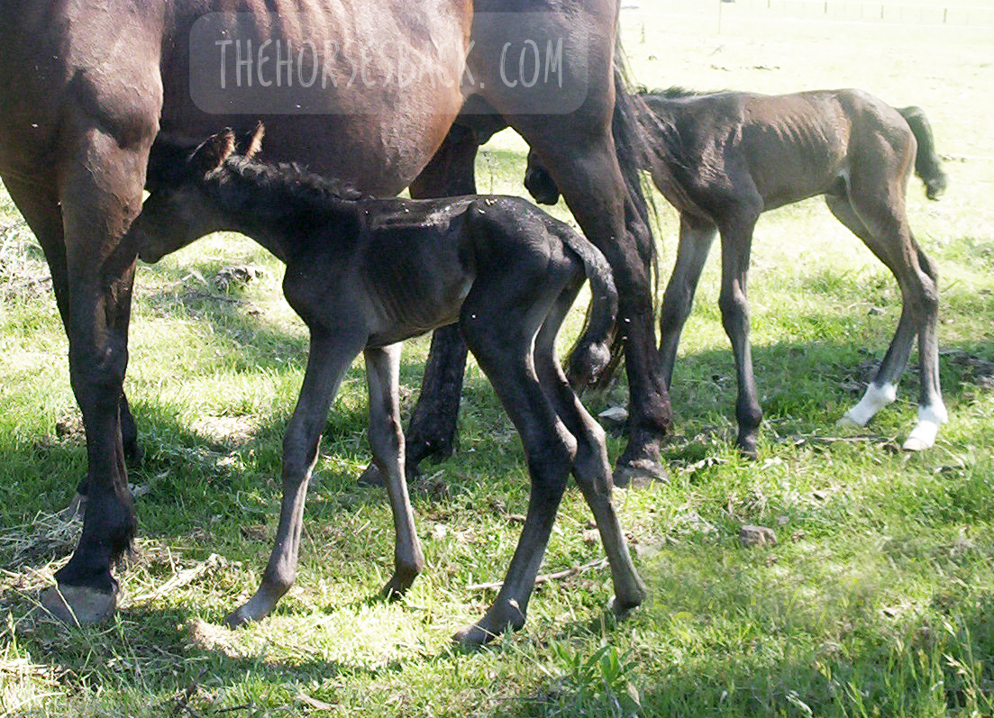
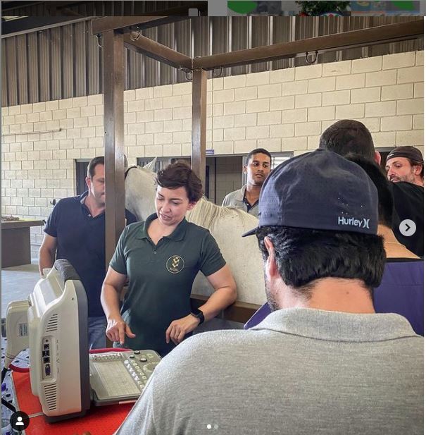
Meet Dr Brunna Fonseca, Associate Professor, educator and specialist in equine orthopedics, focusing on the spine and nervous system. She’s based in São Paulo, Brazil.

I’ve been following her Instagram for a while, because she posts brilliant videos and photos explaining what she does, and how, and why.

I was delighted to see a recent post on imaging for a transitional vertebra, which included fantastic visuals. Such a great communicator!

Dr Brunna has kindly given me permission to repost her images and descriptions here. So without further ado…

  • All images copyright of Axial Vet

Ultrasonograms

Ultrasonography for transitional vertebrae
Angle of transducer. Image: Equine Neck and Back Pathology: Diagnosis and Treatment, 2nd Edn. Ed. Frances M.D. Henson. © 2018 John Wiley & Sons, Ltd.

The following ultronographic images are each a composite of two images, one showing the left side and the other the right.

This textbook illustration helps to show the angle the image is taken at. This angle is usually used for imaging the articular facets of the vertebrae.

Additionally, the image at the top of this article shows a transitional vertebra at T18, like the mare being diagnosed by Dr Brunna.


1.  Can we recognise transitional vertebrae?

The first image shows two sides of a mare’s body. The hand icon gives us a strong hint of where to look… This appearance is very similar to that of the TB mare in my previous post.

Dr Brunna writes, “This mare has the T18 transitional vertebra, presenting a transverse process similar to the lumbar vertebrae on the right side, which causes the appearance of the horse to have the most visible rib on that side.

The occurrence of transactional vertebrae in the horse is not uncommon, especially in the thoracolumbar transition, which can occur in T18 or L1.”


2. Section of a thoracic vertrebra

This image is from a different horse showing a normal rib head and its joint with the vertebra.

Dr Brunna writes, “This is the image of a thoracic vertebra, showing the costotransverse joint.”


3. Image of a normal vertebra

Dr Brunna writes, “This is a T17 ultrasound image, where we can see the image of the normal costotransverse joints.”

This is the bay mare again.

As with the previous cross section, the red pins which show the facet joint between rib head and vertebra.


4. Section of a lumbar vertebra

This is cross section is of a normal lumbar vertebra from a different horse.

As you can see,  there is no joint between the  transverse process and the vertebral body.

The process is wide and flat, and integral to the vertebra.


5. Image of a lumbar vertebra

Here’s an ultrasound of the first lumbar vertebra (L1) in the bay mare.

As in the above cross section (picture 4), there is no joint between the transverse processes and the vertebral body.

We now have ultrasound images of the normal T17 and normal L1. As we will see, the transitional vertebra mixes elements from both.


6. Imaging transitional vertebrae

“This is an ultrasound image of T18, where we can see the image of the costotransverse joint on the left side (red pin) and image of the transverse process on the right side.”

So here’s the underlying skeletal issue in the bay mare.

The left side is a normal joint, being the same as the T17 thoracic vertebra (picture 3).

The right side is similar to the previous image of the lumbar vertebra (picture 5).

It is not identical, for while the process-like rib is joined to the vertebra, it is not the same shape and does not lie as flat as the lumbar process.


Want to Hear More From Dr Brunna Fonesca?

You can follow her Axial Vet Instagram page to see examples of her equine cases and their assessment, in images and videos.

An increasing number of captions are now translated into English.


Filed Under: Bodywork Tagged With: Anatomy, equine anatomy, equine bodywork, equine malformation, equine skeleton, GA, horse anatomy, transitional vertebra, transitional vertebrae

An Unwelcome Side Effect: Transitional Vertebrae in Horses

May 1, 2018 by Jane @ THB 24 Comments


They can lead to scoliosis, spinal arthritis, flexion and straightness problems, saddle fit issues, secondary lameness, hoof problems and soft tissue trauma. So, what on earth are transitional vertebrae, and why haven’t we heard more about them?

To answer the first part of that question, transitional vertebrae are hybrids that appear where one group of vertebrae changes to another. They show mixed features of each group.

They can be found along the spine, where:

  • the cervical (neck) meet the thoracic vertebrae,
  • the thoracic meet the lumbar vertebrae,
  • the lumbar meet the sacral vertebrae (sacrum),
  • where the sacrum meets the caudal vertebrae (tail bones).

As for why we’ve not heard much about them, the answer is probably that they’re rarely identified while a horse is alive.

However, they can lead to some very real problems in the living horse due to the asymmetry they cause along the spine – and they’re far more common than you might think.

A transitional vertebra at L1. (c) J. Clothier

 The affected process or rib can hurt when the horse bends into it, as the abnormal rib/process is literally ‘stabbing’ into soft tissue.”

© All text copyright of the author, Jane Clothier, https://thehorsesback.com.


Thoracic and lumbar transitional vertebrae

Here are the three main types of variation, as shown in this diagram from one of the few research papers to mention this issue.

Here, we’re going to look at the first two – labeled A and B – which are the most common manifestations.

The three kinds of thoracolumbar transitional vertebrae. (c) American Journal of Veterinary Research. (Annotated in green by J. Clothier) Haussler, K.K., Stover, S.M., Willits, N.H. Developmental variation in lumbosacropelvic anatomy of Thoroughbred racehorses (1997); American Journal of Veterinary Research, 58 (10), pp. 1083-1091

A ‘process-like rib’ at T18

Labeled ‘A’ in the above diagram, this is a transitional vertebra at T18 (the last thoracic vertebrae) – a rib that thinks it might be a transverse process, lacking an articulation or joint with the vertebral body.

A normal facet on one side, a non-articulated process-like rib on the other (c) J. Clothier

Instead, the process-like rib is solidly attached, meaning there is no independent movement whatsoever. At its end point, it’s joined by costal cartilage to the preceding rib, partially restricting that rib’s movement, too.

This is a problem, as the caudal ribs are not directly attached to the sternum because they need to move more.

The abnormality can be on one or both sides of the vertebra, although single side is most common.

A ‘rib-like process’ at L1

Labeled ‘B’ in the above diagram, this is a transitional vertebra at L1 (the first lumbar vertebrae). Again, it’s usually one-sided, although two sides also occur.

Here, we’re looking at a transverse process that rather than being fairly short, wide and flat, instead extends outwards like a misshapen rib. There’s no articulation with the vertebral body.

The first lumbar vertebrae (L1) of this Quarter Horse mare is a transitional vertebra. (c) Melissa Longhurst, www.equinebodybalance.com.au 

The above image shows an abnormal L1 found in a Quarter Horse mare. This mare was asymmetric throughout her body, and had a history of unsoundness both fore and rear throughout her lifetime.

Effect on the horse

Scoliosis is the major effect of transitional vertebrae. It’s an asymmetry that in these cases can be lifelong and permanent.

I’ve seen it a few times now in skeletons and on horses that have subsequently been euthanized for unrelated reasons – the spine curves in the affected direction, ie. the horse’s ‘short side’ is the same as the abnormal rib/process that is causing restriction.

The above bones were from a TB gelding who was in his late teens. Over his lifetime, the additional pressure on the side of the abnormal L1 had caused greater bone development in the vertebra further forward. In this photo, T18, the last thoracic vertebra, has been cut to show this impact.

Cases are highly individual and the degree of impact depends on how abnormal the vertebra is, plus other factors affecting the horse’s musculoskeletal balance – including tack and riders. However, we can consider the following points.

There can be an obvious localized effect:

  • The affected process or rib can hurt when the horse bends into it, as it is literally ‘stabbing’ into soft tissue.
  • The attachments of the deep, short muscles involved in segmental stabilization at L1 and T18 are affected, also affecting proprioception and posture.
  • The abdominal muscles involved in breathing and flexion during locomotion are restricted over an affected T18.
  • The diaphragm inserts onto T18, meaning its function is also affected.
L1 transitional vertebra on the left side causing scoliosis along the spine, including the sacrum. (c) Melissa Longhurst, www.equinebodybalance.com.au


This can affect overall spinal health and biomechanics:

  • Scoliosis means that bending to the affected side can be uncomfortable, while bending to the opposite side can be highly limited.
  • Achieving straightness may be impossible. Scoliosis can extend through the withers and into the neck.
  • Impinging transverse processes and vertebral arthrosis at other vertebral joints further limit movement.
  • These restrictions make lifting the back problematic. 

And then there can be a host of secondary effects:

  • In the heavily pregnant mare, existing discomfort due to a T18 may worsen.
  • Achieving saddle fit is difficult on an asymmetric horse with scoliosis.
  • Abnormal loading can lead to recurrent lameness and persistent hoof issues.
  • Unrelated pathologies can scale up uncontrollably, as the horse cannot compensate effectively.


More on this Topic

Take a closer look at the vertebrae featured in this article (Equine Healthworks is my practice page in NSW, Australia – also on Facebook.)


Questions, thoughts or comments? Join us at The Horse’s Back Facebook Group. 


Can we spot transitional vertebrae in the living horse?

Yes, sometimes.

Unilateral transitional vertebra at T18. (c) J. Clothier

As this TB mare (above and below) was unable to maintain weight due to the physical stresses she was experiencing, her rib outline was fairly clear.

In her case, the last rib felt wider and flatter than the other ribs. The space between the rib and the point of hip was also noticeably narrower on the affected side (although this would be true of any horse with scoliosis, it’s a matter of putting the picture together, sign by sign).

The problem is visible here. This mare’s body condition and tension reflects the stresses caused by the T18 transitional vertebra, which was later confirmed at necropsy. (c) J. Clothier

There were other reasons for suspicion. Even when all the surrounding tissue was relaxed, there was no ‘spring’ when the rib was palpated with a flat hand. That’s not definitive, but it’s a cause for concern.

Do something that most people never do – stand on a fence or mounting block and take a photo down the horse’s spine, when it’s standing square…”

This veteran grey Arabian, below, is one I’d also consider a suspect. Again, we can see a very obvious protruding last rib on the offside and a lack of straightness. Even with musculoskeletal bodywork and spinal mobilization, the rib remained just as pronounced.

Arabian mare with a suspect rib. Photo: J. Clothier

Incidentally, I’ve also worked on this horse’s offspring, and the younger horse has the same profile to the ribs, on the same side, accompanied by a history of inexplicable back pain – and lack of straightness. 

Note: It’s important to eliminate other causes first, as horses will often have this appearance at the last rib, without it being caused by a transitional vertebra. What’s happening is that the rib is protruding because the vertebra is immobilised in a rotated position. When chiropractic, osteopathy or bodywork restores mobility to the spine, the rib returns to its normal position. 


Ongoing hoof issues

In the bay TB mare, spinal asymmetry (scoliosis, with bend to the right) had led to excessive loading of the near fore. This was no doubt compounded by constantly training and racing in a clockwise direction, plus the classic long toe/low heel frequently found in ex-racehorses.

As a result, her near fore had constant hoof wall separation, bacterial infection (seedy toe / white line disease) and a deep P3 problem that would never come right.

Here’s the hoof capsule and P3. Yes, the poor girl suffered, despite extensive efforts to reconstruct that hoof.

P3 and hoof capsule, near fore, TB mare. Photo: J. Clothier


Patreon members can view videos of this mare and further photos. Go to: www.patreon.com/thehorsesback for more details.


The TB gelding mentioned earlier also had chronic issues in the opposing fore hoof, with wall separation, damage to P3 and evidence of earlier laminitis.


How many horses are affected?

Who knows? The study mentioned earlier (Haussler et al, 1997) found that 22% of Thoroughbreds examined at necropsy, having died or been euthanized at the racetrack, had thoracolumbar transitional vertebrae.

Transitional vertebra at T18 (above ground skeleton, damaged by scavengers)    (c) J. Clothier


To date, I’ve come across 3 in above-ground skeletons (2 x T18, 1 x L1), plus one in a horse later euthanized (1 x T18). These were TBs and Australian Stock Horses.

And as mentioned, I’ve suspected the T18 issue here and there amongst clients’ horses.

Although found mostly in Thoroughbreds, transitional vertebrae are seen across a range of breeds. And certainly, with equine dissection having taken off in quite a big way in the equine care industry, more and more of these anomalies are being observed.


Questions, thoughts or comments? Join us at The Horse’s Back Facebook Group. 


Should we be concerned?

The answer is, inevitably, both yes and no.

On the positive side, if the numbers harbouring this problem are as high as it seems, we have to assume that many horses are coping just fine. 

For as with any musculoskeletal anomaly, horses can compensate very well.

However, when another problem is added to the mix, things can head south very quickly indeed.

And it can all happen without us ever knowing that a skeletal anomaly is an underlying factor. When this happens, owners often have a lot of unanswered questions about their horses – and often large vets bills.

Transitional vertebrae at T18. (c) J. Clothier

It’s the TB or TB-derived breed horse that is most likely to present this (although not exclusively). If you’re buying one and you view a horse with an obvious T18 that really stands out, you might want to get that checked.

At the very least, do something that most people never do – stand on a fence or mounting block and take a photo down the horse’s spine, when it’s standing square or close to square.

If there’s a clear scoliosis along the spine, be cautious (this is a good rule of thumb anyway, no matter what the cause is). If you see an overly pronounced rib on the concave side, be doubly cautious.

And if you believe your horse may have one, the answer is the same as always: be aware, take a 360 degree approach in ensuring that hooves, tack, training and riding are as good as they can be, and your horse will have the best possible chance of functioning well without cause for concern.

(c) Melissa Longhurst, www.equinebodybalance.com.au


Filed Under: Bodywork Tagged With: equine anatomy, equine bodywork, equine vertebrae, GA, Thoroughbred, transitional vertebrae

Meet Spinalis, the Forgotten Muscle in Saddle Fitting

December 20, 2016 by Jane @ THB 43 Comments

Spinalis Header

It’s barely mentioned in saddle fit or anatomy books, yet the muscle Spinalis cervicis can hugely impact on the spinal health and movement of the horse, particularly with poor tack fit.

Meet muscle Spinalis cervicis et thoracis, a far more important muscle than is generally realized. As a deep muscle, it’s influential in mobilizing and stabilizing that hidden area of the spine at the base of the neck, the cervico-thoracic junction, deep between the scapulae.


Where to Find this Muscle

As part of the deeper musculature, Spinalis is as hidden in books as it is in life. Usually, it’s a single entry in the index.

Spinalis Standard
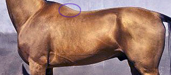
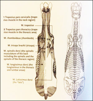
At best, it has no more than a bit part in anatomical illustrations,  usually as a small triangular area at the base of the withers. This is also where we can palpate it.

The reality is quite a bit more interesting. It’s actually a muscle of three parts – dorsalis, thoracis and cervicis. These names denote its many insertions, for it links the spinous processes of the lumbar, thoracic and cervical vertebrae.

  • Bradley_2.1
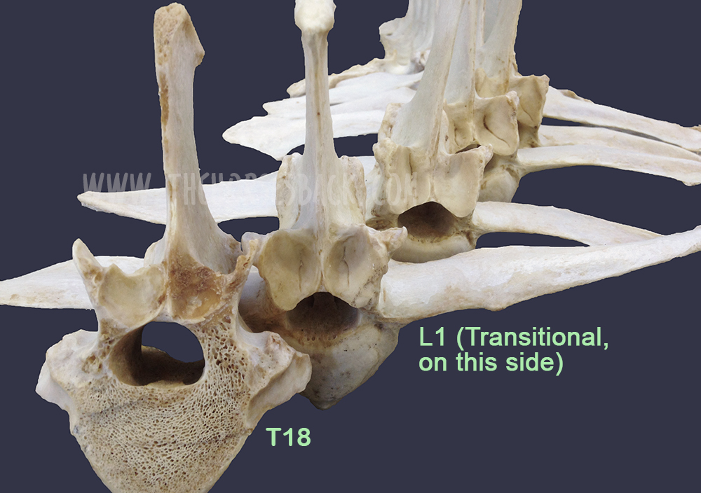
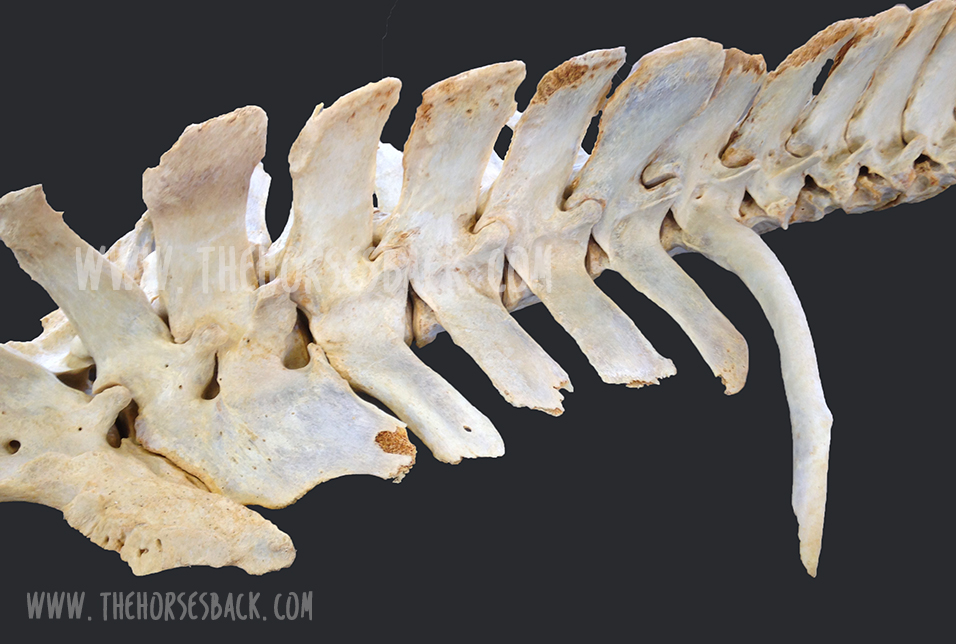
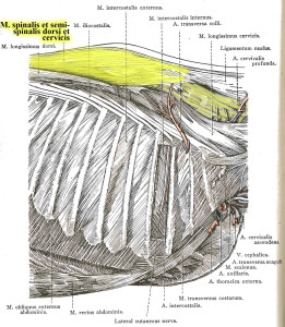
Further back along the spine, it lies medially to Longissimus dorsi, and in fact integrates with this larger, better known muscle, attaching to the processes of the lumbar and thoracic vertebrae.
  • When it reaches the withers, it becomes more independent, attaching to the processes of the first half dozen thoracic vertebrae (T1-T6). Here, the cervical and thoracic portions overlap and integrate to share a common attachment. (The part we palpate, at the base of the withers, is the thoracic section.)
  • Heading into the neck, as Spinalis cervicis, it attches to the last 4 or 5 cervical vertebrae (C3/C4-C7). Only the lamellar portion of the nuchal ligament runs deeper than this muscle.

Dissection 2
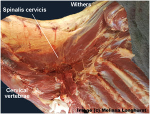
Its integration with other muscles is complex, and its close relationship with Longissimus dorsi partially explains why it doesn’t get much consideration as a muscle in its own right.

It is the more independent section, Spinalis cervicis, between withers and neck, that we are interested in, although its influence is present along the entire spine.

© All text copyright of the author, Jane Clothier, https://thehorsesback.com. 

What Does Spinalis Do? 

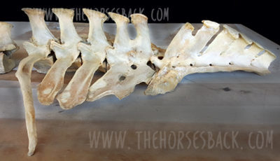
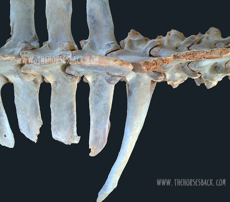
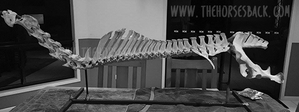
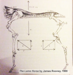
In his 1980s’ Guide to Lameness videos, Dr. James Rooney, first director of the Gluck Equine Research Center, University of Kentucky, referred to Spinalis as part of the suspension bridge of muscles supporting the spine (Longissimus dorsi achoring from the lumbosacral vertebrae, Spinalis thoracis et dorsalis from the upper thoracics). He also refers to this extensively in The Lame Horse (1988).

In fact, the suspension bridge analogy only really makes sense if Spinalis dorsi is considered.

Spinalis cervicis is usually credited with a role in turning the head to left to right, and raising the head.

Bradley Spinalis-1Older texts, such as Bradley’s 1922 veterinary dissection guide, Topographical Anatomy of the Horse, mention its role in stabilizing the spine.

This creates a point of interest. Given that the nuchal ligament (lamellar portion) doesn’t attach to C6 and frequently only weakly with C5 (see the findings of anatomist Sharon May-Davis, in this earlier article ), Spinalis cervicis suddenly appears pretty important in stabilizing and lifting the base of the neck, particularly as it does so at the point of greatest lateral bending.


Spinalis and Poor Saddle Fit

Anyone who has been involved in close examination of the horse’s back will recognize Spinalis thoracis where it surfaces close to the skin, on either side of the withers.

When a horse has been ridden in an overly tight saddle, this small area of muscle can become pretty hypertrophic – raised and hardened. Typically, the neighbouring muscles are atrophied. When Spinalis is palpated, the horse often gives an intense pain response, flinching down and raising the head.

GerdHeuschman
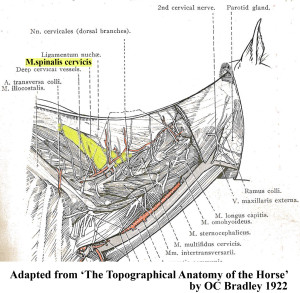
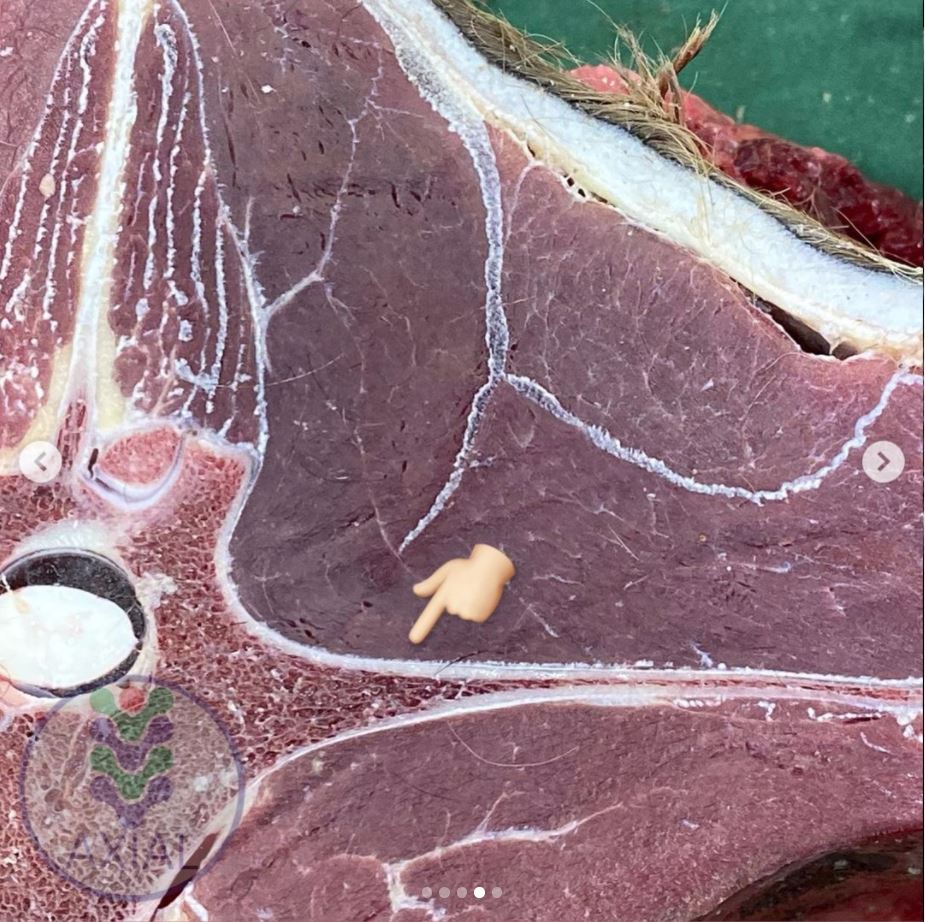
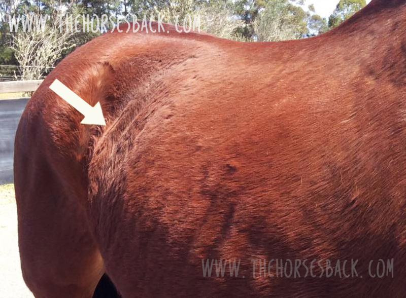
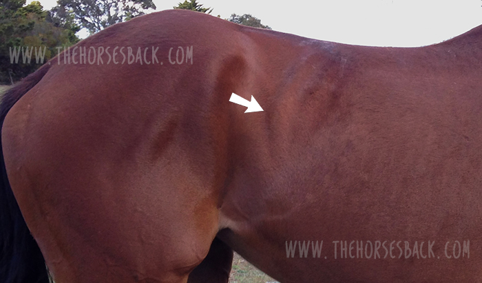
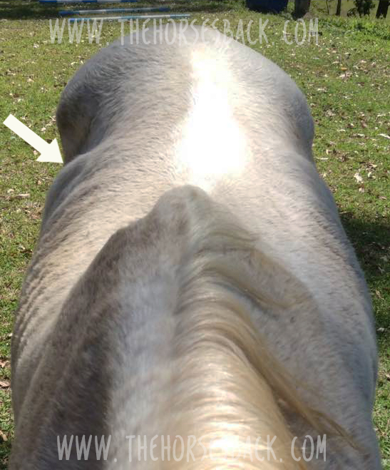
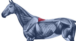
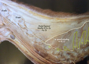
What often happens is this. An overtight saddle fits over the base of the withers like a clothes peg, pinching Trapezius thoracis and  Longissimus dorsi. However, it frequently misses Spinalis thoracis where it surfaces, wholly or partially within the gullet space. Often, the muscle is partially affected.

It’s as if the neighbouring muscles are under lockdown. Free movement of the shoulder is restricted and the horse’s ability to bear weight efficiently while moving is impeded. In response to the surrounding restriction and its own limitation, this muscle starts to overwork.

Result? The horse, which was probably already moving with an incorrect posture, hollows its back even further, shortening the neck and raising its head.  As this becomes even more of a biomechanical necessity, all the muscles work even harder to maintain this ability to move, despite the compromised biomechanics.

Working harder and compensating for its neighbours, Spinalis becomes hypertrophic. It is doing what it was designed to do, but it’s now overdoing it and failing to release. We now have a rather nasty vicious circle.


Spinalis photo

Here, Spinalis thoracis stands out due to atrophy of the surrounding musculature. In this TB, a clearly audible adjustment occurred in the C4-C5 area after the muscle was addressed. 


Vicious CircleThe Inverted Posture and Asymmetry

Of course, saddle fit is not the only cause of an inverted posture. However, any horse that holds its head and neck high for natural or unnatural reasons is more vulnerable to saddle fit issues, thus starting a cascade effect of problems.

Are there further effects of this hypertrophy? Consider the connections.

  • When saddles are too tight, they’re often tighter on one side than the other. This can be due to existing asymmetry in the horse, such as uneven shoulders, uneven hindquarters, scoliosis, etc.
  • On the side with greater restriction, the muscle becomes more more hypertrophic.
  • With its attachment to the spinous processes of the lower cervical vertebrae, there is an unequal muscular tension affecting the spine.
  • Without inherent stability, the neck and head are constantly being pulled more to one side than the other, with the lower curve of the spine also affected.
  • Base of neck asymmetry affects the rest of the spine in both directions and compromises the horses ability to work with straightness or elevation.
  • There is also asymmetric loading into the forefeet.
  • We haven’t even started looking at neurological effects…

This isn’t speculation. I have seen this pattern in horses I’ve worked on, many times over.


So, How Do We Help?

In working with saddle fit problems, the saddle refit may be enough to help the horse, if the riding is appropriate to restoring correct carriage and movement. Obviously, the horse’s musculoskeletal system is complex and no muscle can be considered in isolation. As other muscles are addressed through therapeutic training approaches, with correct lateral and vertical flexion achieved, M. spinalis will be lengthened along with the surrounding musculature.

I hold with a restorative approach:

  1. Refit the saddle, preferably with the help of a trained professional,
  2. Remedial bodywork, to support recovery from the physical damage,
  3. Rest the horse, to enable healing of damaged tissue and lowering of inflammation, and
  4. Rehabilitate the horse, through the appropriate correct training that elevates the upper thoracics while improving lateral mobility.

This is particularly important where saddle fit has been a major contributor to the problem. I have frequently found that in these cases,correction will take longer to achieve, as the debilitating effects of poor saddle fit (especially long-standing issues) can long outlast the change to a new, better-fitting saddle. In bodywork terms, the hypertrophic M. spinalis cervicis is often the last affected muscle to let go.

It’s as if Spinalis cervicis is the emergency worker who will not leave until everyone else is safe.


Bodywork Notes

I am fortunate, in that my modalities enable the gentle release of joints through a non-invasive, neuromuscular approach.  The responses I’ve had from horses when M. spinalis cervicis et thoracis has been addressed in isolation have been hugely informative.


** Questions, thoughts or comments? Join us at The Horse’s Back Facebook Group. 


Appendix: Spinalis in the Textbooks

I’m going to add Spinalis references to this post on a regular basis, as I come across them. It’s interesting to see how much, or how little, the muscle is referenced in various textbooks.


Equine Back Pathology

This image, from Equine Back Pathology, ed. F Henson 2009, shows acute atrophy of Longissimus dorsi due to neurological damage. It’s still possible to see the raised attachment/origin of Spinalis cervicis et thoracis – the highlighting is mine. Spinalis does not appear in the book’s index. (added 23 Dec 2016)


nuchal and spinousI have also altered this image, in order to show M. spinalis cervicis more clearly. This is Fig 2.16 from Colour Atlas of Veterinary Anatomy Vol 2, The Horse, R Ashdown and S Done. Spinalis cervicis is within the bounded area and it’s possible to see how it overlies the lamellar part of the nuchal ligament, lamellar portion. (added 23 Dec 2016)


S&G
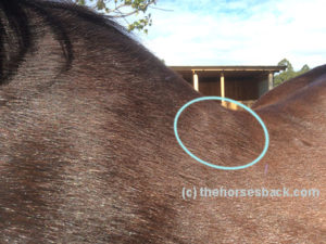
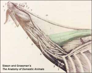
The muscle is tinted green in this image from Sisson and Grossman’s The Anatomy of Domestic Animals, Volume 1, fifth edition 1975.  Here, it is labelled Spinalis et semi-spinalis cervicis. This anatomical figure is credited to an earlier text, Ellenberger and Baum, 1908. (added 23 Dec 2016)


James Roony dedicates two pages to the ‘suspension bridge’ theory of the vertebral column in The Lame Horse (1988). His interest is in Spinalis dorsii section of the muscle and its effect behind the withers, in conjunction with  Longissimus dorsii. (added 4 Jan 2017)


Schleese diagram
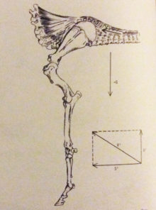
Master Saddler Jochen Schleese refers to Spinalis dorsi and its function in stabilizing the withers in Suffering in Silence, his passionate book about saddle fitting from 2014. “This muscle area is especially prone to significant development – especially with jumpers – because it is continually contracted to accommodate the shock of landing”. The surface area of the muscle is indicated in the anatomical figure, reproduced here. (added 23 Dec 2016)


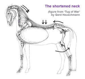
In his seminal text addressing issues of modern dressage training, Tug of War, 2007, Gerd Heuschmann includes Spinalis cervicis in the triangle formed by the rear of the rear of the cervical spine, the withers, and the shoulder blades, “… an extensive connection between the head-neck axis and the truck… it explains how the position and length of the horse’s neck directly affects the biomechanics of the back.” (added 31 Dec 2016)


Filed Under: Bodywork, Saddle Fit Tagged With: Anatomy, equine bodywork, forgotton muscle, GA, homepage, saddle fit, saddle fitting, slider, spinalis, spinalis cervicis, spinalis dorsalis, spinalis thoracis

  • « Go to Previous Page
  • Page 1
  • Page 2
  • Page 3
  • Go to Next Page »

Footer

Contact Me

Send your questions this way…

Get in Touch

  • Store Support
  • Shipping
  • Returns
  • Contact

Copyright © 2026 · Parallax Pro on Genesis Framework · WordPress · Log in