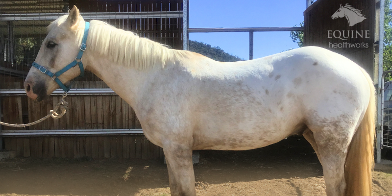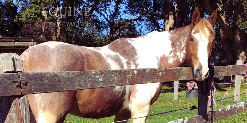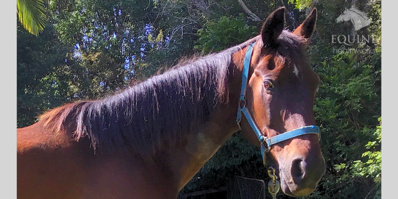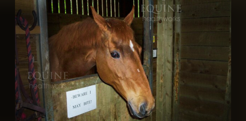
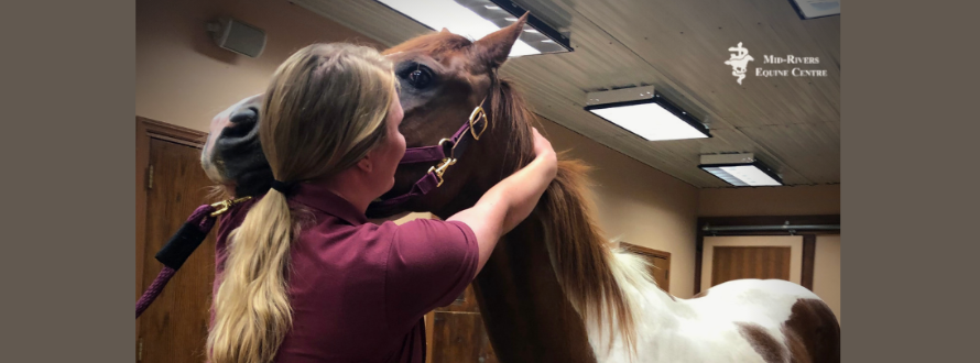
When you’re hiring an equine chiropractor, it’s really important to ask some questions first. And I don’t mean just the usual ‘what do you charge’ and ‘do you work on Saturdays’, although you can obviously ask those too.
You see, there are chiros and then there are chiros. One group consists of equine chiropractors who are genuinely qualified and skilled, while the other includes chiropractors who are not.
Unfortunately, when it comes to hiring a correctly trained chiropractor, word of mouth recommendation isn’t always enough. (I look at some of the issues in an earlier post, Why Do Horse Owners Keep Hiring Unqualified Equine Chiros?)
And unfortunately, it’s often hard for horse owners to ask the right questions of professionals, because the fact is that many of us don’t know enough to know what to ask.

Why does this matter?
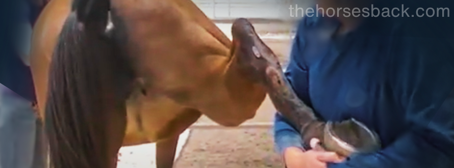
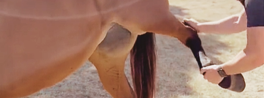
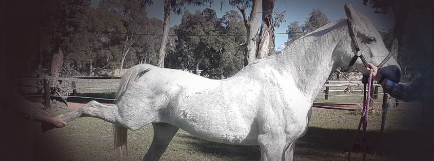
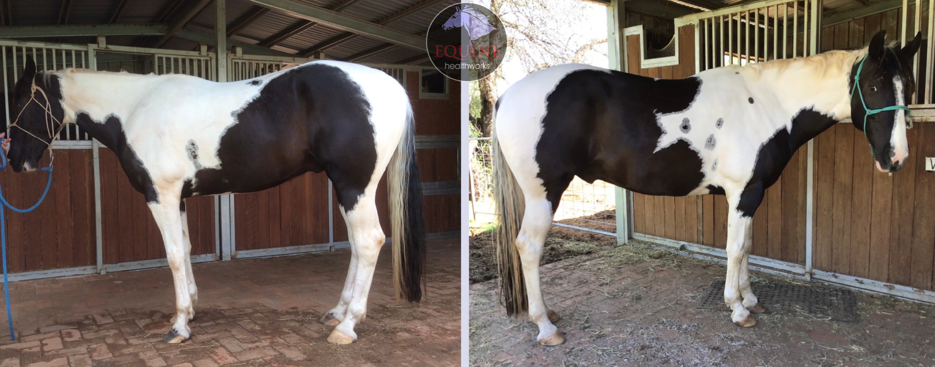
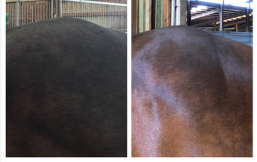
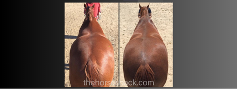
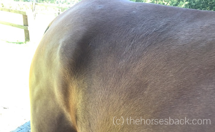
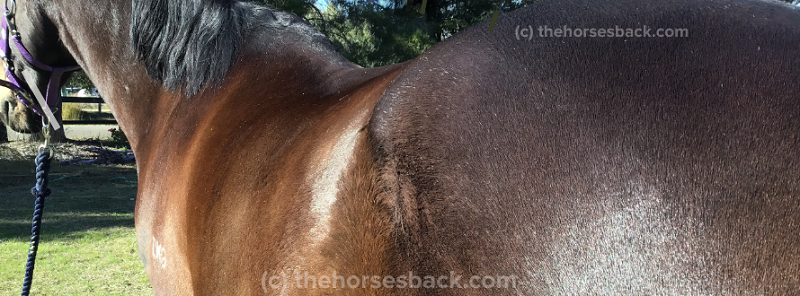
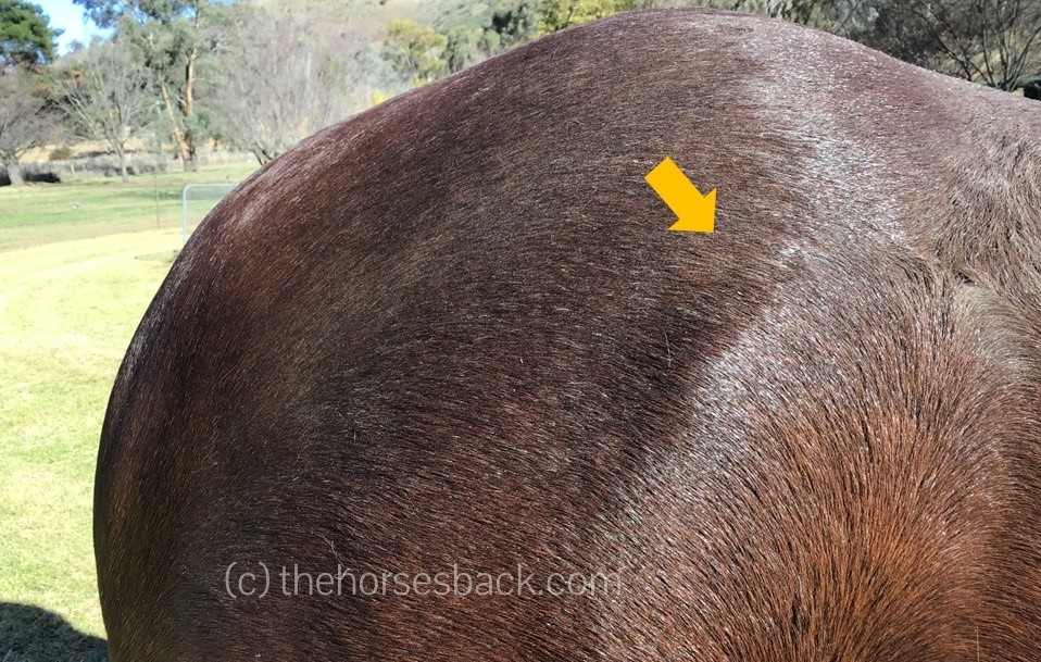
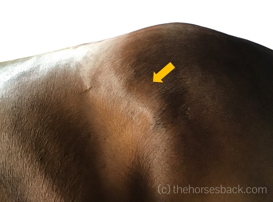
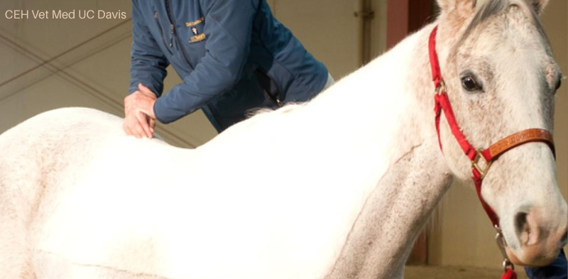
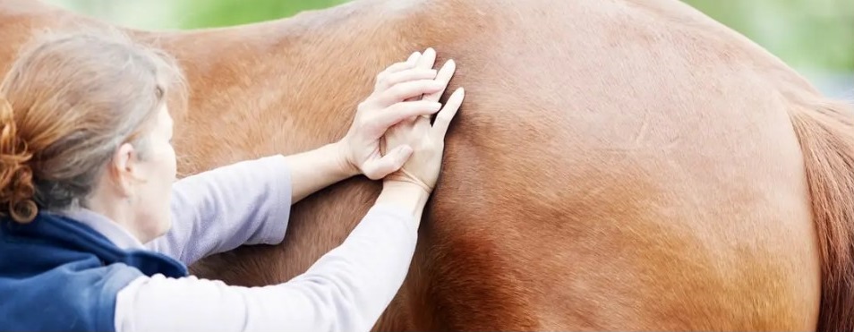
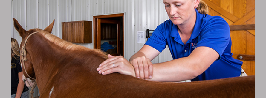
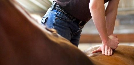
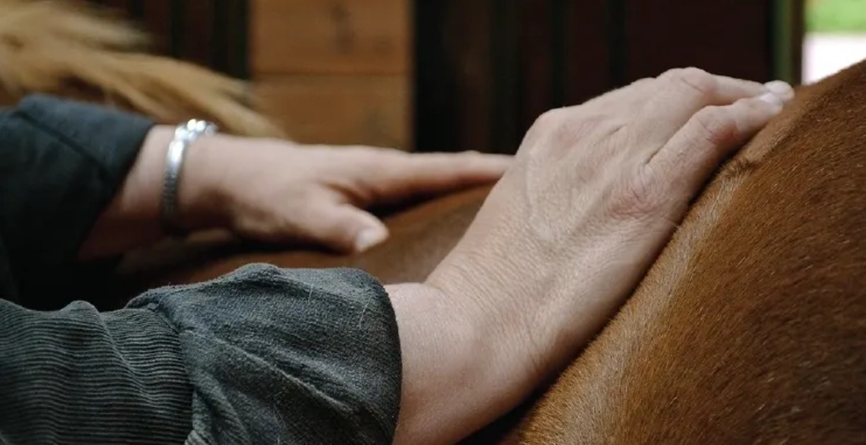
Equine chiropractic is one that’s called an invasive approach. This means it frequently involves the use of high pressure to get results (here’s a good university overview).
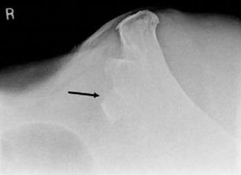
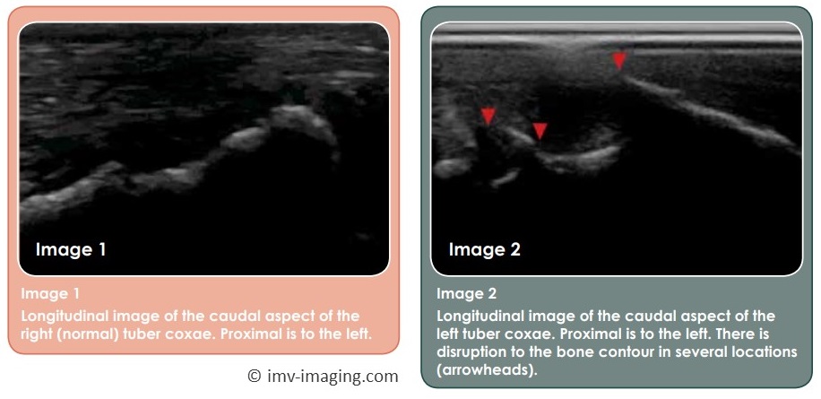
And the thing with approaches that use high pressure is that everything is fine – until it isn’t. That one mistake can be catastrophic, because a high-pressure move was applied in the wrong place or at the wrong time on a particular horse. This is very well expressed in this article by a veterinarian.
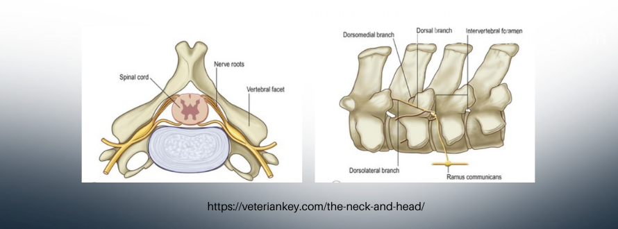
Note that some non-chiro equine vets are sceptical about the benefits of chiropractic. Some of this comes down to different uses of the word ‘subluxation’ – if you’re interested, holistic vet Dr Madalyn Ward DVM explains this aspect very well.
Questions you can ask a horse chiropractor
The following 10 questions may be helpful if you’re thinking of hiring an equine chiropractor.
Some of the answers may be on their website, so do take a look. If there’s no website, ask when you speak to them.
A qualified professional will not mind being asked at all, although do remember they may be busy. But with non-vet chiros, I’d count an irritated response to being asked as a potential red flag 🚩
By the way, if you’re thinking of hiring a chiropractor who’s a veterinarian, then you can be reassured that there’s a high level of professional regulation in place. This protects your horse and you as a customer. This also applies if you live in a region where all equine therapists are heavily regulated. In such cases, you may not need to ask more questions at all.
Otherwise, here’s where you can start.
1. “What is your qualification?”
The answer to this does not always tell the whole story, but is definitely a good starting point. You’ll most likely find it on the website.
It’s not always true that big qualifications make good practitioners. However, when it comes to invasive practices – i.e. those that use a higher level of pressure, meaning moves will have an impact, come what may – solid qualifications are essential. No qualification is a cause for concern 🚩
2. “Where did you train?”
It follows that their qualification needs to come from a reputable training organisation. Find out the answer (it’s likely to be on the website), and then look it up. Is it accredited? Does it come with approvals from regulatory bodies? If not, 🚩
3. “Does your work involve pressure?”
This follow-up question might relate to the practitioner’s understanding of their work and its effect. This is a roundabout way of identifying the possible use of high pressure or force – read this post about Unqualified Equine Chiros for more on this.
If the question can’t be answered, the chiropractor may not even understand what it means, or why it’s important. And if they don’t the answer, how much are they thinking about your horse’s experience of their work? Red flag alert🚩
4. “How exactly do you get results?”
Again, if joints are simply being pushed and pulled, or if the practitioner learned just by watching someone else, this question may be tricky to answer 🚩
On the other hand, if the chiropractor answers articulately, you may not understand what they are on about – anatomy and physiology can certainly be hard to dip into. If you’re not clear, try a follow-up question.
5. “Is there somewhere I can read about that?”
This is a follow-up question if you’re feeling bamboozled by science. Every practitioner should be able to point to a source of information, somewhere, or offer to send a link through later. A vet may be busy, but they’ll at least mention their professional association’s website. Other qualified professionals will appreciate their customers being interested.
If you’re brushed off or the subject gets changed, take note 🚩
6. “Will you be able to tell me what’s wrong with my horse?”
This question is a bit of a bear trap. Diagnosing is telling you specifically what a condition is and trained professionals should know that only a vet can diagnose. So, if a non-vet chiropractor answers ‘yes’, you may have a red flag answer 🚩
7. “Does your approach always work?”
The answer should be that there’s never a guarantee, that some conditions can’t be helped (although the secondary effects can), that veterinary diagnosis and/or intervention may be required, or that not every approach works best for every horse.
If the practitioner says yes, they are either wearing rose-tinted glasses, don’t know as much as they should do, or are over-selling what they do 🚩
8. “How do horses respond to your work?”
If you care about the experience your horse is being lined up for, this is an important one to ask. “They like it” is not an adequate answer 🚩 You definitely need to hear a bit more about what happens with the horse after the session and what you can expect.
9. “Are you insured?”
This one can elicit an interesting answer, given that association membership and up-to-date qualifications are usually required for chiropractic due to its invasive nature (regulations vary by country and region).
I’d recommend that you NEVER hire a chiro who isn’t insured 🚩
10. “How many sessions do you recommend – and why?”
If you hear that all will be sorted out in one session, do not believe them 🚩 If they suggest that your horse should be treated every time they visit the area, hide your horse and padlock your wallet 🚩🚩
To sum up…
Now I realise that not every practitioner is super-articulate. In any group of professionals, there are those who speak well and those who are more introverted.
Hopefully, the information you’re looking for will be readily available on a website if not in conversation.
Yet as with any group of professionals, even with all questions answered positively, there’s no guarantee of high-level skills or ability – or sensitivity to your horses’ responses.
But you will be dramatically lowering the chance of hiring a poorly trained and less knowledgeable chiropractor who could be unwittingly putting your horse at risk.