• Skip to main content
  • Skip to footer

The Horse's Back

Body Talk for Thinking Owners

  • Home
  • Articles
  • BEMER
  • Store
    • Store
    • Shipping & Returns
    • Support
  • Equine Healthworks
    • Bodywork for Horses
    • Reviews
    • Leave a Review
  • About & Contact
    • About
    • Contact

equine anatomy

More Than a Blemish: How a Knocked Down Hip Can Affect Horses

August 2, 2023 by Jane @ THB 13 Comments

Appaloosa with severe fracture of tuber coxae

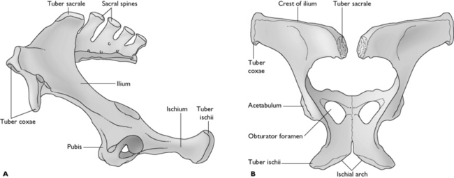
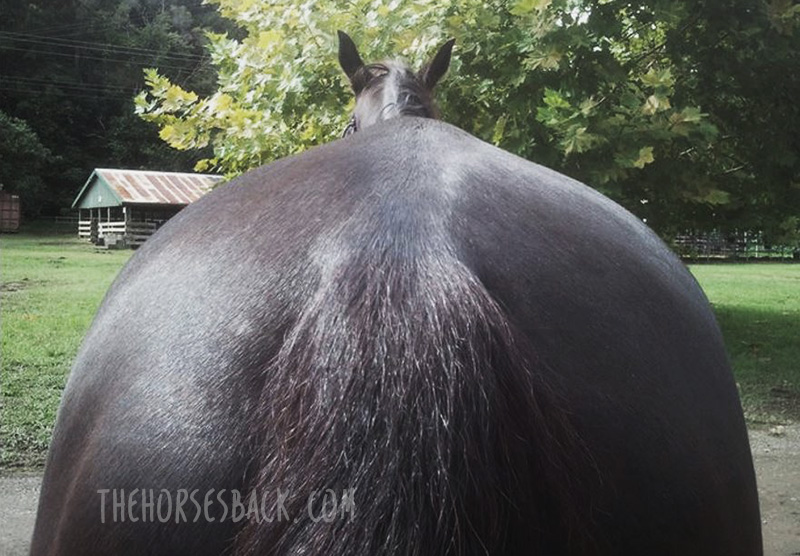
‘Knocked down hip’ is a common name for a fracture of the tuber coxae (point of hip). This is usually the result of a horse having an impact with a gate post, fence or trees, a barrel in a race, another horse’s hoof, or the ground.

A piece of bone is often completely displaced, disappearing downwards and forwards into the paralumbar fossa – the space between the transverse processes of the lumbar vertebrae, internal abdominal oblique muscle, and the last rib.

This displacement can then change the outline of the pelvis on the damaged side. That said, it isn’t always visually obvious, and many horse owners often don’t even know it has happened.

It’s the least damaging of pelvic fractures as it doesn’t involve a joint or compromise the pelvic structure, and for this reason it’s often said that its long term effects are purely cosmetic.

But does that really mean it has no lasting effects whatsoever?

This post presents a range of potential issues, from none whatsoever to an altered gait that affects performance, and explains why you should always thoroughly assess your horse for effects after a tuber coxae fracture.

(c) Jane Clothier, thehorsesback.com. With thanks to Dr Sharon May-Davis for checking this post and catching my errors.

The elusive nature of the problem

I know a horse whose owner reported a succession of minor come-and-go issues in the hindlimb.

These either failed to warrant veterinary attention (there being no lameness) and with no diagnoses made when a vet was consulted. The problem was intermittent and elusive.

Older Arabian with a recent tuber coxae fracture
A recent fracture in a veteran Arabian, showing some swelling. (c) J Clothier, thehorsesback.com

There was no visible asymmetry and no muscle atrophy.

However, the horse was outwardly rotating the right stifle and swinging the leg outwards in the forwards (cranial) phase of the stride.

When I palpated the right tuber coxae, it had a section knocked away halfway up the bone.

Could there be a connection? Very likely!

Let’s take a closer look at the tuber coxae and how a fracture can affect the action of the hind limb on that side.

Why is the knocked down hip so common?

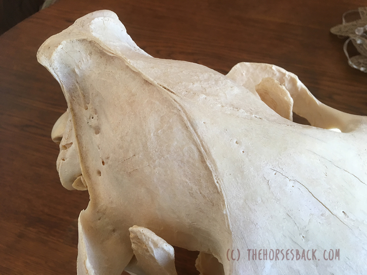
Besides being the widest point of the pelvis and therefore vulnerable to traumatic injuries of all varieties, the horse’s point of hip has another key weakness.

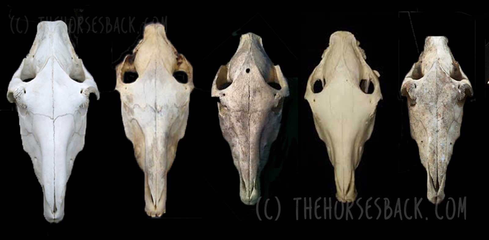
It’s part of a growth plate. It’s an epiphyseal plate, which means it’s separated from the main body of the pelvis by a physis, the line where bone is produced during growth in early life.

Growth plate on TB pelvis
9-yo TB pelvis, showing remaining growth plate at the tuber coxae (Note: some has broken away during cleaning). (c) J Clothier, thehorsesback.com

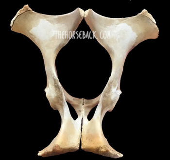
This means we’re looking at the ‘cap’ of the tuber coxae, which is a weaker cap of bone until at least 5 years of age, and often later depending on the breed.

These are vulnerable to getting displaced – the growth plate of the elbow (ulna) being another example.

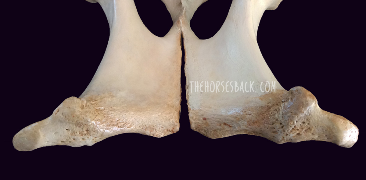
Skeletal specimens such as the one in the image show us that even beyond 5 years of age, the growth plate may  not be fully attached and is a weaker structure. The tuber coxae in the mature horse still has a roughened surface due to muscle attachments, which gives it a rather open honeycomb structure. This makes it far easier to damage.


More than a cosmetic feature

The clue as to why this fracture can be damaging, even though it doesn’t affect the structural integrity of the pelvis, lies in its function.

Yes, its function. Even though it’s not a moving part, the tuber coxae is much more than a cosmetic feature. Like the withers and the elbow/ulna, its function is to provide an anchor for some important muscles.

It serves as the attachment point for muscles involved in flexing the hip and extending the stifle to move the entire hindlimb forwards (more on those later). For this reason, the attachment point needs to be extremely stable.

Anglo with tuber coxae fracture
Anglo gelding with tuber coxae fracture (c) J Clothier thehorsesback.com

The tuber coxae must serve as an anchor point in this way while powerful propulsive forces are conveyed via the pelvis to the spine.

That’s why the tuber coxae are tuberosities – they sit wide and proud and, like handlebars on a motorbike, remain relatively stable in spite of the forces being transmitted from the hind legs forward along the spine.

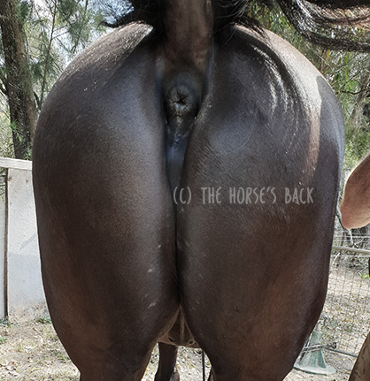
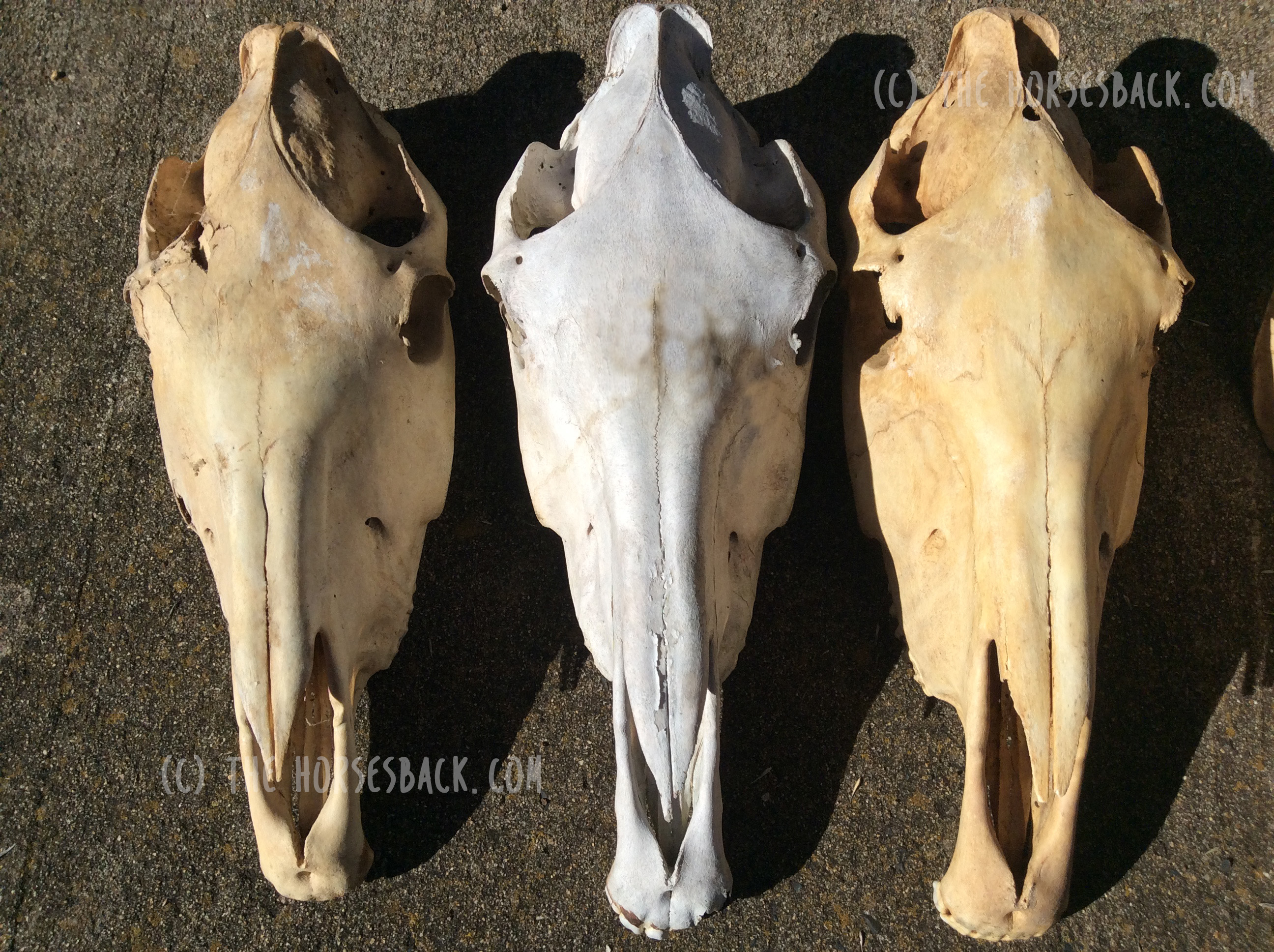
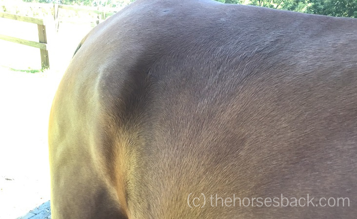
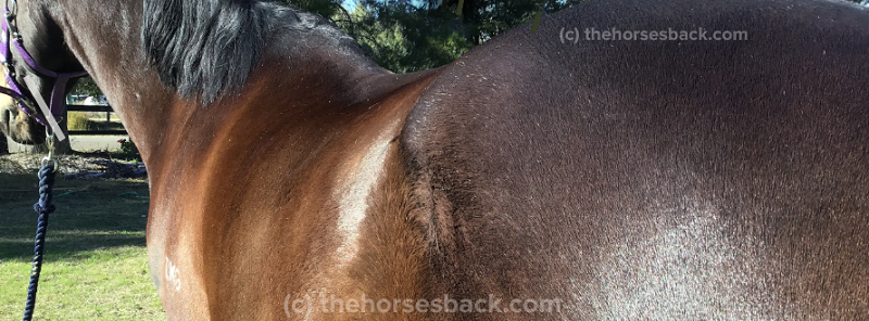
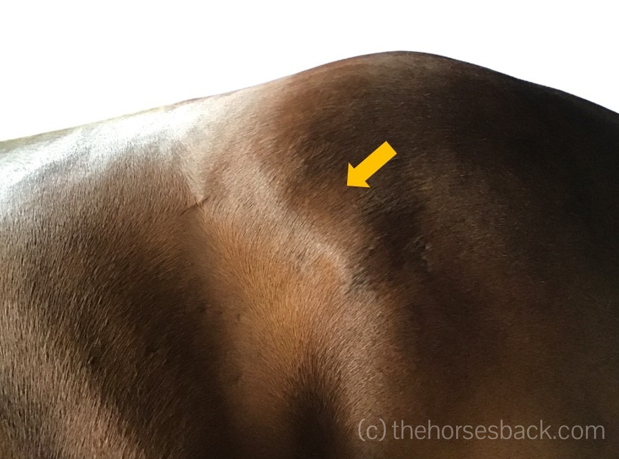
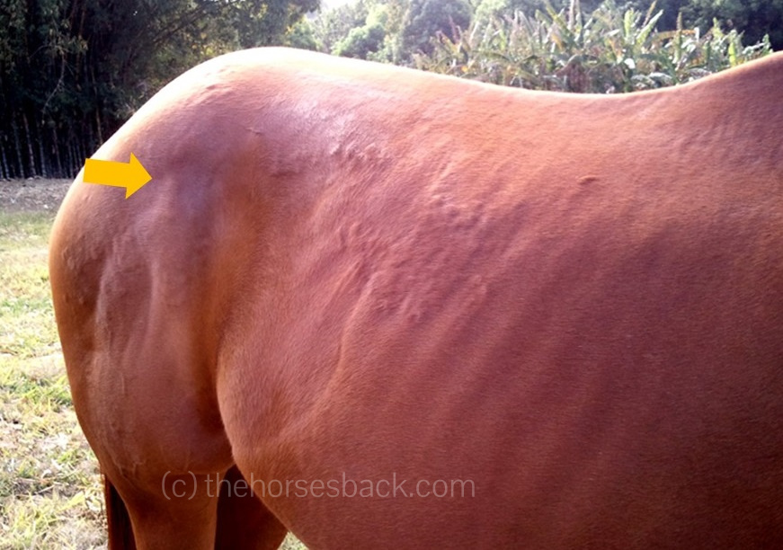
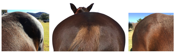
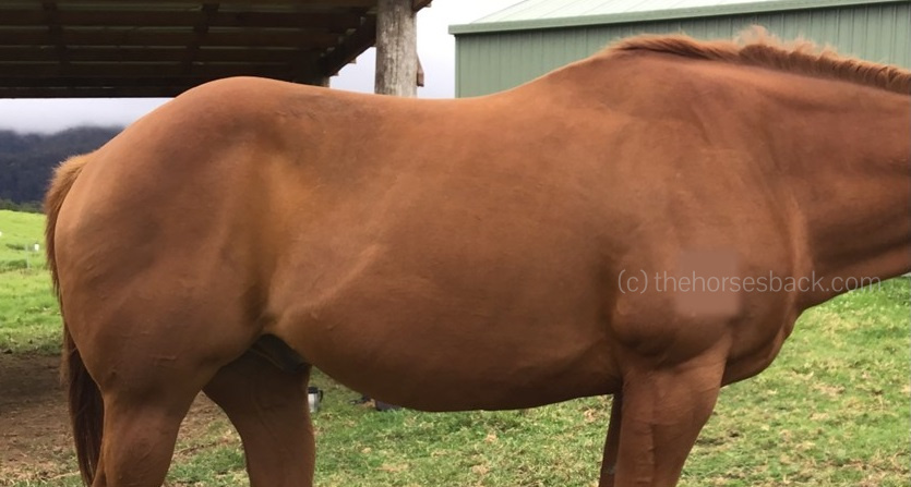
The location of the fracture

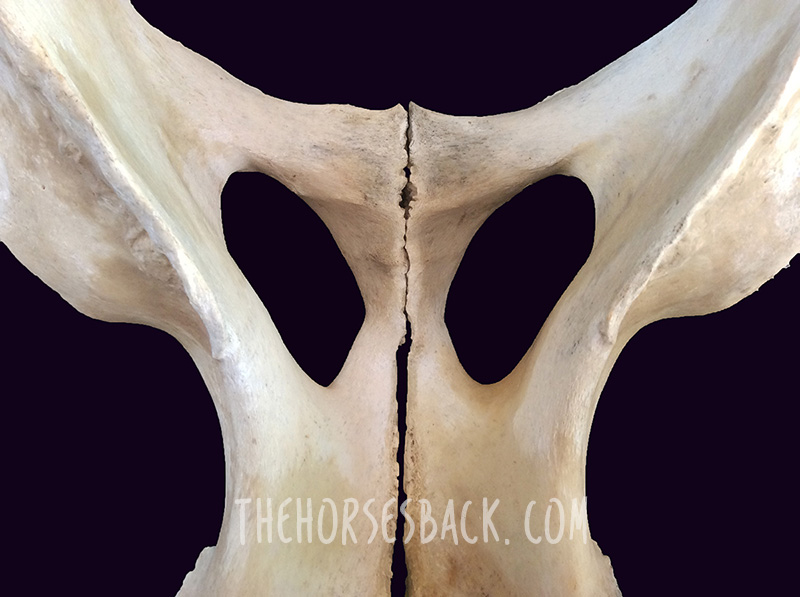
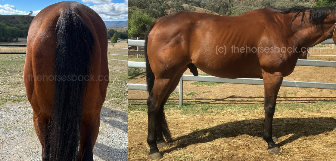
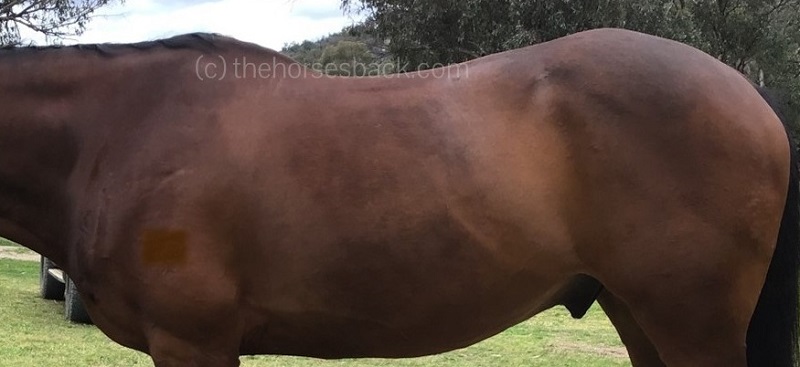
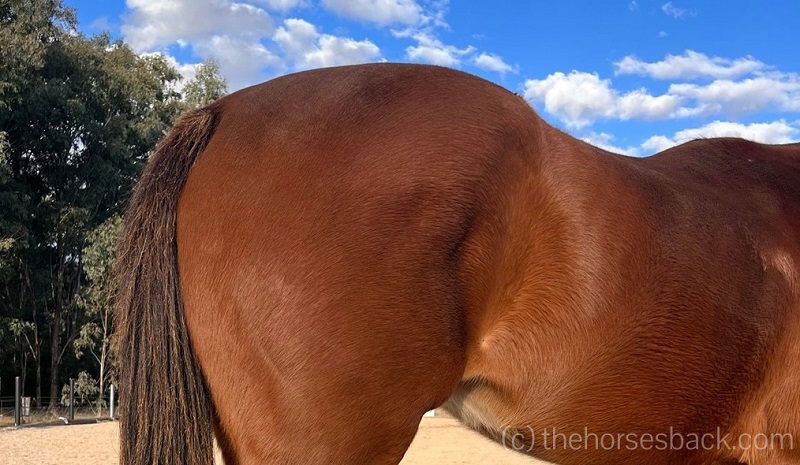
Although generally similar, tuber coxae vary in shape from horse to horse. Fortunately, as they come in matching pairs, it’s possible to identify damage by comparing one side to the other.

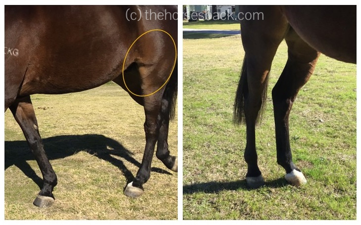
Visually, it can be easy to spot a horse with an old tuber coxae fracture due to its pelvic asymmetry. How visible generally depends on the fracture’s severity.

Alternatively, good old manual palpation can reveal its presence.

Here are the main possibilities. 

  1. Upper piece of bone is displaced
QH tuber coxae fracture
Quarter Horse with old tuber coxae fracture affecting the upper section of the tuberosity. (c) J Clothier, thehorsesback.com

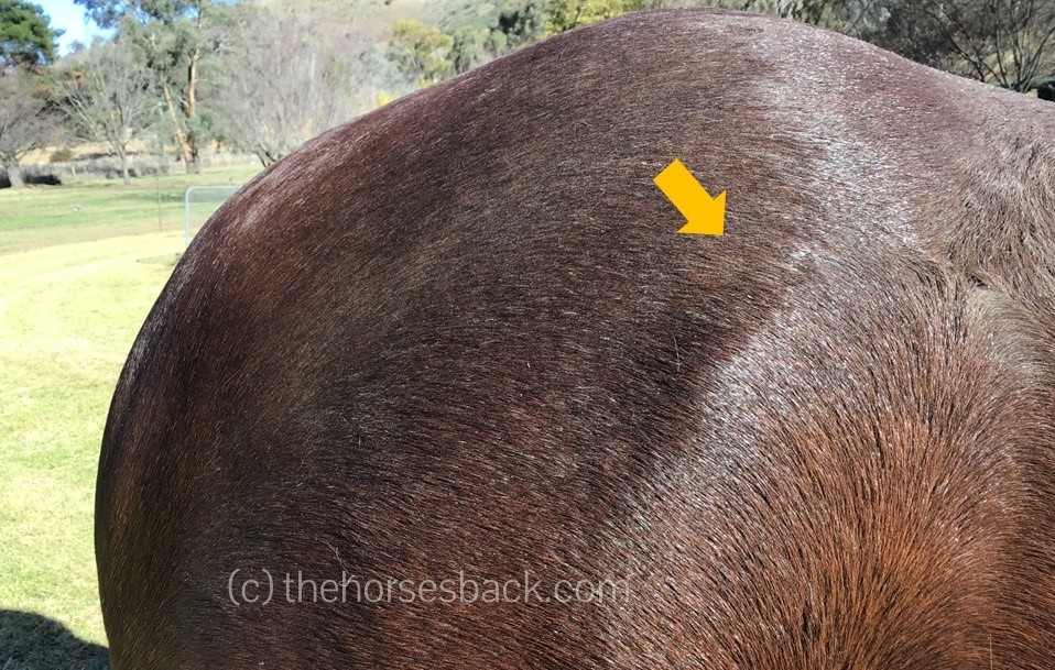
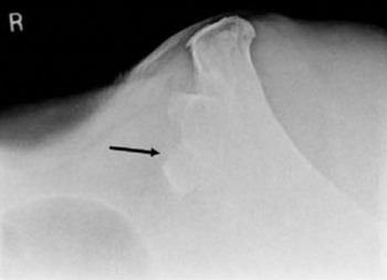
The upper part of the tuber coxae (ie. dorsomedial aspect) may be chipped off. This is usually, but not always, a visible type of fracture, as the point of hip acquires a rounded appearance. This is generally what gives the knocked down hip its name. 

  1. Central piece of bone is displaced
Tuber coxae fracture WB gelding
Tuber coxae fracture showing loss of bone in the centre of the tuberosity (c) J Clothier, thehorsesback.com

In this case, both ends of the tuber coxae are intact, so the overall shape isn’t much different.

However, if you palpate carefully, there’s a divot in the middle where a chip of bone has been dislodged. Identifying this involves comparing both sides of the horse.

  1. Lower piece of bone is displaced
OTTB with old tuber coxae fracture
Ex-racehorse with displaced lower part of the tuber coxae. (c) J Clothier, thehorsesback.com

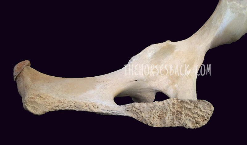
Only the lower part of the bone (ie. ventrolateral aspect) may be missing.

This is harder to see, but easy to feel: the tuber coxae’s ‘ledge’ is noticeably shorter.

This variation of the fracture is common as it’s the outer corner, the widest point, that’s been knocked off.

One study of 29 horses reported that when only the lower, outer aspect of the bone (caudolateral) was fractured, injured horses returned to work in around 3.5 months. [1]

  1. Whole tuber coxae is displaced
Appaloosa tuber coxae fracture
The entire tuber coxae is displaced and the horse has experienced severe muscle loss. (c) J Clothier, thehorsesback.com

In severe cases, the entire tuber coxae is displaced. The visual asymmetry is more obvious. It can also be more serious: with palpation, it may be possible to feel the wider, deeper section of bone remaining, where the ilial shaft becomes the ilial wing.

In the study, when the entire tuber coxae was fractured, the recovery period was longer at around 6.5 months from injury. [1]

Could there be more damage?

If the horse has fallen hard enough to fracture the tuber coxae, it may have damaged other areas of its pelvis too. The various sacroiliac ligaments are prime contenders, as is the sacroiliac joint itself, along with the lumbosacral joint.

Then there are the structures of the actual hip joint (acetabulum and coxofemoral) and head of the femur.

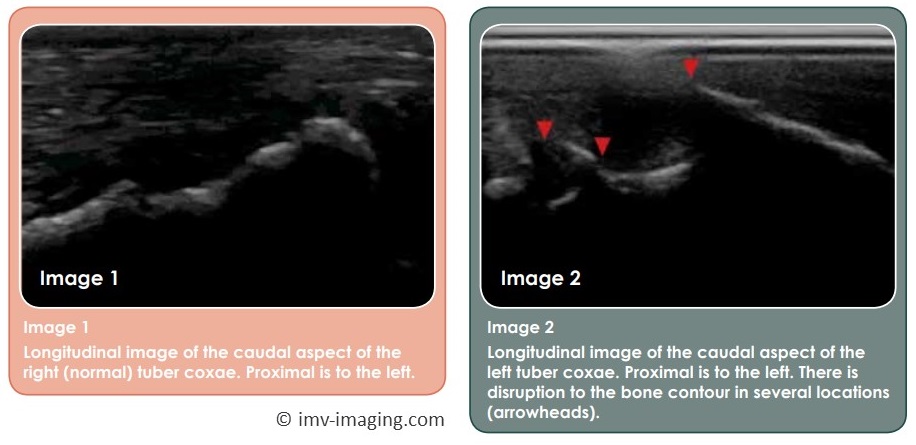
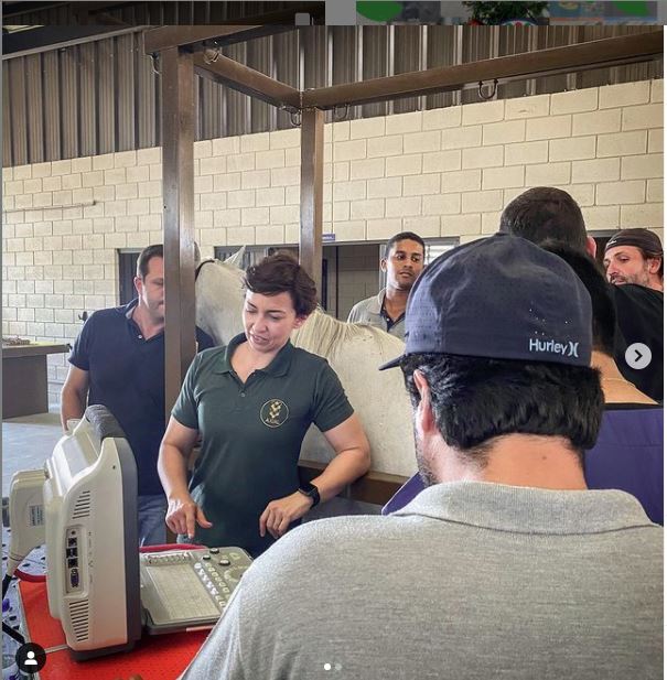
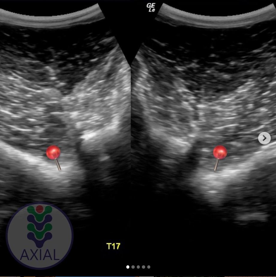
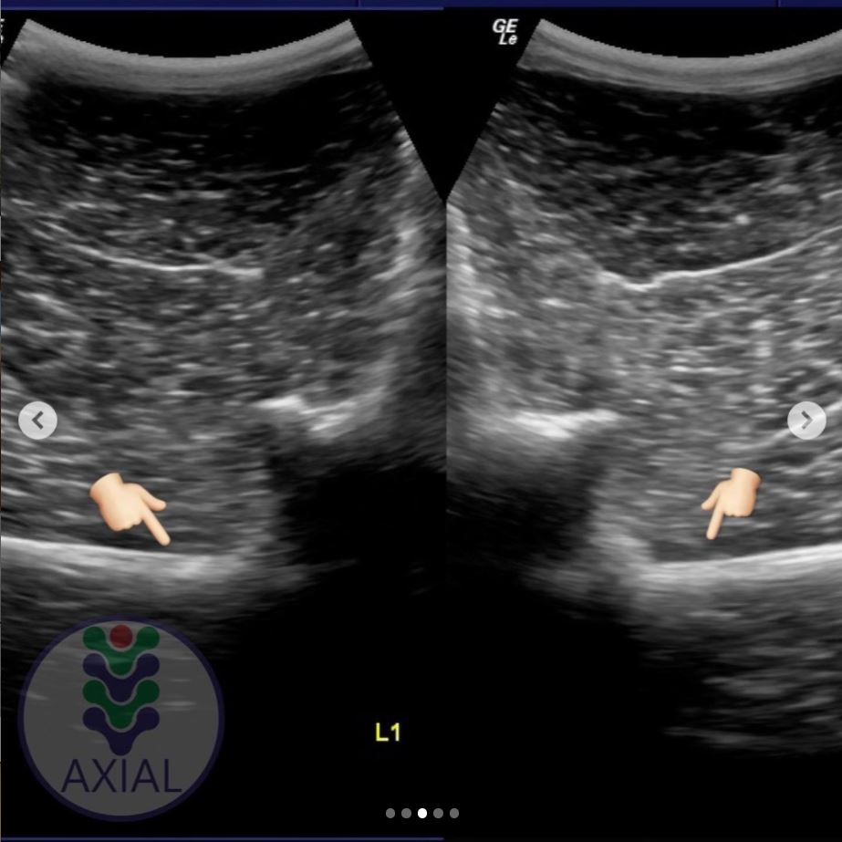
There may even be an incomplete fracture of the ilium that we don’t know about, or separation at the pelvic/pubic symphysis. A full veterinary assessment should always be sought if your horse is lame and you suspect a trauma. Radiographs, ultrasound and scintigraphy have been used to image the tuber coxae and identify the extent of the damage. [2]

Sometimes, palpation and left to right comparison is the only way to identify the damage to the tuber coxae, in this case on the right side. It’s possible (r) to see the dip in the bone once you know it’s there. (c) J Clothier, thehorsesback.com

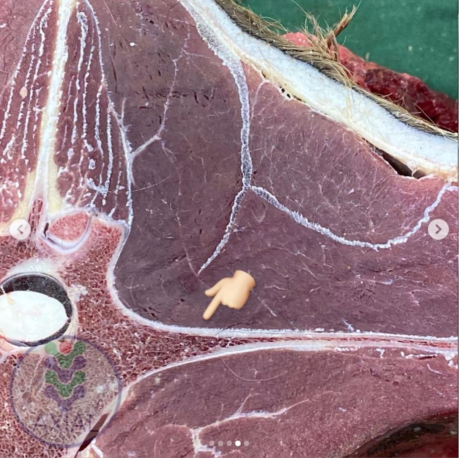
On a more superficial level, a ridge of what feels like soft tissue can sometimes be moved over the remaining tuber coxae – this is where a muscle attachment or its associated tendon or fascia has been damaged.

Small masses may also be palpated, which may be displaced bone chips. Small avulsion fractures have been observed in young racehorses, at the attachment of the Superficial gluteal muscle.

If there’s a pain response when you palpate these, bear in mind that it may be a sequestrum – ie. a bone fragment that has caused a lingering infection. This needs veterinary attention.

Alternatively, as Dr Sharon May-Davis explains (having examined fractures in dissections), these may also be ‘repair jobs’ that have happened as a result of bleeds. Smaller lentil-sized masses are calculi, ie. mineralized lumps, while the larger grape-like masses are lipomas, caused when a specialized fat is deposited in an effort to repair damaged tissue.

Thoroughbred gelding ex racehorse
Off the track Thoroughbred with an old tuber coxae fracture. With more heavily built horses, the bone loss may be still harder to identify visually. [Brand obscured.] (c) J Clothier, thehorsesback.com

What are the lasting effects of a tuber coxae fracture?

If there are no further pelvic issues, the general view is that following recovery, the lasting effect of a tuber coxae fracture is little more than a cosmetic blemish.

Radiographic view of tuber coxae fracture
Dorsomedial-ventrolateral 50° oblique radiographic view of the tuber coxa of the ilium in a horse. Large bone fragement (arrow) displaced ventrally. (c) JAVMA, 234. 10.

Our study of 29 horses says that:

“The majority of horses in this study did have muscle atrophy or abnormal bony flattening over the affected tuber coxae. Horses with tuber coxae fractures have an excellent prognosis for returning to athletic use but will most likely have a permanent blemish associated with the area.” [1]

So, a couple of things here.

First, the horses in the study were sufficiently injured, ie. lame, for veterinary attention to be sought. This suggests the fractures were fairly severe, and it therefore follows that the fracture site would be noticeably different upon healing.

But second, what we don’t know about is the quality of the horses’ work post-recovery. They returned to their previous athletic work, but were they the same as before?

Ultrasound images of a fractured vs normal tuber coxae in the same horse. (c) IMV Imaging


One lameness text states that this may only affect the careers of dressage horses, because they look asymmetrical, and judges may mark down due to this imprecision. [3]

Otherwise, the assumption tends to be that the localised trauma is no more than a superficial injury that leaves only a visual blemish, with occasional minor changes in gait quality.

As to whether horses are left with just that, quite a few people believe otherwise.

In fact, the rise of manual therapies has led to a lot more observation of gait changes and anomalies, such as those in the horse mentioned earlier.

Even while writing this, I went out and placed my hands on two more horses with this fracture and a noticeable effect on their movement.

WB gelding with smaller fracture in the centre of the tuber coxae. [Brand obscured]   (c) J Clothier, thehorsesback.com

The most affected muscles

Down to the nitty gritty – why my concern?

Let’s take a look at the effect on some significant muscles that have origins on the tuber coxae, and the related effect on movement if these attachments are damaged through the fracture.

  • Tensor fasciae late

The Tensor fasciae late muscle originates on the tuber coxae and the gluteal fascia. It has multiple insertion points, including (via fascial connections) the crest of the tibia bone and the lateral patellar ligament.

Its job involves flexing the hip, while extending the stifle (femeropatellar joint) as the hindlimb comes forwards.

It has a stabilising effect on the stifle through its connection to the patella ligament in front and the tibia.

This muscle does lose function when its attachment is permanently damaged. Even if it’s a small area of bone that’s lost up there, the effect lower down is broader.

  • Superficial gluteal

This muscle originates on the rear side of the tuber coxae (caudal) and the gluteal fascia.

It inserts onto the femur at the third trochanter. Its job is also to help flex the hip, while adducting the limb (bringing it inwards).

  • Internal abdominal oblique

The Internal abdominal oblique muscle originates from the tuber coxae and the inguinal ligament, and inserts onto the cartilages of the last 4 or 5 ribs, the linea alba, and the prepubic tendon.

Dr Sharon May-Davis writes that this muscle becomes overworked when engaged in supporting a hind limb lameness, and hypertonic when “excessively aiding pelvic engagement or [adopting] a supportive role in hind limb lameness.” [5]

[Biomechanical issues follow below.]

Ex-racehorse with severe tuber coxae fracture
The Tensor fasciae lata muscle is compromised in this ex-racehorse with a tuber coxae fracture. (c) J Clothier, thehorsesback.com


The biomechanical issues…

I’m sure you can see where this is heading now.

If muscles dedicated to flexing the hip suffer from impaired function due to a tuber coxae fracture, there’s going to be a negative effect on movement.

The horse can still move its hindlimb and flex the hip, of course, but an element of fine tuning is going to be lost. At least.

There’s also going to be compensation from other muscles, a functional asymmetry, and some stress in joints.

Generally speaking, the more bone that’s lost, the larger the negative effect on hindlimb control on that side.

Here’s what I’ve observed in various horses on the side affected by a tuber coxae fracture.

1. Outward rotation of the femur

The stifle is angled outwards, and the foot lands toe out.

The horse is less comfortable working in the same direction as the fracture, ie with the fractured tuber coxae on the inside of a circle. I’m presuming there’s a lack of stabilisation from the Tensor fasciae late and reduced function in this muscle.

    Outward rotation of femur in Anglo gelding with fractured left tuber coxae. (c) thehorsesback.com
2. Overdevelopment of the Rectus femoris muscle

This is the largest muscle of the quadriceps and the only one that attaches to the pelvis. It’s responsible for flexing the hip and extending the stifle. I presume this hypertrophy is due to compensatory action.

3. Reduced function in the Superficial gluteal

This muscle is also responsible for flexing the hip. In cases with full tuber coxae fractures, this muscle is atrophied behind the point of hip, adding to the change in the outline of the hindquarters on that side.

4. Tension in the Iliacus muscle

Along with Psoas major, this muscle forms the Iliopsoas. I’ve presumed this tension from the positive responses to corrective moves for the Iliopsoas muscles on this side.

5. Lumbar imbalances

I’ve observed vertebral rotation and restriction, and painful tension in the caudal Longissimus dorsii muscles.

I assume this is due to the compensatory ‘swing and haul’ action required for protracting the hindlimb when there’s insufficient controlled flexion at hip level. Again, this would depend on the extent of the tuber coxae fracture.

Muscles that may also be directly affected

Depending on the anatomy of the individual horse, other muscles may also be affected. How much so depends on the extent of the fracture and the anatomical variations between individuals.

  • Iliacus muscle

Forming the Iliopsoas along with Psoas major, Iliacus is responsible for flexing and rotating the hip. It has an origin under the ilium and insertion on the lesser trochanter of the femur, along with the tendon of Psoas major.

  • Middle gluteal muscle

This massive muscle has multiple origins, including the ilium, and inserts onto the greater trochanter of the femur.

In some horses, the lateral edge of its origin on the ilium is close to the tuber coxae, and may be affected by a fracture. This major muscle is largely responsible for extending and abducting the hind limb (ie. moving it outwards, away from the body).

  • Accessory gluteal muscle

This smaller muscle is below the Middle gluteal, and works with it so closely that some texts describe it as part of the bigger muscle.

However, it is largely separate and has its own flat tendon that attaches to the greater trochanter of the femur. This insertion means that it also aids in abduction of the hind limb (ie. moves it outwards, away from the body).

Assessing the individual

In many cases, the apparently quick recovery from lameness can cause the horse’s owner to believe the fracture of a tuber coxae is of little consequence.

It may be true for some, but for others it may be more serious.

The only way to tell is to examine the horse as an individual, starting with a visual assessment and palpation with the hands, comparing one tuber coxae to the other.

The horse’s hind limb action should also be assessed for balance and evenness. The horse may not be lame, but may have limitations in its movement on the side of the fracture.

If muscles flexing the hip aren’t working as they should, other muscles may be compensating, and these in turn can lead to secondary pain.

If the effect on hindlimb action appears to be significant, it is worth considering veterinary imaging to measure the extent of the damage (although this can usually be felt).

At any rate, some rehabilitative work is probably going to be needed so that better muscle condition and strength can be developed on the fracture side.

Ultimately, in severe cases, we have to remember that ‘we can’t put back what’s gone’. This injury may well mean the horse is unsuited to certain sports.

But please, never assume that this is simply a cosmetic blemish.


References
[1] Dabareiner, R. M. and R. C. Cole (2009). Fractures of the tuber coxa of the ilium in horses: 29 cases (1996-2007). Journal of the American Veterinary Medical Association 234 10: 1303-1307.
[2] Pilsworth, R. C. (2003). Chapter 51 – Diagnosis and Management of Pelvic Fractures in the Thoroughbred Racehorse, Diagnosis and Management of Lameness in the Horse. M. W. Ross and S. J. Dyson. Saint Louis, W.B. Saunders: 484-490.
[3] Van Wessum, R. (2020). Lameness Associated with the Axial Skeleton. Adams and Stashak’s Lameness in Horses: 763-800.
[4] Ashdown, R. R.; Done, S. H.; Evans, S. A. (2000). Color Atlas of Veterinary Anatomy: Vol. 2: The Horse, 2nd ed. Mosby Elsevier: Edinburgh.
[5] May-Davis, S. (2023). Dissecting Out The Facts. Author’s workshop manual.


Filed Under: Bodywork Tagged With: equine anatomy, equine bodywork, equine knocked down hip, equine pelvis, equine skeleton, equine tensor fasciae lata, equine tensor fasciae late, equine tuber coxa, GA, horse pelvis, horse skeleton, horse tensor fasciae late, ilium fracture, knocked down hip, knocked down hip horses, pelvic fracture, pelvic fracture horses, point of hip, tuber coxa horses, tuber coxae

Here’s a Round Up of My Premature and Dysmature Foal Research

May 26, 2022 by Jane @ THB Leave a Comment

Here are abstracts, downloads and links for my research into the ongoing effects or premature or dysmature birth in horses.

These are the publicly available details of my thesis (full download) and published, peer-reviewed journal articles. The articles aren’t open access, but if you really want to read something, please contact me.

As always, huge thanks are due to the breeder owners who so very kindly allowed me to study their horses, and who provided such valuable images. Together, you’ve helped me to learn a lot and reach initial findings that I now hope to pass on.


Beyond the Miracle Foal: A Study into the Persistent Effects of Gestational Immaturity in Horses 

PhD Thesis, University of New England and CSIRO

Abstract

Breeding horses can be a financially and emotionally expensive undertaking, particularly when a foal is born prematurely, or full term but dysmature, showing signs normally associated with prematurity. In humans, a syndrome of gestational immaturity is now emerging, with associated long-term sequelae, including metabolic syndrome, growth abnormalities and behavioural problems.

If a similar syndrome exists in the equine and can be characterised, opportunities for early identification of at-risk individuals emerge, and early intervention strategies can be developed. This thesis explores the persistent effects of gestational immaturity manifest as adrenocortical, orthopaedic and behavioural adaptation in the horse.

Basal diurnal cortisol levels do not differ from healthy, term controls, but when subjected to a low dose ACTH challenge, gestationally immature horses presented a depressed or elevated salivary cortisol response, suggesting bilateral adaptation of the adrenocortical response. This may be reflected in behavioural reactivity, but the outcomes from a startle test were inconclusive.

A survey of horse owners indicated that gestationally immature horses tended to be more aggressive and active than controls, aggression being displayed mostly in families of Arabian horses. Case horses also tended to be more active, intolerant, and untrusting.

Gestationally immature horses have restricted growth distal to the carpal and tarsal joints, and this results in a more ‘rectangular’ conformation in adulthood compared to controls. They also often present with angular limb deformities that adversely affect lying behaviour and recumbent rest. This, however, can be mitigated using analgesic therapy, suggesting chronic discomfort.

Based on these findings, it is reasonable to postulate that a syndrome of gestational immaturity may persist, both clinically and sub-clinically, in affected adult horses. Further work is required to fully characterise this syndrome and validate the outcomes in larger populations, thereby providing a foundation for interventions applicable in the equine breeding industry.

The entire PhD thesis can be downloaded here. This is a 236-page PDF.

Clothier, Jane  (author); Brown, Wendy  (supervisor); Small, Alison (supervisor); Hinch, Geoff  (supervisor)


Equine Gestational Length and Location: Is There More That The Research Could Be Telling Us?

Australian Veterinary Journal

Abstract

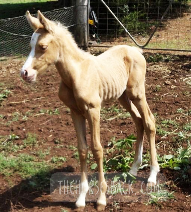
Clear definitions of ‘normal’ equine gestation length (GL) are elusive, with GL being subject to a considerable number of internal and external variables that have confounded interpretation and estimation of GL for over 50 years. Consequently, the mean GL of 340 days first established by Rossdale in 1967 for Thoroughbred horses in northern Europe continues to be the benchmark value referenced by veterinarians, breeders and researchers worldwide. Application of a 95% confidence limit to reported GL range values indicates a possible connection between geographic location and GL.

Improved knowledge of this variable may help in assessing the degree of the neonate’s prematurity and dysmaturity at or soon after birth, and identification of conditions such as incomplete ossification of the carpal and tarsal bones. Associated pathologies such as bone malformation and fracture, angular limb deformity and degenerative joint disease can cause chronic unsoundness, rendering horses unsuitable for athletic purpose and shortening ridden careers.

This review will examine both the factors contributing to GL variation and the published data to determine whether there is potential to refine our understanding of GL by establishing a more accurate and regionally relevant GL range based on a 95% confidence limit. This may benefit both equine industry economics and equine welfare by improving early identification of skeletally immature neonates, so that appropriate intervention may be considered.

The paper can be accessed here.

Clothier, J., Hinch, G., Brown, W. and Small, A. (2017), Equine gestational length and location: is there more that the research could be telling us?. Aust Vet J, 95: 454-461. https://doi.org/10.1111/avj.12653

Using Movement Sensors to Assess Lying Time in Horses With and Without Angular Limb Deformities 

Journal of Equine Veterinary Science

Abstract

Chronic musculoskeletal pathologies are common in horses, however, identifying related effects can be challenging. This study tested the hypothesis that movement sensors and analgesics could be used in combination to confirm the presence of restrictive pathologies by assessing lying time. Four horses presenting a range of angular limb deformities (ALDs) and four non-affected controls were used.

The study comprised two trials at separate paddock locations. Trial A consisted of a 3-day baseline phase and 2 × 3-day treatment phases, during which two analgesics were administered to two ALD horses and two controls in a standard crossover design. Trial B replicated trial A, except that as no difference between analgesics had been evident in trial A, only one analgesic was tested. Movement sensors were used to measure the horses’ lying time and lying bouts.

In trial A, ALD horses’ basal mean lying time was significantly less than controls (means ± SD for ALD horses 213 ± 1.4 minutes and for controls 408 ± 46.7 minutes, P = .007); with analgesic administration, the difference became nonsignificant. In trial B, ALD horses’ basal mean lying time was also significantly less than controls (ALD horses 179 ± 110.3 minutes; controls 422.5 ± 40.3 minutes, P < .001), again becoming nonsignificant with analgesic administration. Given the increases in ALD horses’ lying time with analgesic administration, it is possible that their shorter basal lying time is associated with musculoskeletal discomfort. Despite the small sample size, movement sensors effectively measured this behavior change, indicating that they could be a useful tool to indirectly assess the impact of chronic musculoskeletal pathologies in horses.

The paper can be accessed here.

Clothier J, Small A, Hinch G, Barwick J, Brown WY. Using Movement Sensors to Assess Lying Time in Horses With and Without Angular Limb Deformities. J Equine Vet Sci. 2019; 75:5559. doi: 10.1016/j.jevs.2019.01.011


Prematurity and Dysmaturity Are Associated With Reduced Height and Shorter Distal Limb Length in Horses 

Journal of Equine Veterinary Science

Abstract

The long-term effects of gestational immaturity in the premature (defined as < 320 days gestation) and dysmature (normal term but showing some signs of prematurity) foal have not been thoroughly investigated. Studies have reported that a high percentage of gestationally immature foals with related orthopedic issues such as incomplete ossification may fail to fulfill their intended athletic purpose, particularly in Thoroughbred racing. In humans, premature birth is associated with shorter stature at maturity and variations in anatomical ratios, linked to alterations in metabolism and timing of physeal closure in the long bones.

We hypothesized that gestational immaturity in horses might similarly be associated with reduced height and different anatomical ratios at maturity. In this preliminary study, the skeletal ratios of horses with a history of gestational immaturity, identified through veterinary and breeder records, were compared with those of unaffected, closely related horses (i.e., sire, dam, sibling).

External measurements were taken from conformation photographs of cases (n = 19) and related horses (n = 28), and these were then combined into indices to evaluate and compare metric properties of conformation. A principal component analysis showed that the first two principal components account for 43.8% of the total conformational variation of the horses’ external features, separating horses with a rectangular conformation (body length > height at the withers), from those that are more square (body length = height at the withers). Varimax rotation of PC1 and analysis of different gestational groups showed a significant effect of gestational immaturity (P = .001), with the premature group being more affected than the dysmature group (P = .009, P = .012). Mean values for the four dominant indices showed that these groups have significantly lower distal limb to body length relationships than controls. The observed differences suggest that gestational immaturity may affect anatomical ratios at maturity, which, in combination with orthopedic issues arising from incomplete ossification, may have a further impact on long-term athletic potential.

The paper can be accessed here.

Clothier J, Small A, Hinch G, Brown WY. Prematurity and Dysmaturity Are Associated With Reduced Height and Shorter Distal Limb Length in Horses. J Equine Vet Sci. 2020 Aug;91:103129. doi: 10.1016/j.jevs.2020.103129. Epub 2020 May 22. PMID: 32684267.


Perinatal Stress in Immature Foals May Lead to Subclinical Adrenocortical Dysregulation in Adult Horses: Pilot Study 

Journal of Equine Veterinary Science

Abstract

The persistent endocrinological effects of perinatal stress due to gestational immaturity in horses are unknown, although effects have been reported in other livestock species. This pilot study tested the hypothesis that persistent adrenocortical dysregulation is present in horses that were gestationally immature at birth by assessing the salivary cortisol response to exogenous ACTH.Case horses (n = 10) were recruited with histories of gestation length < 315 d or dysmaturity observable through neonatal signs. Positive controls (n = 7) and negative controls (n = 5) were recruited where possible from related horses at the same locations.

Cases and positive controls received an intramuscular, low-dose (0.1 ug/kg) of synthetic ACTH (Tetracosactrin 250 mg/mL, Synacthen); negative controls received no ACTH. Saliva samples were collected from all horses at baseline T = 0 and at 30 min intervals post injection from T = 30 to T = 150. These were assayed for salivary cortisol concentration (SCC) using a commercially available ELISA kit (Salimetrics).All baseline values (T = 0) were within normal published ranges. Peak and AUC values (corrected for baseline) for case horses were significantly different (ANOVA P < .001) to positive controls, with either higher (H-cases) or lower (L-cases) SCC values, outside the 95% Confidence Interval of the reference population.

There was no significant effect of breed, age, sex, test month, or location on results. The results suggest that gestational immaturity may lead to subclinical adrenocortical dysregulation, with affected horses presenting an elevated or blunted response to a low-dose ACTH stimulation, despite normal basal levels.

The paper can be accessed here.

Clothier J, Small A, Hinch G, Brown WY. Perinatal Stress in Immature Foals May Lead to Subclinical Adrenocortical Dysregulation in Adult Horses: Pilot Study. J Equine Vet Sci. 2022 Apr;111:103869. doi: 10.1016/j.jevs.2022.103869. Epub 2022 Jan 21. PMID: 35074402.

Filed Under: Bodywork, Foals Tagged With: dysmature foals, equine anatomy, equine bodywork, equine dysmaturity, equine prematurity, GA, horse anatomy, immature foals, Premature foals

Yes, We Can Image for Transitional Vertebrae in Horses

September 23, 2021 by Jane @ THB 7 Comments

It’s been a question of mine for a while. Can diagnostic imaging show the presence of transitional vertebrae?

We’re seeing many bone samples from dissections, as shown in my previous article on transitional vertebrae.

But if we’re to help our horses that live with this issue, we need to identify it before they’re dead. (Yes, right?!)

Allow me to introduce a practicing vet and educator who is doing just that.


Imaging for Transitional Vertebrae 

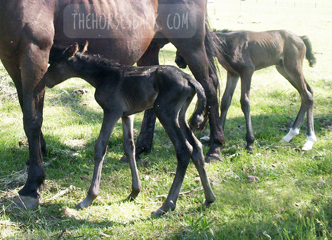
Meet Dr Brunna Fonseca, Associate Professor, educator and specialist in equine orthopedics, focusing on the spine and nervous system. She’s based in S
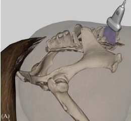
ão Paulo, Brazil.

I’ve been following her Instagram for a while, because she posts brilliant videos and photos explaining what she does, and how, and why.

I was delighted to see a recent post on imaging for a transitional vertebra, which included fantastic visuals. Such a great communicator!

Dr Brunna has kindly given me permission to repost her images and descriptions here. So without further ado…

  • All images copyright of Axial Vet

Ultrasonograms

Ultrasonography for transitional vertebrae
Angle of transducer. Image: Equine Neck and Back Pathology: Diagnosis and Treatment, 2nd Edn. Ed. Frances M.D. Henson. © 2018 John Wiley & Sons, Ltd.

The following ultronographic images are each a composite of two images, one showing the left side and the other the right.

This textbook illustration helps to show the angle the image is taken at. This angle is usually used for imaging the articular facets of the vertebrae.

Additionally, the image at the top of this article shows a transitional vertebra at T18, like the mare being diagnosed by Dr Brunna.


1.  Can we recognise transitional vertebrae?

The first image shows two sides of a mare’s body. The hand icon gives us a strong hint of where to look… This appearance is very similar to that of the TB mare in my previous post.

Dr Brunna writes, “This mare has the T18 transitional vertebra, presenting a transverse process similar to the lumbar vertebrae on the right side, which causes the appearance of the horse to have the most visible rib on that side.

The occurrence of transactional vertebrae in the horse is not uncommon, especially in the thoracolumbar transition, which can occur in T18 or L1.”


2. Section of a thoracic vertrebra

This image is from a different horse showing a normal rib head and its joint with the vertebra.

Dr Brunna writes, “This is the image of a thoracic vertebra, showing the costotransverse joint.”


3. Image of a normal vertebra

Dr Brunna writes, “This is a T17 ultrasound image, where we can see the image of the normal costotransverse joints.”

This is the bay mare again.

As with the previous cross section, the red pins which show the facet joint between rib head and vertebra.


4. Section of a lumbar vertebra

This is cross section is of a normal lumbar vertebra from a different horse.

As you can see,  there is no joint between the  transverse process and the vertebral body.

The process is wide and flat, and integral to the vertebra.


5. Image of a lumbar vertebra

Here’s an ultrasound of the first lumbar vertebra (L1) in the bay mare.

As in the above cross section (picture 4), there is no joint between the transverse processes and the vertebral body.

We now have ultrasound images of the normal T17 and normal L1. As we will see, the transitional vertebra mixes elements from both.


6. Imaging transitional vertebrae

“This is an ultrasound image of T18, where we can see the image of the costotransverse joint on the left side (red pin) and image of the transverse process on the right side.”

So here’s the underlying skeletal issue in the bay mare.

The left side is a normal joint, being the same as the T17 thoracic vertebra (picture 3).

The right side is similar to the previous image of the lumbar vertebra (picture 5).

It is not identical, for while the process-like rib is joined to the vertebra, it is not the same shape and does not lie as flat as the lumbar process.


Want to Hear More From Dr Brunna Fonesca?

You can follow her Axial Vet Instagram page to see examples of her equine cases and their assessment, in images and videos.

An increasing number of captions are now translated into English.


Filed Under: Bodywork Tagged With: Anatomy, equine anatomy, equine bodywork, equine malformation, equine skeleton, GA, horse anatomy, transitional vertebra, transitional vertebrae

‘The Size of a Walnut’ – Does Equine Brain Size Matter?

November 5, 2019 by Jane @ THB 7 Comments

There seem to be quite a few social media posts about the equine brain of late – and that’s no bad thing. 

In some ways, the brain is simply the latest part of the equine anatomy to come under the spot light. It’s being subject to statements about welfare, training and psychology – and that’s definitely a good thing (here’s one from Hippologic.)

However, I want to add something to this equine brain discussion. I just happened to run in to it when I went down a research rabbit hole a couple of years back.

We often hear how brain size is not directly linked to an individual’s intelligence. At the same time, a relatively large brain is said to signify intelligence in humans, while that of the horse, popularly said to be the size of a (large) walnut, is said to account for their lack of intelligence.

Vintage anatomy print showing relatively small size of equine brain to body size.

This falls down once we look at elephants, which have relatively small brains yet are pretty cluey.

In horses, innovative behaviors without evolutionary basis are often used as a measure of intelligence (read more here about unlatching gates). Leaving behaviour-based measurement methods aside for the moment, let’s ask: how do we figure out if there’s an association between different brain sizes and intelligence levels?

(Note: if you’re a neuroscientist of any description, look away now. What follows is a highly simplistic overview of this incredibly complex subject area.)

© All text copyright of the author, Jane Clothier, www.thehorsesback.com. No reproduction of partial or entire text without permission. Sharing the link back to this page is fine. Please contact me for more information. Thank you!


Measuring Equine Brain Mass and Body Mass 

In zoology, the starting point isn’t about brain size, but brain mass compared with body mass or weight.

Even then, it’s not a matter of separating the brain from the body and then weighing both. The most accurate way of measuring this accounts for several anatomical, physiological factors, including the amount of water in the brain.

The result is a single figure that is called the encephalization quotient (EQ). The EQ for a species is arrived at after researchers have performed the calculation for dozens of animals.

The parietal bones form the domed ‘cranial vault’ of the skull.


So How Does This Look for the Equine Brain?

Only a handful of equine researchers have delved into EQs, as this is mostly an area of zoological neuroanatomy.

In this study by Cozzi et al (2014), the brains of 131 mixed breed adult horses (no ponies) were collected and weighed.[1] Researchers found first that the adult horse’s brain weighs 600 – 700 g. The average brain weight for horses aged 2 years and over was 606 g, while the average bodyweight was 535.22 kg.

This meant the horses in this study had an EQ of 0.78.

Here are the EQs for some of the large mammals: Cow – 0.55, Pig – 0.6, Camel – 0.61, Horse – 0.78, Goat – 0.8, Wolf – 0.9, Domestic Cat – 1.00, African Elephant – 1.67, Gorilla – 1.76, Human – 6.62.

And if you’re really interested, here’s the calculation used in the equine paper. Other scientists use different calculations – there is no standard approach.

EQ = E i / 0.12 P2/3Ea/Ee


So, Are Horses Intelligent – or Not?

A larger brain mass compared with body mass is often associated with better cognitive functioning, but that does not mean it causes it.

Brain size is therefore a very general measure for intelligence. What actually matters are the specific areas of the brain and their relative sizes.

The bigger the frontal lobes, the more capable the species is of ‘goal directed’ behaviors – that is, the ability to analyse information and act accordingly, planning ahead. [2]

Here we hit an issue. The frontal lobes are either relatively small in the horse, or non-existent – and this is a matter of contention. Some published veterinary researchers maintain that they do, as shown below.

Rough comparison of the frontal lobes of the horse (left) and human brains.

However, researcher and author of Horse Brain, Human Brain Janet Jones PhD writes, “Basic anatomy shows that horses have no frontal lobes and no prefrontal cortex. No qualified PhD trained in neuroscience disputes this anatomy.”[3]

Whichever is true, the take home for both is that the horse is more likely to react in the moment. This is not to say that horses lack intelligence, but that they think and respond differently.


The brain’s fissures are also important. These are the wrinkles and grooves, known as sulci (sunken inwards) and gyri (protrude outwards). They’re standard within species, although the brains of some species have more complex surfaces than others.

Rats, considered to be on the lower end of the intelligence scale of mammals (although rat owners will surely disagree), have smoother brain surfaces than horses. In turn, horses have fewer fissures in their brains than primates.

The area contained within the cranium is the ‘cranial vault’. Its inner surface perfectly matches the outer surface of the brain, as they develop together as the animal grows.  If you could look inside this part of the skull, you would see a perfect mould of the fissures.

More recent research also links the organization of neurons (nerve cells) and synapses in the brain to intelligence.


Surely There’s a Difference Between Breeds?

Different breeds of horses certainly have differences in the shapes of their heads.

However, these differences are slight overall. In a study of TBs, STBs and Arabians, the relative proportions of the ‘neurocranium’ – the area above the frontonasal suture, including the cranium – were reasonably similar between breeds.

It was the lower part of the skull, primarily the nasal bones and the maxilla, that varied most and gave the breeds their different looks [4]. The study did not measure the cranium itself.

The neurocranium aligns with the ends of the frontonasal suture and includes the temporal and parietal bones, ending at the occiput.

This suggests that while some breeds may look extremely different – take the Welsh Cob and the TB, for instance – the neurocranium may be nearly square in all, at least when viewed from the front.

And even though some breeds may have proportionately larger heads, all (excluding ponies) will have EQs grouped around the average score of 0.78 mentioned earlier.

Small and wide ponies, incidentally, often have quite large parietal domes (or tuberosities, as they should be known), but the jury is out as to whether this makes them more intelligent… The fact is that we don’t know.


A Little More on Equine Brain Size

There are a few other differences that aren’t documented. Comparing horse skulls, we can see that some have a cranium that is narrower in relation to overall skull width than others. They also vary in shape: some are very full and round, while others are more teardrop shaped.

You can see this when you look at the spaces to either side of the parietal ‘dome’ and temporal bones, where the coronoid processes (tips) of the mandible protrude behind the zygomatic arch.

This may be due to breed or it may be individual. Our own skulls vary from person to person, with some aspects being just how we are, while others may be more developmental.

We can see this in horses too. Dwarf horses can have domed heads, as can horses that have been born prematurely.

This can affect intelligence – researchers have found that in humans, when the brain is smaller due to development delays, the intelligence can be lower. If it is smaller without any developmental delay, it makes no difference at all. [5]

Interestingly, a new study in humans shows that the longer the time Romanian orphans spent in the institutions as babies, the smaller their total brain volume, with these changes being associated with a lower IQ. You can read more about that study here. [6]

Personally, I would love to know more about this, as I’ve been researching the developmental effects of gestational problems in horses, including the effects of premature birth (my PhD thesis lives here). It’s the same old problem though: once a horse is at the stage where we can examine its skull, its early history is usually lost in the mists of time.

Ultimately, as with humans, what is going to make the most difference to us as horse owners is the individual’s learning experiences at different stages of its life. This is also where equine personality comes in, and the methods of training used, but those are different subject areas altogether.


[1] Cozzi et al., The Brain of the Horse: Weight and Cephalization Quotients, Brain Behav. Evol., 2014; 83:9-16

[2] McGreevy, P., Equine Behavior – A Guide for Veterinarians and Equine Scientists, Elsevier, 2012.

[3] Janet Jones – Horse Brains Facebook page

[4] Evans KE, McGreevy, PD., Conformation of the Equine Skull: a Morphometric Study, Anat. Histol. Embryol., 2006, 35(4): 221-7

[5] de Bie H. et al. Brain Development, Intelligence and Cognitive Outcome in Children Born Small for Gestational Age. Horm Res Paediatr 2010, (73)6-14.

[6] Mackes, NK. et al.,  on behalf of the E. Y. A. F. (2020). Early childhood deprivation is associated with alterations in adult brain structure despite subsequent environmental enrichment. Proceedings of the National Academy of Sciences.

Filed Under: Bodywork Tagged With: Anatomy, equine anatomy, equine brain, equine skull, GA, horse anatomy, horse brain, horse skull

An Unwelcome Side Effect: Transitional Vertebrae in Horses

May 1, 2018 by Jane @ THB 24 Comments


They can lead to scoliosis, spinal arthritis, flexion and straightness problems, saddle fit issues, secondary lameness, hoof problems and soft tissue trauma. So, what on earth are transitional vertebrae, and why haven’t we heard more about them?

To answer the first part of that question, transitional vertebrae are hybrids that appear where one group of vertebrae changes to another. They show mixed features of each group.

They can be found along the spine, where:

  • the cervical (neck) meet the thoracic vertebrae,
  • the thoracic meet the lumbar vertebrae,
  • the lumbar meet the sacral vertebrae (sacrum),
  • where the sacrum meets the caudal vertebrae (tail bones).

As for why we’ve not heard much about them, the answer is probably that they’re rarely identified while a horse is alive.

However, they can lead to some very real problems in the living horse due to the asymmetry they cause along the spine – and they’re far more common than you might think.

A transitional vertebra at L1. (c) J. Clothier

 The affected process or rib can hurt when the horse bends into it, as the abnormal rib/process is literally ‘stabbing’ into soft tissue.”


© All text copyright of the author, Jane Clothier, https://thehorsesback.com.


Thoracic and lumbar transitional vertebrae

Here are the three main types of variation, as shown in this diagram from one of the few research papers to mention this issue.

Here, we’re going to look at the first two – labeled A and B – which are the most common manifestations.

The three kinds of thoracolumbar transitional vertebrae. (c) American Journal of Veterinary Research. (Annotated in green by J. Clothier) Haussler, K.K., Stover, S.M., Willits, N.H. Developmental variation in lumbosacropelvic anatomy of Thoroughbred racehorses (1997); American Journal of Veterinary Research, 58 (10), pp. 1083-1091

A ‘process-like rib’ at T18

Labeled ‘A’ in the above diagram, this is a transitional vertebra at T18 (the last thoracic vertebrae) – a rib that thinks it might be a transverse process, lacking an articulation or joint with the vertebral body.

A normal facet on one side, a non-articulated process-like rib on the other (c) J. Clothier

Instead, the process-like rib is solidly attached, meaning there is no independent movement whatsoever. At its end point, it’s joined by costal cartilage to the preceding rib, partially restricting that rib’s movement, too.

This is a problem, as the caudal ribs are not directly attached to the sternum because they need to move more.

The abnormality can be on one or both sides of the vertebra, although single side is most common.

A ‘rib-like process’ at L1

Labeled ‘B’ in the above diagram, this is a transitional vertebra at L1 (the first lumbar vertebrae). Again, it’s usually one-sided, although two sides also occur.

Here, we’re looking at a transverse process that rather than being fairly short, wide and flat, instead extends outwards like a misshapen rib. There’s no articulation with the vertebral body.

The first lumbar vertebrae (L1) of this Quarter Horse mare is a transitional vertebra. (c) Melissa Longhurst, www.equinebodybalance.com.au 

The above image shows an abnormal L1 found in a Quarter Horse mare. This mare was asymmetric throughout her body, and had a history of unsoundness both fore and rear throughout her lifetime.

Effect on the horse

Scoliosis is the major effect of transitional vertebrae. It’s an asymmetry that in these cases can be lifelong and permanent.

I’ve seen it a few times now in skeletons and on horses that have subsequently been euthanized for unrelated reasons – the spine curves in the affected direction, ie. the horse’s ‘short side’ is the same as the abnormal rib/process that is causing restriction.

The above bones were from a TB gelding who was in his late teens. Over his lifetime, the additional pressure on the side of the abnormal L1 had caused greater bone development in the vertebra further forward. In this photo, T18, the last thoracic vertebra, has been cut to show this impact.

Cases are highly individual and the degree of impact depends on how abnormal the vertebra is, plus other factors affecting the horse’s musculoskeletal balance – including tack and riders. However, we can consider the following points.

There can be an obvious localized effect:

  • The affected process or rib can hurt when the horse bends into it, as it is literally ‘stabbing’ into soft tissue.
  • The attachments of the deep, short muscles involved in segmental stabilization at L1 and T18 are affected, also affecting proprioception and posture.
  • The abdominal muscles involved in breathing and flexion during locomotion are restricted over an affected T18.
  • The diaphragm inserts onto T18, meaning its function is also affected.
L1 transitional vertebra on the left side causing scoliosis along the spine, including the sacrum. (c) Melissa Longhurst, www.equinebodybalance.com.au


This can affect overall spinal health and biomechanics:

  • Scoliosis means that bending to the affected side can be uncomfortable, while bending to the opposite side can be highly limited.
  • Achieving straightness may be impossible. Scoliosis can extend through the withers and into the neck.
  • Impinging transverse processes and vertebral arthrosis at other vertebral joints further limit movement.
  • These restrictions make lifting the back problematic. 

And then there can be a host of secondary effects:

  • In the heavily pregnant mare, existing discomfort due to a T18 may worsen.
  • Achieving saddle fit is difficult on an asymmetric horse with scoliosis.
  • Abnormal loading can lead to recurrent lameness and persistent hoof issues.
  • Unrelated pathologies can scale up uncontrollably, as the horse cannot compensate effectively.


More on this Topic

Take a closer look at the vertebrae featured in this article (Equine Healthworks is my practice page in NSW, Australia – also on Facebook.)


Questions, thoughts or comments? Join us at The Horse’s Back Facebook Group. 


Can we spot transitional vertebrae in the living horse?

Yes, sometimes.

Unilateral transitional vertebra at T18. (c) J. Clothier

As this TB mare (above and below) was unable to maintain weight due to the physical stresses she was experiencing, her rib outline was fairly clear.

In her case, the last rib felt wider and flatter than the other ribs. The space between the rib and the point of hip was also noticeably narrower on the affected side (although this would be true of any horse with scoliosis, it’s a matter of putting the picture together, sign by sign).

The problem is visible here. This mare’s body condition and tension reflects the stresses caused by the T18 transitional vertebra, which was later confirmed at necropsy. (c) J. Clothier

There were other reasons for suspicion. Even when all the surrounding tissue was relaxed, there was no ‘spring’ when the rib was palpated with a flat hand. That’s not definitive, but it’s a cause for concern.

Do something that most people never do – stand on a fence or mounting block and take a photo down the horse’s spine, when it’s standing square…”

This veteran grey Arabian, below, is one I’d also consider a suspect. Again, we can see a very obvious protruding last rib on the offside and a lack of straightness. Even with musculoskeletal bodywork and spinal mobilization, the rib remained just as pronounced.

Arabian mare with a suspect rib. Photo: J. Clothier

Incidentally, I’ve also worked on this horse’s offspring, and the younger horse has the same profile to the ribs, on the same side, accompanied by a history of inexplicable back pain – and lack of straightness. 

Note: It’s important to eliminate other causes first, as horses will often have this appearance at the last rib, without it being caused by a transitional vertebra. What’s happening is that the rib is protruding because the vertebra is immobilised in a rotated position. When chiropractic, osteopathy or bodywork restores mobility to the spine, the rib returns to its normal position. 


Ongoing hoof issues

In the bay TB mare, spinal asymmetry (scoliosis, with bend to the right) had led to excessive loading of the near fore. This was no doubt compounded by constantly training and racing in a clockwise direction, plus the classic long toe/low heel frequently found in ex-racehorses.

As a result, her near fore had constant hoof wall separation, bacterial infection (seedy toe / white line disease) and a deep P3 problem that would never come right.

Here’s the hoof capsule and P3. Yes, the poor girl suffered, despite extensive efforts to reconstruct that hoof.

P3 and hoof capsule, near fore, TB mare. Photo: J. Clothier


Patreon members can view videos of this mare and further photos. Go to: www.patreon.com/thehorsesback for more details.


The TB gelding mentioned earlier also had chronic issues in the opposing fore hoof, with wall separation, damage to P3 and evidence of earlier laminitis.


How many horses are affected?

Who knows? The study mentioned earlier (Haussler et al, 1997) found that 22% of Thoroughbreds examined at necropsy, having died or been euthanized at the racetrack, had thoracolumbar transitional vertebrae.

Transitional vertebra at T18 (above ground skeleton, damaged by scavengers)    (c) J. Clothier


To date, I’ve come across 3 in above-ground skeletons (2 x T18, 1 x L1), plus one in a horse later euthanized (1 x T18). These were TBs and Australian Stock Horses.

And as mentioned, I’ve suspected the T18 issue here and there amongst clients’ horses.

Although found mostly in Thoroughbreds, transitional vertebrae are seen across a range of breeds. And certainly, with equine dissection having taken off in quite a big way in the equine care industry, more and more of these anomalies are being observed.


Questions, thoughts or comments? Join us at The Horse’s Back Facebook Group. 


Should we be concerned?

The answer is, inevitably, both yes and no.

On the positive side, if the numbers harbouring this problem are as high as it seems, we have to assume that many horses are coping just fine. 

For as with any musculoskeletal anomaly, horses can compensate very well.

However, when another problem is added to the mix, things can head south very quickly indeed.

And it can all happen without us ever knowing that a skeletal anomaly is an underlying factor. When this happens, owners often have a lot of unanswered questions about their horses – and often large vets bills.

Transitional vertebrae at T18. (c) J. Clothier

It’s the TB or TB-derived breed horse that is most likely to present this (although not exclusively). If you’re buying one and you view a horse with an obvious T18 that really stands out, you might want to get that checked.

At the very least, do something that most people never do – stand on a fence or mounting block and take a photo down the horse’s spine, when it’s standing square or close to square.

If there’s a clear scoliosis along the spine, be cautious (this is a good rule of thumb anyway, no matter what the cause is). If you see an overly pronounced rib on the concave side, be doubly cautious.

And if you believe your horse may have one, the answer is the same as always: be aware, take a 360 degree approach in ensuring that hooves, tack, training and riding are as good as they can be, and your horse will have the best possible chance of functioning well without cause for concern.

(c) Melissa Longhurst, www.equinebodybalance.com.au


Filed Under: Bodywork Tagged With: equine anatomy, equine bodywork, equine vertebrae, GA, Thoroughbred, transitional vertebrae

  • Page 1
  • Page 2
  • Go to Next Page »

Footer

Contact Me

Send your questions this way…

Get in Touch

  • Store Support
  • Shipping & Returns
  • Contact

Copyright © 2025 · Parallax Pro on Genesis Framework · WordPress · Log in

MAY Red Light Products Now In Stock! Dismiss