Sometimes, a person from outside a profession successfully identifies something that has been unnoticed, overlooked or wrongly assessed for a long, long time. Coming from another direction, they see something that has been hidden in plain sight, simply because nobody looked there before.
Update: this post was written with Sharon’s support in 2013. Finally, in 2022, Dr May-Davis is publishing her findings. This article will be updated with full details once her paper is in print in a peer-reviewed journal.
© All text copyright of the author, Jane Clothier, https://thehorsesback.com. No reproduction of partial or entire text without permission. Sharing the link back to this page is fine. Please contact me for more information. Thank you!
One lady who’s working in the field
If you’re looking for a different set of eyes for equine musculoskeletal pathologies, they don’t come much sharper than those of Dr Sharon May-Davis. Few people have the razor sharp eye she has for a hidden pathology or condition in the horse.

Sharon is also a biomechanics expert and – significantly – a practical anatomist. She has been conducting private equine autopsies for many years – it’s not for nothing that she’s been labeled The Bone Lady and Equine CSI.
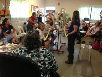
She also uses these 2-3 day dissection workshops to teach equine professionals and horse owners more about how their horses move and the damage their bodies can incur as a result of breeding, illness, injury or work.
Sharon is therefore uniquely placed to provide a source of raw data that is all but unparalleled.
Evidence from the dissection table
Some years ago, Sharon noticed an unusual action in the elbow of horses. She mentioned this to qualified practitioners and was informed that this action was quite normal. Not convinced, she began videoing horses prior to dissection and, within a short period of time, was able to match this action to a change in the elbow.

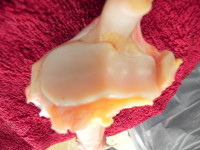
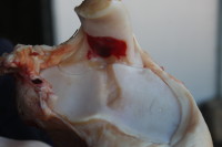
Not to beat around the bush, it’s an unusual form of degeneration in the horse’s elbow joint that involves all three bones. It’s a form of osteoarthritis that strikes the humeroradial joint and the ulna, causing deep and dramatic gouges into the cartilage, and eventually eroding bone.
When the joint is opened up, blood is frequently found in the synovial fluid (haemarthrosis). The fluid also displays decreased viscosity.
This is more than a little bit odd, as arthritis of the elbow is supposed to be rare in the horse.
Yet Sharon has found it to be present in numerous horses that have been euthanized under veterinary supervision for completely unrelated reasons.
Note: that’s not just some horses, but many.
Do you know where the nuchal ligament attaches on the cervical vertebrae? You think so? Evidence from the dissection table might prove you wrong – read more about Sharon’s findings on the nuchal ligament’s lamellar attachments…
This equine arthritis is visible in the living horse
The vital connection from video to dissection has enabled Sharon to indicate the presence of the elbow osteoarthritis in the horses she had been treating as an equine therapist.
It’s easy to spot, being a noticeable jarring in the elbow as the horse moves downhill – a kind of double action. Significantly, it’s what can be termed a gait anomaly, rather than lameness.
Does it sound – and look – familiar? It’s very likely that you’ve seen it in horses before and wondered what it was. The fact is that it’s so common, many people think it’s a normal action. It’s not. It’s a form of equine arthritis.
Sharon tells us she has seen the elbow problem in all types, breeds, sizes and ages of horses. Some affected horses have been elite dressage and eventing competitors. Interestingly, the problem is only presenting in ridden and driven horses.
If never worked, horses appear to remain forever free of this particular joint change.
Why the fuss – isn’t this just regular arthritis?
No. Arthritis of the horse’s elbow is considered to be rare in equine veterinary medicine.

The key to why it doesn’t often get diagnosed and is considered rare could be the absence of visible lameness. The arthritis identified by Sharon does not cause a distinctive lameness in the horse, although it does bring on a notable gait change, with the double step in the joint’s motion on the downhill.
Riders of such horses often just feel that their horse is a bit ‘off’, feeling a hesitation in the movement, but without being able to define the point of origin.
There are a couple more reasons why it’s not very visible: first, the action of the elbow is highly integrated with the overall shoulder action, and second, the massive triceps muscle has a further stabilizing effect on the joint.

And even if the elbow is explored, the relatively tight joint space means that degenerative problems are rarely seen in diagnostic imaging, although inflammation can show up in thermographic images.
When, unusually, a problem has been recognized and vets have attempted a corticosteroid injection of the joint (which happens to be the most difficult joint to access), blood has been found to be present.
A closer look at Sharon’s findings
Sharon May-Davis first presented some of her findings into elbow arthritis at a conference in Australia in February 2013: the Bowker Lectures at the Australian College of Equine Podiatherapy. Presenting alongside Prof Robert Bowker and Dr Bruce Nock amongst others, she discussed the club foot in the horse, and noted how the elbow degeneration she observes on the dissection table is always worse in the forelimb with the more upright hoof.
If the condition is bilaterally present, it unfortunately appears worse on the side with the slightly upright or higher hoof. What’s more, and according to Sharon, this also applies to the limb where an inferior check ligament desmotomy (surgery undertaken with the aim of correcting an upright hoof) has taken place and the ligament has later reconnected.
She has, as already mentioned, since established that it can occur in any ridden or driven horse. Here, she describes the problem in her own words.
“The action looks like a slip and or clunk into the shoulder or a shudder or a sliding / slipping action. It depends upon your perspective. The actual change in the action begins when the foreleg is in the ‘Stance Phase’ during the stride as the limb goes into the posterior phase of the stride. It is more obvious going down a hill.
“So far, 100% of ridden horses exhibit this condition to a varying degree (under dissection). Horses not ridden and with no abnormalities do not exhibit this condition (under dissection). Horses in harness also exhibit this condition.
“What does the joint look like? There appears under dissection to be substantial degradation in the cartilage of the humerus, radius and ulna.
“Most horses appear to handle this condition and continue with a normal life if not pushed to extremes. Although this sounds career-ending, in fact it is not. Once the horse gets through the worst of the wear pattern they re-settle in the joint and continue on with work.
“High level competitors require joint support to help sustain the elbow and other joints that may compensate for the change in action.
“Horses that jump are more inclined to land with straighter forelimbs. Be mindful that jumping and downhill work could possibly make the condition worse.
“Riders often feel instability in the horse’s forelimbs when traveling downhill and some even question the horse’s proprioception.
“Bodyworkers massaging the triceps (particularly the lateral triceps) actually exacerbate the condition as the massage releases the cast-like formation that this muscle provides.”
Here are some more examples of the elbow in action.
More research is needed, but so is support

Despite finding and documenting a huge number of dissection cases involving this particular issue, all unaided and unfunded by outside bodies, Sharon has consistently met with brick walls and skeptical responses when she has put the information forward to relevant authorities.
Why? It’s not as if she’s new to this. She has previously identified congenital malformations in the caudal cervical vertebrae of thoroughbreds, and in the atlas of Spanish Mustangs, as well as asymmetries in the femur structures of racehorses due to racing (published in the Australian Veterinary Journal).
She isn’t looking for funding (although she obviously wouldn’t say no), but would like to have this research taken up for the benefit of all ridden and driven horses. The sooner the problem is recognized and investigated, the sooner that episodic pain in the horse can be recognized, with appropriate joint support or rest given where appropriate.
And the sooner we can all learn more in our great drive towards improved equine health, the better. As Sharon says,
“In truth, we are still in the dark. Seeing it is one thing, analyzing it and providing a preventative program is something totally different.”
Questions, thoughts or comments? Join us at The Horse’s Back Facebook group.
More Information
Sharon May-Davis, PhD, M. App. Sc. (Ag and Rural), B. App. Sc. (Equine), ACHM, EBW, EMR was the Equine Therapist for the Modern Pentathlon Horses and the Australian Reining Team at the Sydney 2000 Olympics. She has worked with the Australian Champion from seven differing disciplines and has a particular interest in researching the musculoskeletal system. She also conducts clinics and seminars in relation to her work and regularly presents in the Northern and Southern hemisphere.
© All text copyright of the author, Jane Clothier, www.thehorsesback.com. No reproduction of partial or entire text without permission. Sharing the link back to this page is fine. Please contact jane@thehorsesback.com for more information. Thank you!
