
Angular Limb Deformities: it’s the technical term for crooked leg problems. The term sounds alarming, but how bad are they and exactly how common?
We’ve all seen shocking pictures of crooked foal legs in groups and forums, but thankfully they stand out exactly because they’re not so common.
However, many horses have mild or moderate versions of these. And what is normal is that many tend to slip under the radar, undetected by owners and breeders. And that’s not a good thing.
The problem is that moderate angular limb deformities (ALDs, as we’ll now call them) can create bigger problems as the horse grows older, with increased risk of lameness, instability, and joint degeneration.
They can cause complications when the horse is injured elsewhere in the body and needs to compensate, but can’t, because the non-straight limbs are already under pressure.
To repeat: many ALDs aren’t an issue. In fact, many are so mild that they’re never a problem.
The thing is, you need to know the difference. And that means knowing what to look for.
Some are obvious, others need veterinary radiographs to be sure. For all of them, the earlier you catch them in foals, the better. Always talk to your vet first.
So without further ado, here are ten ALDs you really ought to know about.
© All text copyright of the author, Jane Clothier, https://thehorsesback.com. No reproduction of partial or entire text without permission. Sharing the link back to this page is fine. Please contact me for more information. Thank you!

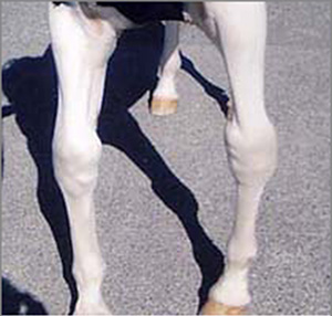
1. Carpal Valgus
‘Knock Knees’
This is the most commonly seen ALD in foals and horses. Technically it is an outward deviation of the lower leg from the mid line. Most of us see the other effect – the knees coming closer together.
In foals, the hoof often points outward. In older horses, the hoof may turn inwards – this is compensation to bring weight bearing back in.
There may have been a fetlock valgus (see #3) lower down, which has then caused the carpal valgus higher up.
This can be congenital. It can also be acquired, as a result of physitis of the radial growth plate immediately above the knee at a few months of age.
Incomplete ossification, when the carpal bones are underdeveloped at birth due to dysmaturity or prematurity, can contribute to this problem.
Key points: ALD of forelegs; knock-kneed appearance; lower leg angled outwards; affects one or both forelegs.
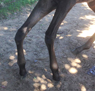
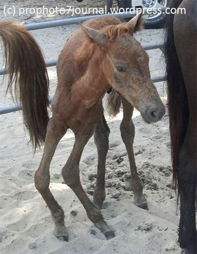
2. Tarsal Valgus
‘Cow Hocks’ (mistaken for)
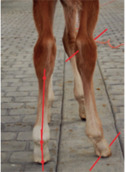
In these limbs, the point of hock angles inwards, while the lower limb angles outwards. Often, the pasterns are also angled outwards, giving a splay-footed look. In walk, the foot will swing in and then land wide.
The entire leg may be rotated out from the midline, as in this image.
In older horses, the medial heel may be crushed or under run.
Key points: ALD of hind legs; lower limb or whole limb angled out; affected hinds land wide; affects one or both limbs; one limb often worse.
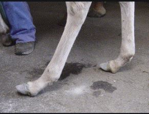
3. Fetlock Valgus
‘Toe Out’
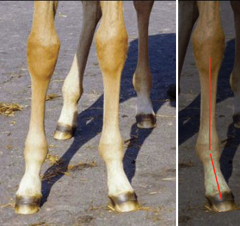
It’s extremely hard to find a picture that shows a fetlock valgus with no other visible limb deformity. Even the mild ones tend to be associated with rotation at knee level (see #10).
This is when the pastern and therefore hoof are angled away from the midline, below the level of the fetlock.
The valgus in this photo is reasonably clear, although there’s some rotation in the knee. Note that the foal is weighting this limb less and an upright foot is already starting to develop.
Key points: usually ALD of forelegs; sometimes with tarsal valgus in hind legs; one or both feet angled outwards; one often worse.
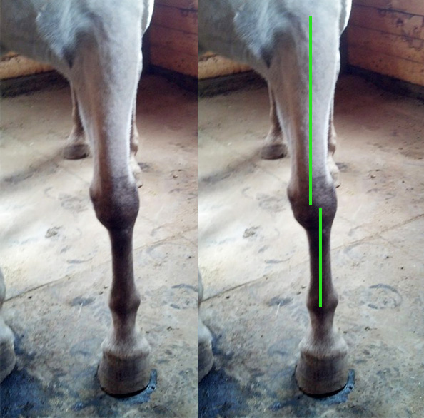
4. Fetlock Varus
‘Toe In, Pigeon-toed’
With this problem, the foot is angled towards the midline below the level of the fetlock,
This problem is often combined with offset canons or rotated carpals (see # 9,10), as the foal is compensating in an effort to bring the weight bearing line back beneath the shoulder joint.
In older horses, the lateral heel may be crushed or wear down quicker. Depending on severity, there may be a tendency to ringbone in later life.
Key points: usually ALD of forelimb; affects one or both legs; often accompanied by a carpal ALD.
5. Carpal Varus
‘Bow Legs’
In a carpal varus deformity, the lower leg is angled inwards, towards the midline. Our eye tends to see it as the joint being angled outwards.
You don’t often hear about bow legs in horses, do you? That’s because it’s rare.
It’s usually seen in older horses, when the problems have layered up. Arthritis may already be present in the joints, while tendons and ligaments may have lesions.
Some may have had lax ligaments at birth, or have ruptured the common digital extensor tendon at a young age, causing misalignment of the carpal bones.
Key points: rare ALD of foreleg; bow-legged look; foot dishes inward; arthritis and lameness likely.
6. Flexural Deformity
‘Contracted Tendons’ (so-called)
This is a common deformity in newborn foals (call your vet).
Sometimes it affects the carpal (knee) joints only and the foal is still able to use the lower leg quite effectively. Or, it affects both the carpal joints and the fetlocks, leading to a more serious situation where they can’t walk on the hoof.
This can also be an acquired deformity related to incorrect nutrition during the first weeks and months of life. In these cases, nutritional measures, remedial hoofcare and even surgery may be required .
The hind legs can also be affected, although this is less serious as the direction of joint flexion encourages correction.
Mild cases in foals can often come right on their own in the first days of life with a little help. However, it is always important to involve a vet at an early stage for monitoring.
Key points: ALD mostly affecting forelegs; either knees only or knees and fetlocks.
7. Hyperextension
‘Lax Tendons’
Again, this is super-common in newborn foals and it can often resolve all on its own in the first few days of life with a mix of confinement and limited exercise.
Occasionally, in the case of dysmature and premature foals, the problem can be more complex. Combined with incomplete ossification and lax ligaments, hyperextension can lead to another level of ALD, as the cuboid bones of the carpals and tarsals become misshapen.
Once more, give the foal a couple of days to straighten up and then talk to your vet if it persists.
Key points: affects forelegs and hindlegs often together; usually resolves with conservative exercise; support for limbs and hooves may be required.
8. ‘Windswept’
This startling looking deviation is often seen in foals that are post-mature – they have had a long gestation and have been restricted within the uterus.
 What we’re looking at is a combination of ALDs: carpal valgus and varus in front, and/or a tarsal valgus and varus behind.
The problem will general resolve within a few days, sometimes with a bit of hoofcare and bodywork to assist.
However, if the foal appears weak despite being ‘over-cooked’, with hyperextension (#7) as well, be sure to monitor it especially carefully during the early weeks and months. This is because incomplete ossification may also be present.
Key points: affects forelimbs and/or hindlimbs; hyperextension may also be present; often resolves in days.
9. Offset Cannons
Strictly speaking, this is a conformational trait, as it often visible in the sire or dam. However, it is still classified as an ALDs on account of the structural weaknesses involved.
In the offset cannon, the cannon bone is positioned wider than the midline of the leg, although it still faces forwards. The hoof is also forward facing.
In one leg, this ALD may have no negative effect on athletic performance. Yet if present in both forelegs, the horse may be less stable in some situations, such as going downhill.
It increases the chances of two things: developing a splint (medial leg) and a tendency for the foot to turn inwards, which could then lead to an acquired fetlock varus (see #4).
In racehorses, it can increase the chance of injury in the medial carpals, while other sports horses may be prone to osteoarthritis in the joint.
Key points: cannon appears shifted sideways; cannon, hoof and knee face forwards; usually inherited conformation; medial splints more likely.
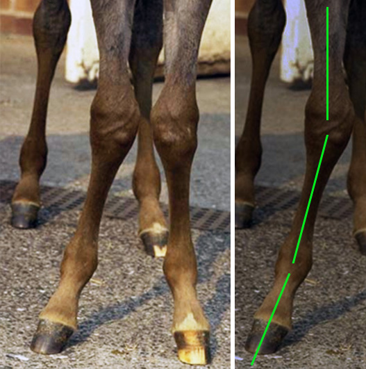
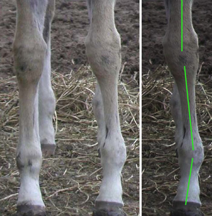
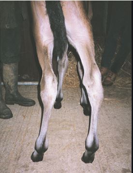
10. Carpal Rotation
‘Bench Knees’
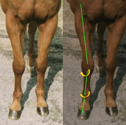
In this ALD, the cannon bones are rotated slightly outwards, as are the lower knee bones and the feet. This sets the horse up for a fetlock valgus (‘toe out’), as can be seen in the left foreleg in this image.
Also a conformational trait, this kind of leg is frequently seen in Quarter Horses. A wide chest and narrow lower limbs can also contribute to a postural rotation of the foreleg.
Similar rotation in the lower forelimb can also be an acquired ALD, if a young horse has to change its weight bearing for a long time, due to (say) an injury in the diagonally opposite hind limb.
From certain angles, this rotation can look like a carpal valgus. Always check from different angles to be sure.
Key point: face of cannon and knee appear rotated outwards; pastern and toe often angled outwards, medial splints more likely; medial hoof wears faster.
Other resources
[1] Redden, RF. How to Evaluate Foot Flight and Leg Alignment
AAEP PROCEEDINGS, Vol. 57, 2011, p.407
[2] O’Grady, SE, Routine Trimming and Therapeutic Farriery in Foals, Th Veterinary Clinics of North America, Equine Practice, 2017