• Skip to main content
  • Skip to footer

The Horse's Back

Body Talk for Thinking Owners

  • Home
  • Articles
  • BEMER
  • Store
    • Store
    • Shipping & Returns
    • Support
  • Equine Healthworks
    • Bodywork for Horses
    • Reviews
    • Leave a Review
  • About & Contact
    • About
    • Contact

Anatomy

Yes, We Can Image for Transitional Vertebrae in Horses

September 23, 2021 by Jane @ THB 7 Comments

It’s been a question of mine for a while. Can diagnostic imaging show the presence of transitional vertebrae?

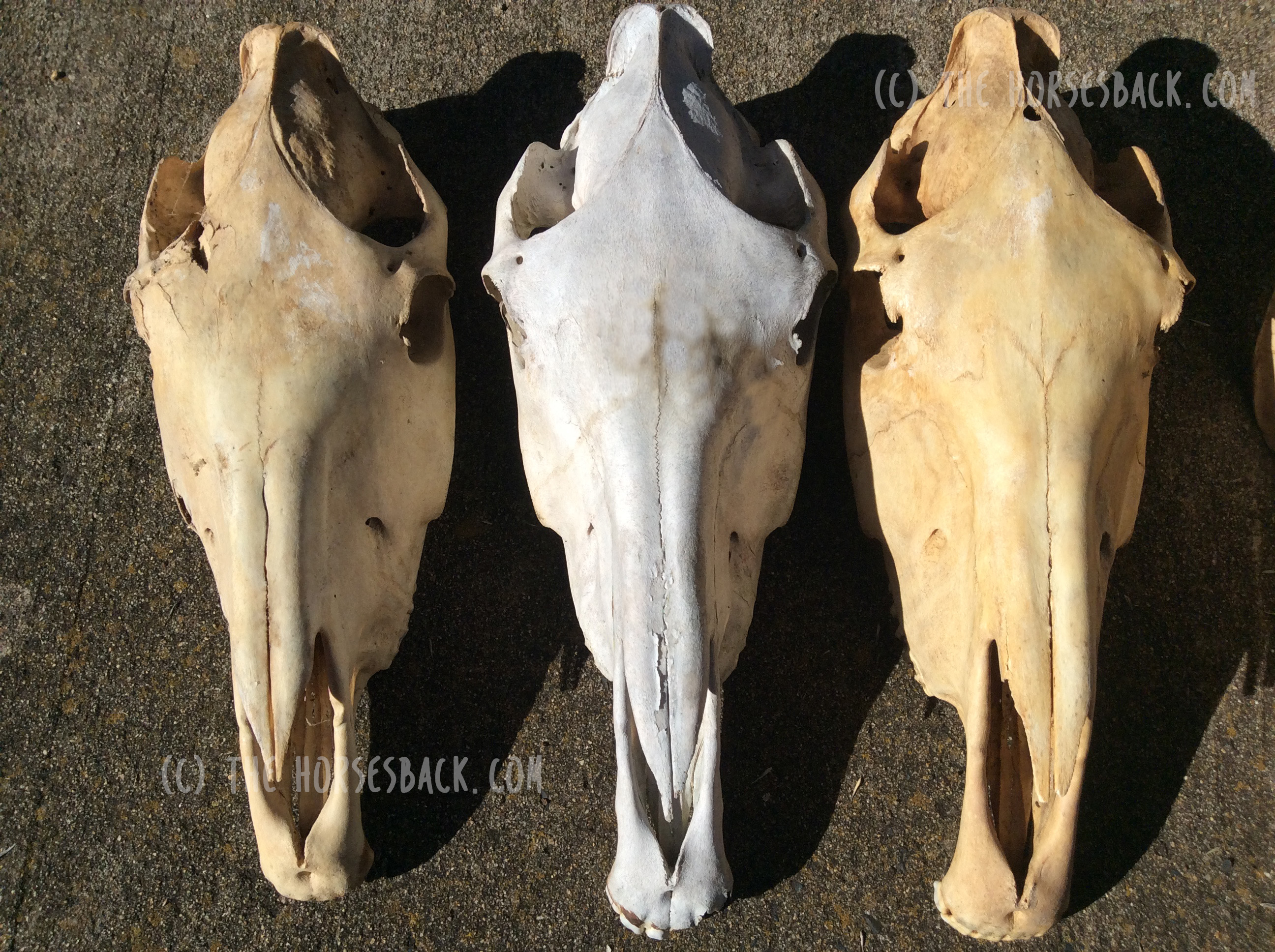
We’re seeing many bone samples from dissections, as shown in my previous article on transitional vertebrae.

But if we’re to help our horses that live with this issue, we need to identify it before they’re dead. (Yes, right?!)

Allow me to introduce a practicing vet and educator who is doing just that.


Imaging for Transitional Vertebrae 

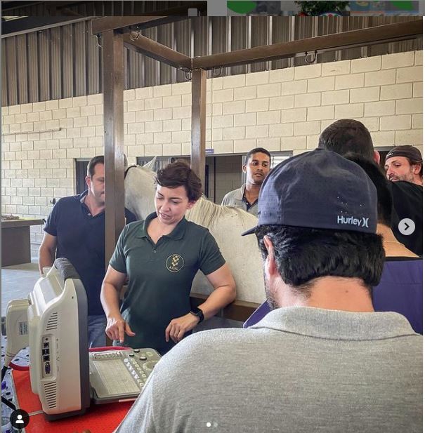
Meet Dr Brunna Fonseca, Associate Professor, educator and specialist in equine orthopedics, focusing on the spine and nervous system. She’s based in São Paulo, Brazil.

I’ve been following her Instagram for a while, because she posts brilliant videos and photos explaining what she does, and how, and why.

I was delighted to see a recent post on imaging for a transitional vertebra, which included fantastic visuals. Such a great communicator!

Dr Brunna has kindly given me permission to repost her images and descriptions here. So without further ado…

  • All images copyright of Axial Vet

Ultrasonograms

Ultrasonography for transitional vertebrae
Angle of transducer. Image: Equine Neck and Back Pathology: Diagnosis and Treatment, 2nd Edn. Ed. Frances M.D. Henson. © 2018 John Wiley & Sons, Ltd.

The following ultronographic images are each a composite of two images, one showing the left side and the other the right.

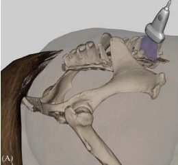
This textbook illustration helps to show the angle the image is taken at. This angle is usually used for imaging the articular facets of the vertebrae.

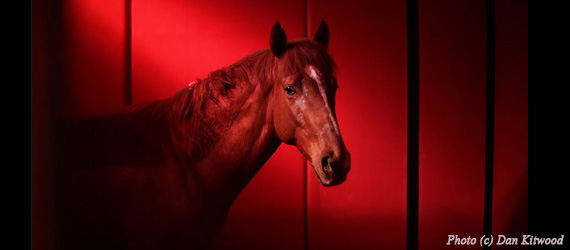
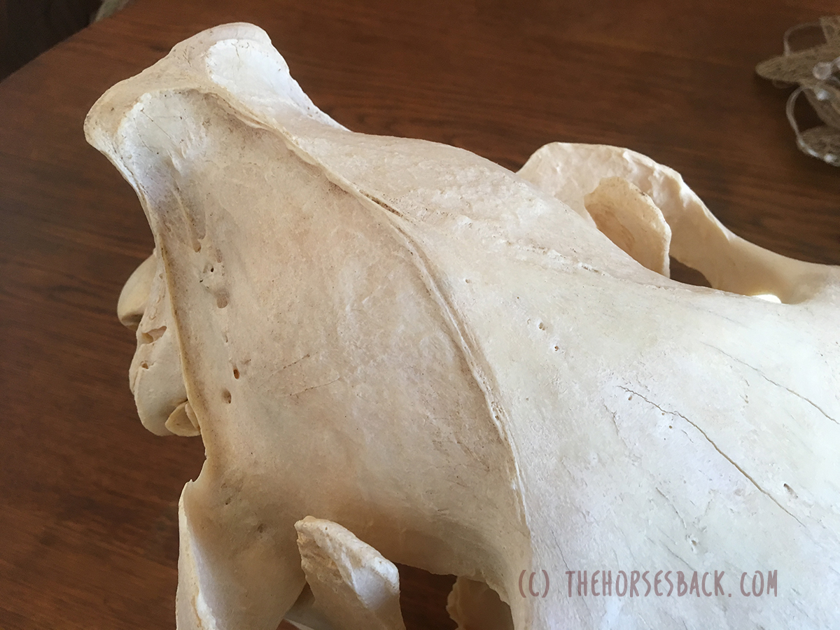
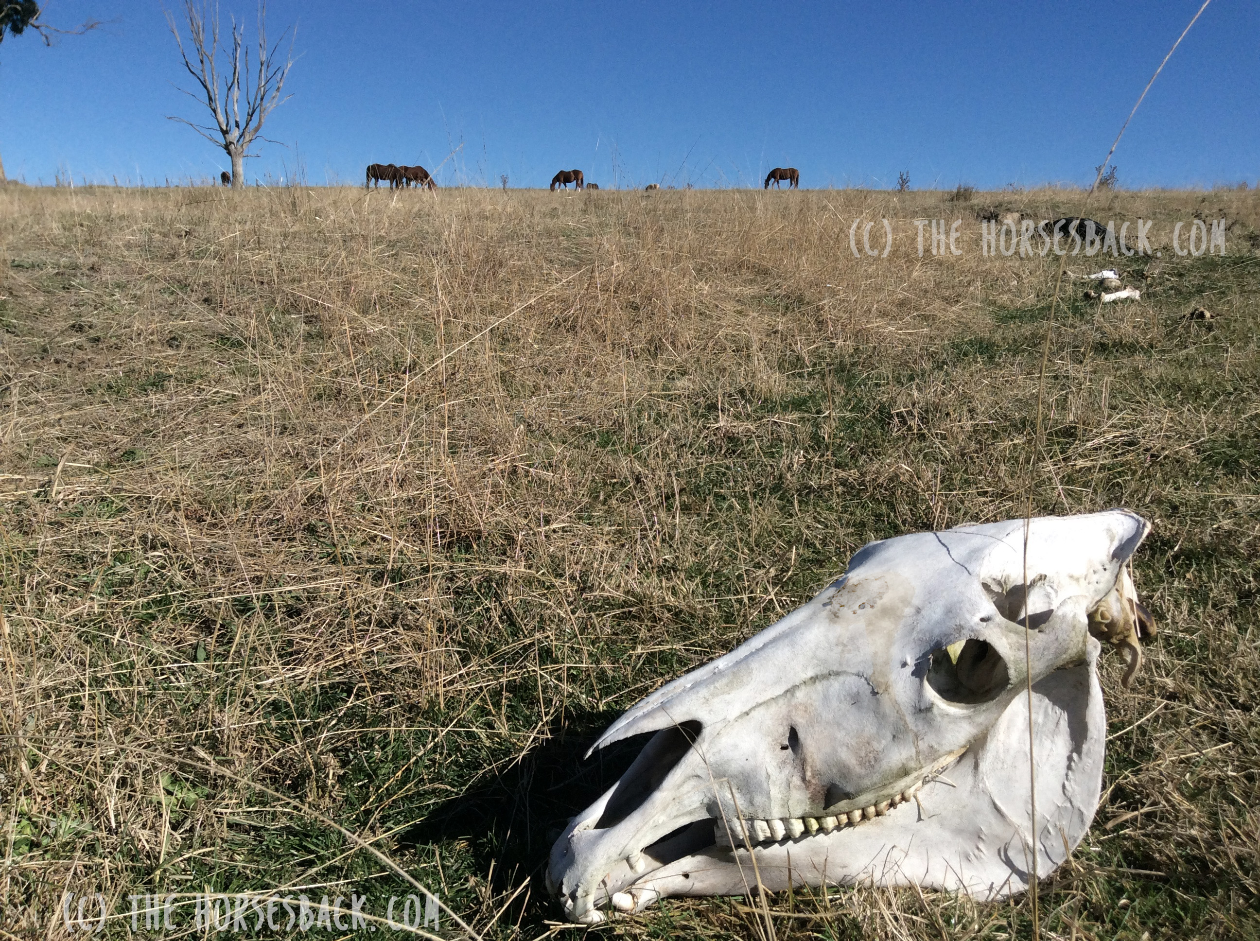
Additionally, the image at the top of this article shows a transitional vertebra at T18, like the mare being diagnosed by Dr Brunna.


1.  Can we recognise transitional vertebrae?

The first image shows two sides of a mare’s body. The hand icon gives us a strong hint of where to look… This appearance is very similar to that of the TB mare in my previous post.

Dr Brunna writes, “This mare has the T18 transitional vertebra, presenting a transverse process similar to the lumbar vertebrae on the right side, which causes the appearance of the horse to have the most visible rib on that side.

The occurrence of transactional vertebrae in the horse is not uncommon, especially in the thoracolumbar transition, which can occur in T18 or L1.”


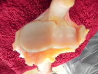
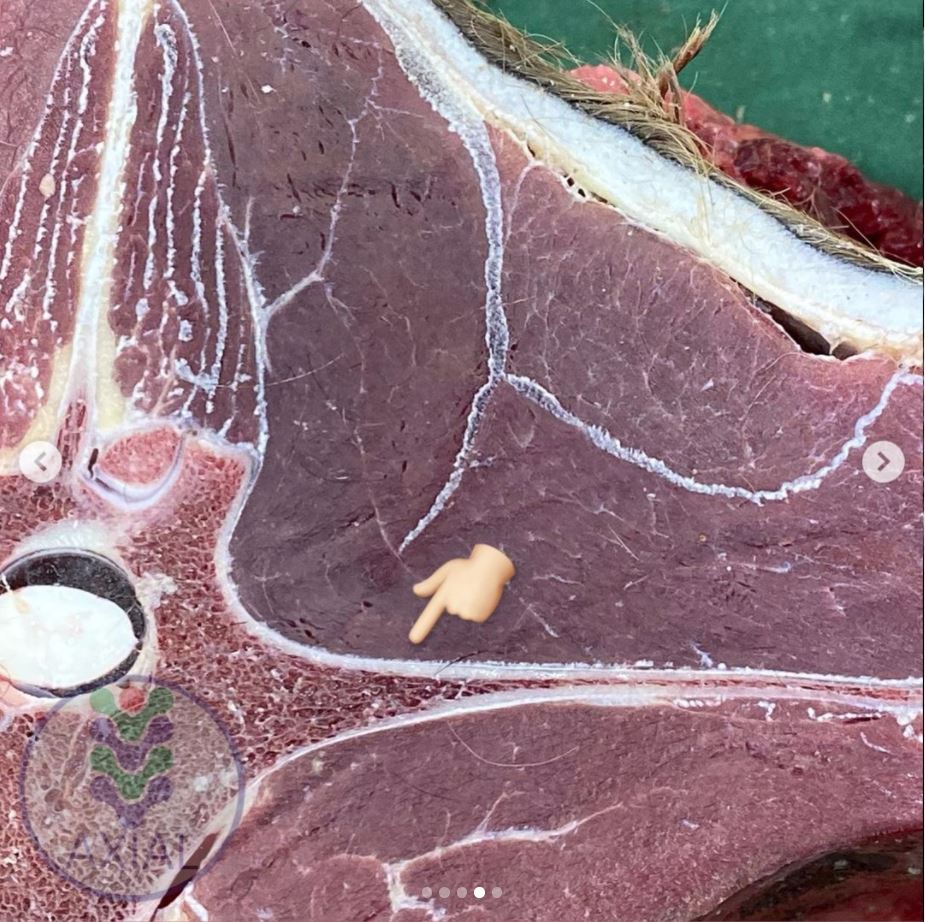
2. Section of a thoracic vertrebra

This image is from a different horse showing a normal rib head and its joint with the vertebra.

Dr Brunna writes, “This is the image of a thoracic vertebra, showing the costotransverse joint.”


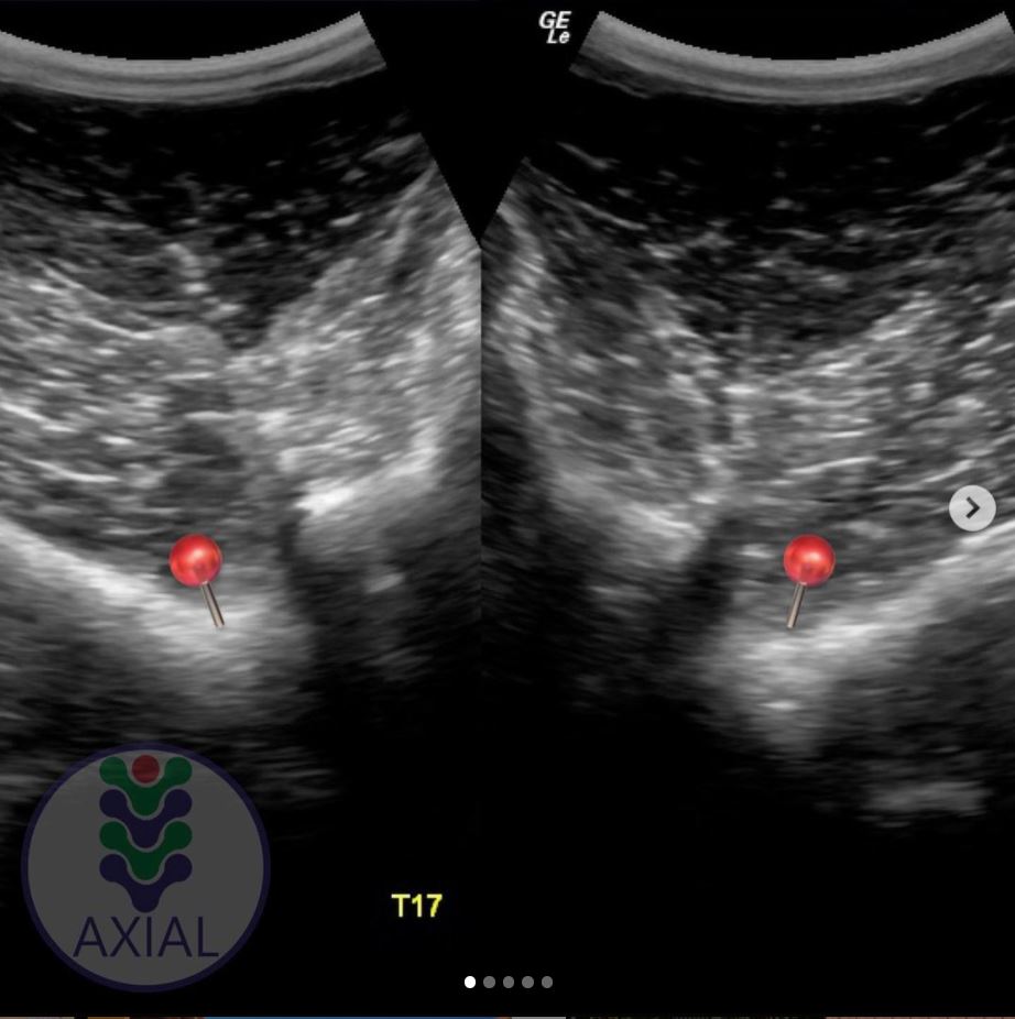
3. Image of a normal vertebra

Dr Brunna writes, “This is a T17 ultrasound image, where we can see the image of the normal costotransverse joints.”

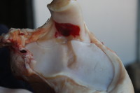
This is the bay mare again.

As with the previous cross section, the red pins which show the facet joint between rib head and vertebra.


4. Section of a lumbar vertebra

This is cross section is of a normal lumbar vertebra from a different horse.

As you can see,  there is no joint between the  transverse process and the vertebral body.

The process is wide and flat, and integral to the vertebra.


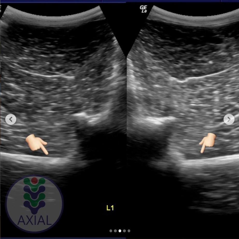
5. Image of a lumbar vertebra

Here’s an ultrasound of the first lumbar vertebra (L1) in the bay mare.

As in the above cross section (picture 4), there is no joint between the transverse processes and the vertebral body.

We now have ultrasound images of the normal T17 and normal L1. As we will see, the transitional vertebra mixes elements from both.


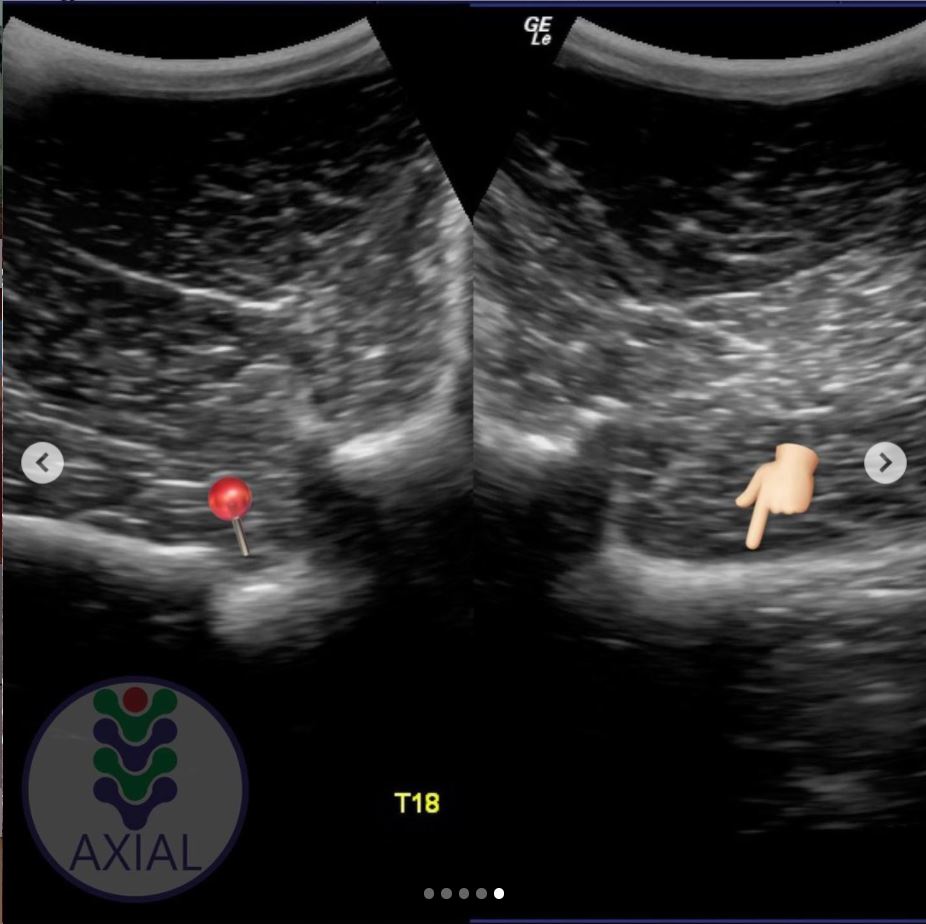
6. Imaging transitional vertebrae

“This is an ultrasound image of T18, where we can see the image of the costotransverse joint on the left side (red pin) and image of the transverse process on the right side.”

So here’s the underlying skeletal issue in the bay mare.

The left side is a normal joint, being the same as the T17 thoracic vertebra (picture 3).

The right side is similar to the previous image of the lumbar vertebra (picture 5).

It is not identical, for while the process-like rib is joined to the vertebra, it is not the same shape and does not lie as flat as the lumbar process.


Want to Hear More From Dr Brunna Fonesca?

You can follow her Axial Vet Instagram page to see examples of her equine cases and their assessment, in images and videos.

An increasing number of captions are now translated into English.


Filed Under: Bodywork Tagged With: Anatomy, equine anatomy, equine bodywork, equine malformation, equine skeleton, GA, horse anatomy, transitional vertebra, transitional vertebrae

‘The Size of a Walnut’ – Does Equine Brain Size Matter?

November 5, 2019 by Jane @ THB 7 Comments

There seem to be quite a few social media posts about the equine brain of late – and that’s no bad thing. 

In some ways, the brain is simply the latest part of the equine anatomy to come under the spot light. It’s being subject to statements about welfare, training and psychology – and that’s definitely a good thing (here’s one from Hippologic.)

However, I want to add something to this equine brain discussion. I just happened to run in to it when I went down a research rabbit hole a couple of years back.

We often hear how brain size is not directly linked to an individual’s intelligence. At the same time, a relatively large brain is said to signify intelligence in humans, while that of the horse, popularly said to be the size of a (large) walnut, is said to account for their lack of intelligence.

Vintage anatomy print showing relatively small size of equine brain to body size.

This falls down once we look at elephants, which have relatively small brains yet are pretty cluey.

In horses, innovative behaviors without evolutionary basis are often used as a measure of intelligence (read more here about unlatching gates). Leaving behaviour-based measurement methods aside for the moment, let’s ask: how do we figure out if there’s an association between different brain sizes and intelligence levels?

(Note: if you’re a neuroscientist of any description, look away now. What follows is a highly simplistic overview of this incredibly complex subject area.)

© All text copyright of the author, Jane Clothier, www.thehorsesback.com. No reproduction of partial or entire text without permission. Sharing the link back to this page is fine. Please contact me for more information. Thank you!


Measuring Equine Brain Mass and Body Mass 

In zoology, the starting point isn’t about brain size, but brain mass compared with body mass or weight.

Even then, it’s not a matter of separating the brain from the body and then weighing both. The most accurate way of measuring this accounts for several anatomical, physiological factors, including the amount of water in the brain.

The result is a single figure that is called the encephalization quotient (EQ). The EQ for a species is arrived at after researchers have performed the calculation for dozens of animals.

The parietal bones form the domed ‘cranial vault’ of the skull.


So How Does This Look for the Equine Brain?

Only a handful of equine researchers have delved into EQs, as this is mostly an area of zoological neuroanatomy.

In this study by Cozzi et al (2014), the brains of 131 mixed breed adult horses (no ponies) were collected and weighed.[1] Researchers found first that the adult horse’s brain weighs 600 – 700 g. The average brain weight for horses aged 2 years and over was 606 g, while the average bodyweight was 535.22 kg.

This meant the horses in this study had an EQ of 0.78.

Here are the EQs for some of the large mammals: Cow – 0.55, Pig – 0.6, Camel – 0.61, Horse – 0.78, Goat – 0.8, Wolf – 0.9, Domestic Cat – 1.00, African Elephant – 1.67, Gorilla – 1.76, Human – 6.62.

And if you’re really interested, here’s the calculation used in the equine paper. Other scientists use different calculations – there is no standard approach.

EQ = E i / 0.12 P2/3Ea/Ee


So, Are Horses Intelligent – or Not?

A larger brain mass compared with body mass is often associated with better cognitive functioning, but that does not mean it causes it.

Brain size is therefore a very general measure for intelligence. What actually matters are the specific areas of the brain and their relative sizes.

The bigger the frontal lobes, the more capable the species is of ‘goal directed’ behaviors – that is, the ability to analyse information and act accordingly, planning ahead. [2]

Here we hit an issue. The frontal lobes are either relatively small in the horse, or non-existent – and this is a matter of contention. Some published veterinary researchers maintain that they do, as shown below.

Rough comparison of the frontal lobes of the horse (left) and human brains.

However, researcher and author of Horse Brain, Human Brain Janet Jones PhD writes, “Basic anatomy shows that horses have no frontal lobes and no prefrontal cortex. No qualified PhD trained in neuroscience disputes this anatomy.”[3]

Whichever is true, the take home for both is that the horse is more likely to react in the moment. This is not to say that horses lack intelligence, but that they think and respond differently.


The brain’s fissures are also important. These are the wrinkles and grooves, known as sulci (sunken inwards) and gyri (protrude outwards). They’re standard within species, although the brains of some species have more complex surfaces than others.

Rats, considered to be on the lower end of the intelligence scale of mammals (although rat owners will surely disagree), have smoother brain surfaces than horses. In turn, horses have fewer fissures in their brains than primates.

The area contained within the cranium is the ‘cranial vault’. Its inner surface perfectly matches the outer surface of the brain, as they develop together as the animal grows.  If you could look inside this part of the skull, you would see a perfect mould of the fissures.

More recent research also links the organization of neurons (nerve cells) and synapses in the brain to intelligence.


Surely There’s a Difference Between Breeds?

Different breeds of horses certainly have differences in the shapes of their heads.

However, these differences are slight overall. In a study of TBs, STBs and Arabians, the relative proportions of the ‘neurocranium’ – the area above the frontonasal suture, including the cranium – were reasonably similar between breeds.

It was the lower part of the skull, primarily the nasal bones and the maxilla, that varied most and gave the breeds their different looks [4]. The study did not measure the cranium itself.

The neurocranium aligns with the ends of the frontonasal suture and includes the temporal and parietal bones, ending at the occiput.

This suggests that while some breeds may look extremely different – take the Welsh Cob and the TB, for instance – the neurocranium may be nearly square in all, at least when viewed from the front.

And even though some breeds may have proportionately larger heads, all (excluding ponies) will have EQs grouped around the average score of 0.78 mentioned earlier.

Small and wide ponies, incidentally, often have quite large parietal domes (or tuberosities, as they should be known), but the jury is out as to whether this makes them more intelligent… The fact is that we don’t know.


A Little More on Equine Brain Size

There are a few other differences that aren’t documented. Comparing horse skulls, we can see that some have a cranium that is narrower in relation to overall skull width than others. They also vary in shape: some are very full and round, while others are more teardrop shaped.

You can see this when you look at the spaces to either side of the parietal ‘dome’ and temporal bones, where the coronoid processes (tips) of the mandible protrude behind the zygomatic arch.

This may be due to breed or it may be individual. Our own skulls vary from person to person, with some aspects being just how we are, while others may be more developmental.

We can see this in horses too. Dwarf horses can have domed heads, as can horses that have been born prematurely.

This can affect intelligence – researchers have found that in humans, when the brain is smaller due to development delays, the intelligence can be lower. If it is smaller without any developmental delay, it makes no difference at all. [5]

Interestingly, a new study in humans shows that the longer the time Romanian orphans spent in the institutions as babies, the smaller their total brain volume, with these changes being associated with a lower IQ. You can read more about that study here. [6]

Personally, I would love to know more about this, as I’ve been researching the developmental effects of gestational problems in horses, including the effects of premature birth (my PhD thesis lives here). It’s the same old problem though: once a horse is at the stage where we can examine its skull, its early history is usually lost in the mists of time.

Ultimately, as with humans, what is going to make the most difference to us as horse owners is the individual’s learning experiences at different stages of its life. This is also where equine personality comes in, and the methods of training used, but those are different subject areas altogether.


[1] Cozzi et al., The Brain of the Horse: Weight and Cephalization Quotients, Brain Behav. Evol., 2014; 83:9-16

[2] McGreevy, P., Equine Behavior – A Guide for Veterinarians and Equine Scientists, Elsevier, 2012.

[3] Janet Jones – Horse Brains Facebook page

[4] Evans KE, McGreevy, PD., Conformation of the Equine Skull: a Morphometric Study, Anat. Histol. Embryol., 2006, 35(4): 221-7

[5] de Bie H. et al. Brain Development, Intelligence and Cognitive Outcome in Children Born Small for Gestational Age. Horm Res Paediatr 2010, (73)6-14.

[6] Mackes, NK. et al.,  on behalf of the E. Y. A. F. (2020). Early childhood deprivation is associated with alterations in adult brain structure despite subsequent environmental enrichment. Proceedings of the National Academy of Sciences.

Filed Under: Bodywork Tagged With: Anatomy, equine anatomy, equine brain, equine skull, GA, horse anatomy, horse brain, horse skull

Why I Now Believe My Warmblood Gelding Had the C6-C7 Problem – Reader Story

November 16, 2017 by Jane @ THB 14 Comments

“I went through three years of horror…”

So speaks a Warmblood owner, reflecting upon a truly dreadful period in her life. Like many other owners, she has gained belated understanding of her horse’s problems after reading about research findings into the C6-C7 malformation (article) led by Australian gross anatomist, Sharon May-Davis.

Sadly, this is a bittersweet moment: there is a temporary relief as everything finally makes sense, yet with it comes the realisation that there is nothing that can be, or could have been, done for a treasured horse bearing the more extreme version of this malformation.

Here, Diana from Michigan, USA, gives her story of the desperation and – ultimately – heartache she experienced when trying to find out what was wrong with her horse of a lifetime, a young Oldenburg gelding, “the kindest horse I ever had”.

Note: We are not identifying the lines of the individual horse in this story. If you’re thinking we should, perhaps remember that it’s a litigious world out there.

But for now, over to Diana.

© All text copyright of Jane Clothier, www.thehorsesback.com. No reproduction of partial or entire text without permission. Sharing the link back to this page is fine. Please contact me for more information. Thank you!


Diana’s Story

I purchased a stunning 3-year-old Oldenburg to fulfill my dream of developing a young horse through the upper levels in dressage. I cashed in the 401K [retirement savings plan] for the horse and bought a Schleese saddle.

For the first two years (ages 3 and 4) we were champion every time out.

At age 5, however, my trainer noticed he began to drag his left hind toe. Then his impulsion began to deteriorate, and months later he became too unstable to safely handle. He was euthanized at 7 years old.

Looking back, I now believe that all his quirks were directly related to this problem. I am going to briefly list the oddities I experienced with this wonderful horse.

I am not a vet or tech, and am only sharing my experiences in the event that it may help someone else, because my journey was one of the most emotionally draining experiences in my life.


Possible Signs of this Warmblood’s C6-C7 Problem

I purchased my gelding on his 3rd birthday and he’d had 30 days under saddle. The oddities of my gelding: he was extremely sweet and willing – the kindest horse I ever had.

He required a tremendous amount of leg every step. Although there seemed to be no ‘self-carriage’, I attributed this to his being green and still learning.

He would buck EVERY TIME he was saddled (I was very conscientious about slowly tightening the girth, tried every type of girth, pads, had adjustable saddle, etc).

Again, I attributed the bucking to youthfulness, and would let him buck out for a few laps, and then wondered if I had created a habit, because this occurred every schooling.

  • If he wasn’t lunged first, he would explode into a non-rideable rodeo with absolutely no warning.
  • He had a very difficult time bending to the left – he would try, but would drift.
  • Two separate times after mounting at the block, he felt like he arched or sucked his back up about 3 inches before walking off.
  • After 25 mins of schooling he would start rooting at the reins, or if the lesson was a short one, as soon as I’d give him his head to cool out.
  • He was a stumbler – not often – again, I attributed it to youth.
  • He could not be shod or trimmed without being sedated.
  • Once, a friend said it looked like he couldn’t put his head down to reach hay. Of course, I thought she was nuts until I witnessed it first hand.

Questions, thoughts or comments? Join us at The Horse’s Back Facebook Group. 


Investigating the C6-C7 Problem

During my discovery phase, he was diagnosed with moderate PSSM. So, he and I spent 9 months testing sugar levels in hay, with turnout in a dry lot.

There were periods he would barely walk forward when leading, and at the time I thought he was tying up, but it didn’t seem like what I had read, and I have never witnessed a horse tying up.

Then in his latter days, his neurological issues became more obvious with each passing day.

I did have a myelogram done on him, and it revealed compression of the spinal cord at C3 and C4.

Vets at Michigan State University did finally identify malformations of both C6 and C7 through radiographs. Their report stated:

The intervertebral foramen is severely reduced at C6-C7. A minimal amount of osseous proliferation is noted along the dorsal aspect of the articular processes at C5-C6.

Interpretation: Multifocal cervical articular degenerative joint disease (C4-T1), most severe at C6-7.”

I had a very difficult time with the decision to euthanize him, because his neurological issues would fluctuate. Some days, no evidence all. On other days, he would fall against the stall wall when turning him around in the stall.

In any event, I euthanized him, believing that he was experiencing severe pain from the narrowed foramen at C6 and C7.

I believe they were the reasons for the bucking, for needing to be sedated for the farrier, for not being able to lower his head, for rooting on the reins, for barely walking forward when I thought he was tying up, and the bizarre lifting of his back. He also had a very difficult time holding his right lead, and would swap unless you really held him.

I am so, so glad I ran across this website and article because I will go to sleep tonight knowing that euthanizing was for sure the right thing to do. Thank you!

Diana


If you’re about to purchase a horse that falls into the ‘at risk’ category for this malformation, do your due diligence as a buyer, and have the caudal cervical vertebrae radiographed as part of your pre-purchase examination. A protocol for radiographing for the malformation is on this site.

The following papers also cover radiographs and CT imaging of the C6 malformation:

Ex Vivo Computated Tomographic Evaluation of Morphology Variations in Equine Cervical Vertebrae, Veraa, S. et al,  Veterinary Radiology & Ultrasound, Vol. 57, Issue 5

Prevalence of Anatomical Variation of the Sixth Cervical Vertebra and Association with Vertebral Canal Stenosis and Articular Process Arthritis in the Horse,
Spriet, M. and M Aleman, Veterinary Radiology & Ultrasound, Vol. 57, Issue 3


If you wish to comment, please feel free to do so below, but please do so with respect for an owner who found herself in a nightmare situation. Alternatively, come over the blog’s discussion group on Facebook.

Filed Under: Bodywork, Sharon May-Davis Tagged With: Anatomy, c6 c7, cervical vertebrae, oldenburg, Sharon May-Davis, warmblood, wobbler syndrome, wobblers

Meet Spinalis, the Forgotten Muscle in Saddle Fitting

December 20, 2016 by Jane @ THB 43 Comments

Spinalis Header

It’s barely mentioned in saddle fit or anatomy books, yet the muscle Spinalis cervicis can hugely impact on the spinal health and movement of the horse, particularly with poor tack fit.

Meet muscle Spinalis cervicis et thoracis, a far more important muscle than is generally realized. As a deep muscle, it’s influential in mobilizing and stabilizing that hidden area of the spine at the base of the neck, the cervico-thoracic junction, deep between the scapulae.


Where to Find this Muscle

As part of the deeper musculature, Spinalis is as hidden in books as it is in life. Usually, it’s a single entry in the index.

Spinalis Standard
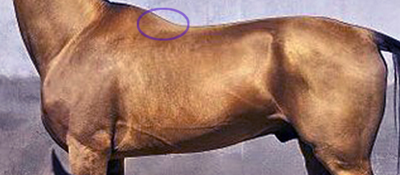
At best, it has no more than a bit part in anatomical illustrations,  usually as a small triangular area at the base of the withers. This is also where we can palpate it.

The reality is quite a bit more interesting. It’s actually a muscle of three parts – dorsalis, thoracis and cervicis. These names denote its many insertions, for it links the spinous processes of the lumbar, thoracic and cervical vertebrae.

  • Bradley_2.1
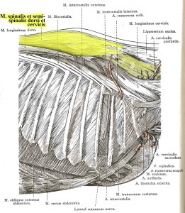
Further back along the spine, it lies medially to Longissimus dorsi, and in fact integrates with this larger, better known muscle, attaching to the processes of the lumbar and thoracic vertebrae.
  • When it reaches the withers, it becomes more independent, attaching to the processes of the first half dozen thoracic vertebrae (T1-T6). Here, the cervical and thoracic portions overlap and integrate to share a common attachment. (The part we palpate, at the base of the withers, is the thoracic section.)
  • Heading into the neck, as Spinalis cervicis, it attches to the last 4 or 5 cervical vertebrae (C3/C4-C7). Only the lamellar portion of the nuchal ligament runs deeper than this muscle.

Dissection 2
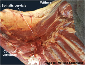
Its integration with other muscles is complex, and its close relationship with Longissimus dorsi partially explains why it doesn’t get much consideration as a muscle in its own right.

It is the more independent section, Spinalis cervicis, between withers and neck, that we are interested in, although its influence is present along the entire spine.

© All text copyright of the author, Jane Clothier, https://thehorsesback.com. 

What Does Spinalis Do? 

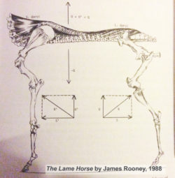
In his 1980s’ Guide to Lameness videos, Dr. James Rooney, first director of the Gluck Equine Research Center, University of Kentucky, referred to Spinalis as part of the suspension bridge of muscles supporting the spine (Longissimus dorsi achoring from the lumbosacral vertebrae, Spinalis thoracis et dorsalis from the upper thoracics). He also refers to this extensively in The Lame Horse (1988).

In fact, the suspension bridge analogy only really makes sense if Spinalis dorsi is considered.

Spinalis cervicis is usually credited with a role in turning the head to left to right, and raising the head.

Bradley Spinalis-1Older texts, such as Bradley’s 1922 veterinary dissection guide, Topographical Anatomy of the Horse, mention its role in stabilizing the spine.

This creates a point of interest. Given that the nuchal ligament (lamellar portion) doesn’t attach to C6 and frequently only weakly with C5 (see the findings of anatomist Sharon May-Davis, in this earlier article ), Spinalis cervicis suddenly appears pretty important in stabilizing and lifting the base of the neck, particularly as it does so at the point of greatest lateral bending.


Spinalis and Poor Saddle Fit

Anyone who has been involved in close examination of the horse’s back will recognize Spinalis thoracis where it surfaces close to the skin, on either side of the withers.

When a horse has been ridden in an overly tight saddle, this small area of muscle can become pretty hypertrophic – raised and hardened. Typically, the neighbouring muscles are atrophied. When Spinalis is palpated, the horse often gives an intense pain response, flinching down and raising the head.

GerdHeuschman
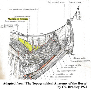
What often happens is this. An overtight saddle fits over the base of the withers like a clothes peg, pinching Trapezius thoracis and  Longissimus dorsi. However, it frequently misses Spinalis thoracis where it surfaces, wholly or partially within the gullet space. Often, the muscle is partially affected.

It’s as if the neighbouring muscles are under lockdown. Free movement of the shoulder is restricted and the horse’s ability to bear weight efficiently while moving is impeded. In response to the surrounding restriction and its own limitation, this muscle starts to overwork.

Result? The horse, which was probably already moving with an incorrect posture, hollows its back even further, shortening the neck and raising its head.  As this becomes even more of a biomechanical necessity, all the muscles work even harder to maintain this ability to move, despite the compromised biomechanics.

Working harder and compensating for its neighbours, Spinalis becomes hypertrophic. It is doing what it was designed to do, but it’s now overdoing it and failing to release. We now have a rather nasty vicious circle.


Spinalis photo

Here, Spinalis thoracis stands out due to atrophy of the surrounding musculature. In this TB, a clearly audible adjustment occurred in the C4-C5 area after the muscle was addressed. 


Vicious CircleThe Inverted Posture and Asymmetry

Of course, saddle fit is not the only cause of an inverted posture. However, any horse that holds its head and neck high for natural or unnatural reasons is more vulnerable to saddle fit issues, thus starting a cascade effect of problems.

Are there further effects of this hypertrophy? Consider the connections.

  • When saddles are too tight, they’re often tighter on one side than the other. This can be due to existing asymmetry in the horse, such as uneven shoulders, uneven hindquarters, scoliosis, etc.
  • On the side with greater restriction, the muscle becomes more more hypertrophic.
  • With its attachment to the spinous processes of the lower cervical vertebrae, there is an unequal muscular tension affecting the spine.
  • Without inherent stability, the neck and head are constantly being pulled more to one side than the other, with the lower curve of the spine also affected.
  • Base of neck asymmetry affects the rest of the spine in both directions and compromises the horses ability to work with straightness or elevation.
  • There is also asymmetric loading into the forefeet.
  • We haven’t even started looking at neurological effects…

This isn’t speculation. I have seen this pattern in horses I’ve worked on, many times over.


So, How Do We Help?

In working with saddle fit problems, the saddle refit may be enough to help the horse, if the riding is appropriate to restoring correct carriage and movement. Obviously, the horse’s musculoskeletal system is complex and no muscle can be considered in isolation. As other muscles are addressed through therapeutic training approaches, with correct lateral and vertical flexion achieved, M. spinalis will be lengthened along with the surrounding musculature.

I hold with a restorative approach:

  1. Refit the saddle, preferably with the help of a trained professional,
  2. Remedial bodywork, to support recovery from the physical damage,
  3. Rest the horse, to enable healing of damaged tissue and lowering of inflammation, and
  4. Rehabilitate the horse, through the appropriate correct training that elevates the upper thoracics while improving lateral mobility.

This is particularly important where saddle fit has been a major contributor to the problem. I have frequently found that in these cases,correction will take longer to achieve, as the debilitating effects of poor saddle fit (especially long-standing issues) can long outlast the change to a new, better-fitting saddle. In bodywork terms, the hypertrophic M. spinalis cervicis is often the last affected muscle to let go.

It’s as if Spinalis cervicis is the emergency worker who will not leave until everyone else is safe.


Bodywork Notes

I am fortunate, in that my modalities enable the gentle release of joints through a non-invasive, neuromuscular approach.  The responses I’ve had from horses when M. spinalis cervicis et thoracis has been addressed in isolation have been hugely informative.


** Questions, thoughts or comments? Join us at The Horse’s Back Facebook Group. 


Appendix: Spinalis in the Textbooks

I’m going to add Spinalis references to this post on a regular basis, as I come across them. It’s interesting to see how much, or how little, the muscle is referenced in various textbooks.


Equine Back Pathology

This image, from Equine Back Pathology, ed. F Henson 2009, shows acute atrophy of Longissimus dorsi due to neurological damage. It’s still possible to see the raised attachment/origin of Spinalis cervicis et thoracis – the highlighting is mine. Spinalis does not appear in the book’s index. (added 23 Dec 2016)


nuchal and spinousI have also altered this image, in order to show M. spinalis cervicis more clearly. This is Fig 2.16 from Colour Atlas of Veterinary Anatomy Vol 2, The Horse, R Ashdown and S Done. Spinalis cervicis is within the bounded area and it’s possible to see how it overlies the lamellar part of the nuchal ligament, lamellar portion. (added 23 Dec 2016)


S&G
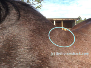
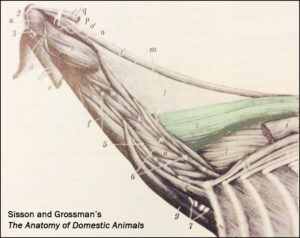
The muscle is tinted green in this image from Sisson and Grossman’s The Anatomy of Domestic Animals, Volume 1, fifth edition 1975.  Here, it is labelled Spinalis et semi-spinalis cervicis. This anatomical figure is credited to an earlier text, Ellenberger and Baum, 1908. (added 23 Dec 2016)


James Roony dedicates two pages to the ‘suspension bridge’ theory of the vertebral column in The Lame Horse (1988). His interest is in Spinalis dorsii section of the muscle and its effect behind the withers, in conjunction with  Longissimus dorsii. (added 4 Jan 2017)


Schleese diagram
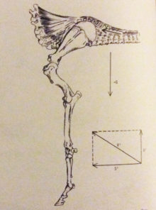
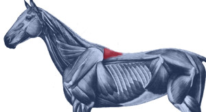
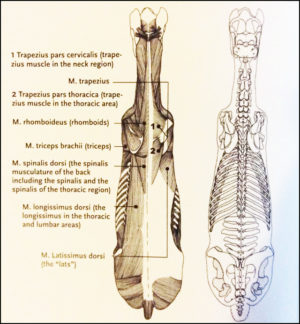
Master Saddler Jochen Schleese refers to Spinalis dorsi and its function in stabilizing the withers in Suffering in Silence, his passionate book about saddle fitting from 2014. “This muscle area is especially prone to significant development – especially with jumpers – because it is continually contracted to accommodate the shock of landing”. The surface area of the muscle is indicated in the anatomical figure, reproduced here. (added 23 Dec 2016)


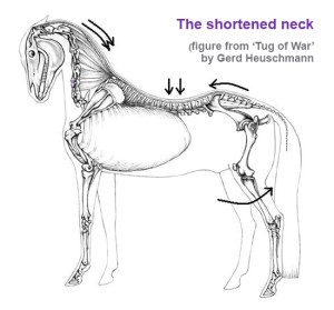
In his seminal text addressing issues of modern dressage training, Tug of War, 2007, Gerd Heuschmann includes Spinalis cervicis in the triangle formed by the rear of the rear of the cervical spine, the withers, and the shoulder blades, “… an extensive connection between the head-neck axis and the truck… it explains how the position and length of the horse’s neck directly affects the biomechanics of the back.” (added 31 Dec 2016)


Filed Under: Bodywork, Saddle Fit Tagged With: Anatomy, equine bodywork, forgotton muscle, GA, homepage, saddle fit, saddle fitting, slider, spinalis, spinalis cervicis, spinalis dorsalis, spinalis thoracis

Revealed: the Common Equine Arthritis You Won’t Read About in Textbooks

November 19, 2013 by Jane @ THB 144 Comments

elbow-header2


Sometimes, a person from outside a profession successfully identifies something that has been unnoticed, overlooked or wrongly assessed for a long, long time. Coming from another direction, they see something that has been hidden in plain sight, simply because nobody looked there before.

Update: this post was written with Sharon’s support in 2013. Finally, in 2022, Dr May-Davis is publishing her findings. This article will be updated with full details once her paper is in print in a peer-reviewed journal.

© All text copyright of the author, Jane Clothier, https://thehorsesback.com. No reproduction of partial or entire text without permission. Sharing the link back to this page is fine. Please contact me for more information. Thank you!


One lady who’s working in the field

If you’re looking for a different set of eyes for equine musculoskeletal pathologies, they don’t come much sharper than those of Dr Sharon May-Davis. Few people have the razor sharp eye she has for a hidden pathology or condition in the horse.

Teaching biomechanics, Sept 2013
Sharon, seated, teaching biomechanics, Sept 2013

Sharon is also a biomechanics expert and – significantly – a practical anatomist. She has been conducting private equine autopsies for many years – it’s not for nothing that she’s been labeled The Bone Lady and Equine CSI.

She also uses these 2-3 day dissection workshops to teach equine professionals and horse owners more about how their horses move and the damage their bodies can incur as a result of breeding, illness, injury or work.

Sharon is therefore uniquely placed to provide a source of raw data that is all but unparalleled.

Evidence from the dissection table

Some years ago, Sharon noticed an unusual action in the elbow of horses. She mentioned this to qualified practitioners and was informed that this action was quite normal. Not convinced, she began videoing horses prior to dissection and, within a short period of time, was able to match this action to a change in the elbow.

Humerus, radius and ulna, showing damage to cartilage

Not to beat around the bush, it’s an unusual form of degeneration in the horse’s elbow joint that involves all three bones. It’s a form of osteoarthritis that strikes the humeroradial joint and the ulna, causing deep and dramatic gouges into the cartilage, and eventually eroding bone.

When the joint is opened up, blood is frequently found in the synovial fluid (haemarthrosis). The fluid also displays decreased viscosity.

This is more than a little bit odd, as arthritis of the elbow is supposed to be rare in the horse.

Yet Sharon has found it to be present in numerous horses that have been euthanized under veterinary supervision for completely unrelated reasons.

Note: that’s not just some horses, but many.

Do you know where the nuchal ligament attaches on the cervical vertebrae? You think so? Evidence from the dissection table might prove you wrong – read more about Sharon’s findings on the nuchal ligament’s lamellar attachments…

This equine arthritis is visible in the living horse

The vital connection from video to dissection has enabled Sharon to indicate the presence of the elbow osteoarthritis in the horses she had been treating as an equine therapist.

It’s easy to spot, being a noticeable jarring in the elbow as the horse moves downhill – a kind of double action. Significantly, it’s what can be termed a gait anomaly, rather than lameness.

Does it sound – and look – familiar? It’s very likely that you’ve seen it in horses before and wondered what it was. The fact is that it’s so common, many people think it’s a normal action. It’s not. It’s a form of equine arthritis.

Sharon tells us she has seen the elbow problem in all types, breeds, sizes and ages of horses. Some affected horses have been elite dressage and eventing competitors. Interestingly, the problem is only presenting in ridden and driven horses.

If never worked, horses appear to remain forever free of this particular joint change.

Why the fuss – isn’t this just regular arthritis?

No. Arthritis of the horse’s elbow is considered to be rare in equine veterinary medicine.

How it should look: healthy radius and ulna (unridden horse)

The key to why it doesn’t often get diagnosed and is considered rare could be the absence of visible lameness. The arthritis identified by Sharon does not cause a distinctive lameness in the horse, although it does bring on a notable gait change, with the double step in the joint’s motion on the downhill.

Riders of such horses often just feel that their horse is a bit ‘off’, feeling a hesitation in the movement, but without being able to define the point of origin.

There are a couple more reasons why it’s not very visible: first, the action of the elbow is highly integrated with the overall shoulder action, and second, the massive triceps muscle has a further stabilizing effect on the joint.

Radius and ulna of ridden horse, showing cartilage wear and blood in joint

And even if the elbow is explored, the relatively tight joint space means that degenerative problems are rarely seen in diagnostic imaging, although inflammation can show up in thermographic images.

When, unusually, a problem has been recognized and vets have attempted a corticosteroid injection of the joint (which happens to be the most difficult joint to access), blood has been found to be present.

A closer look at Sharon’s findings

Sharon May-Davis first presented some of her findings into elbow arthritis at a conference in Australia in February 2013: the Bowker Lectures at the Australian College of Equine Podiatherapy. Presenting alongside Prof Robert Bowker and Dr Bruce Nock amongst others, she discussed the club foot in the horse, and noted how the elbow degeneration she observes on the dissection table is always worse in the forelimb with the more upright hoof.

If the condition is bilaterally present, it unfortunately appears worse on the side with the slightly upright or higher hoof. What’s more, and according to Sharon, this also applies to the limb where an inferior check ligament desmotomy (surgery undertaken with the aim of correcting an upright hoof) has taken place and the ligament has later reconnected.

She has, as already mentioned, since established that it can occur in any ridden or driven horse. Here, she describes the problem in her own words.

“The action looks like a slip and or clunk into the shoulder or a shudder or a sliding / slipping action. It depends upon your perspective. The actual change in the action begins when the foreleg is in the ‘Stance Phase’ during the stride as the limb goes into the posterior phase of the stride. It is more obvious going down a hill.

“So far, 100% of ridden horses exhibit this condition to a varying degree (under dissection). Horses not ridden and with no abnormalities do not exhibit this condition (under dissection). Horses in harness also exhibit this condition.

“What does the joint look like? There appears under dissection to be substantial degradation in the cartilage of the humerus, radius and ulna.

“Most horses appear to handle this condition and continue with a normal life if not pushed to extremes. Although this sounds career-ending, in fact it is not. Once the horse gets through the worst of the wear pattern they re-settle in the joint and continue on with work.

“High level competitors require joint support to help sustain the elbow and other joints that may compensate for the change in action.

“Horses that jump are more inclined to land with straighter forelimbs. Be mindful that jumping and downhill work could possibly make the condition worse.

“Riders often feel instability in the horse’s forelimbs when traveling downhill and some even question the horse’s proprioception.

“Bodyworkers massaging the triceps (particularly the lateral triceps) actually exacerbate the condition as the massage releases the cast-like formation that this muscle provides.”

Here are some more examples of the elbow in action.

More research is needed, but so is support

Humerus and radial bones of ex-racehorse, showing arthritic wear
Humerus and radial bones showing arthritic wear

Despite finding and documenting a huge number of dissection cases involving this particular issue, all unaided and unfunded by outside bodies, Sharon has consistently met with brick walls and skeptical responses when she has put the information forward to relevant authorities.

Why? It’s not as if she’s new to this. She has previously identified congenital malformations in the caudal cervical vertebrae of thoroughbreds, and in the atlas of Spanish Mustangs, as well as asymmetries in the femur structures of racehorses due to racing (published in the Australian Veterinary Journal).

She isn’t looking for funding (although she obviously wouldn’t say no), but would like to have this research taken up for the benefit of all ridden and driven horses. The sooner the problem is recognized and investigated, the sooner that episodic pain in the horse can be recognized, with appropriate joint support or rest given where appropriate.

And the sooner we can all learn more in our great drive towards improved equine health, the better. As Sharon says,

“In truth, we are still in the dark. Seeing it is one thing, analyzing it and providing a preventative program is something totally different.”


Questions, thoughts or comments? Join us at The Horse’s Back Facebook group. 


More Information

Shop for the Best Discounted Pet, Equine, & Livestock Supplies!Sharon May-Davis, PhD, M. App. Sc. (Ag and Rural), B. App. Sc. (Equine), ACHM, EBW, EMR was the Equine Therapist for the Modern Pentathlon Horses and the Australian Reining Team at the Sydney 2000 Olympics. She has worked with the Australian Champion from seven differing disciplines and has a particular interest in researching the musculoskeletal system. She also conducts clinics and seminars in relation to her work and regularly presents in the Northern and Southern hemisphere.


 © All text copyright of the author, Jane Clothier, www.thehorsesback.com. No reproduction of partial or entire text without permission. Sharing the link back to this page is fine. Please contact jane@thehorsesback.com for more information. Thank you!


Filed Under: Bodywork Tagged With: Anatomy, elbow arthritis, equine anatomy, Equine arthritis, equine bodywork, GA, horse dissection, Sharon May-Davis

Footer

Contact Me

Send your questions this way…

Get in Touch

  • Store Support
  • Shipping & Returns
  • Contact

Copyright © 2025 · Parallax Pro on Genesis Framework · WordPress · Log in

MAY Red Light Products Now In Stock! Dismiss