
The nuchal ligament is a soft tissue structure that is widely discussed in dressage circles. Unsurprisingly, given its deep location, relatively few of us get to cast eyes on it or feel it directly under our hands.
It’s equally unsurprising, then, that most of us don’t realize that the image we hold in our heads is somewhat different to the reality of the ligament inside our horse.
© All text copyright of the author, Jane Clothier, www.thehorsesback.com. No reproduction of images, partial or entire text without permission. Sharing the link back to this page is fine. Please contact me for more information. Thank you!
I have recently been fortunate enough to attend another dissection with renowned Australian gross anatomist (and she will point out repeatedly that despite this title, she is not gross – or, at least, not that often), Sharon May-Davis.
In this dissection workshop, Sharon had yet another opportunity to show us that an aspect of textbook anatomy is incorrect.
Yes, apparently there are many points where this is the case.
Where the nuchal ligament is and what it connects
The structure in question is the nuchal ligament, or the nuchal ligament lamellae to be exact.
To quickly explain, the funicular part of the nuchal ligament is the cord-like part that runs from the withers to the occiput (back of skull). The lamellae is the fibrous sheet-like part that extends from the funicular part to the cervical (neck) vertebrae.
According to the majority of anatomy diagrams and textbooks, it extends down to attach to the cervical vertebrae, from C2 to C7.
According to Sharon, it doesn’t. And here’s why.
Findings on the nuchal ligament’s true location
In this study of 35 horses on the dissection table, Sharon found:
- No cases where the attachments were from C2 to C7.
- No horses where the attachments were from C2 to C6.
- In all 35 horses, the attachments were from C2 to C5.
- And in 9 of the 35, the attachments to C5 consisted of thin and feeble fibers.
- The horses were of a mixture of identifiable breeds, aged 2 to 28 years old.
So, why do the majority of anatomical drawings of the deeper structures of the horse show something different?
When received knowledge can be a problem
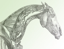
![Nuchal ligament, 5yo TB [click to enlarge]](https://thehorsesback.com/wp-content/uploads/2014/07/Nuchal-Ligament-Tb-5yr.jpg)
(The header image for this site’s most viewed post, The Disturbing Truth About Neck Threadworms and Your Itchy Horse, shows an inaccurate rendering of this ligament, as do most of the other illustrations I used. Dang!)
Inaccuracy is a recognized problem when it comes to received knowledge – was this anomaly due to an earlier artist’s error, or was it a characteristic of some 17th century horses that has been progressively bred out over subsequent centuries?
- And this raises the question of which structure, exactly, is supporting the base of the neck of the horse in motion? Read more about m. Spinalis cervicis in this post, Meet Spinalis, the Forgotten Muscle in Saddle Fitting.
- And how does this awareness inform current training approaches that require horses to raise themselves into self-carriage?
The findings from this study are in a peer-reviewed paper by Sharon May-Davis and Janeen Kleine currently in press with the Journal of Equine Veterinary Science. The paper includes a detailed review of illustrations in equine anatomy literature, an explanation of the study, and a thought-provoking discussion on the implications for our understanding of equine biomechanics.
Variations and implications of the gross anatomy in the equine nuchal ligament lamellae, Sharon May-Davis, Janeen Kleine, Journal of Equine Veterinary Science 30 June 2014 (Article in Press DOI: 10.1016/j.jevs.2014.06.018)
Have you read about Sharon’s findings on arthritis of the humeroradial (elbow) joint in all ridden or driven horses?
Questions, thoughts or comments? Join us at The Horse’s Back Facebook Group.
Yay! How good is it that this stuff is finally getting out there! Lets hope we see much more published by Sharon in the near future! Thanks for the article!
good morning, i read your article and as a saddle fitter my mind wanders and surmises how saddle fit will affect this area of the neck and shoulders. but could you please humor me and give me your diagnosis and a summary on this topic of saddle fitting in relation to this ligament?
thank you,
‘juli ulm
Lebanon oregon
Is this one a question for Sharon? If so, I’ll pass it on – let me know.
Finally this wonderful piece of information of going to print. Well done Sharon. Lets hope more o her findings finally get to the press.
Mary
South Australia
Before I read this article, I didn´t know where exactly the Ligamentum nuchae is fixed – and I never thought about it. So thanks for that. To my opinion, the base of the neck gets its support from serratus ventralis cervicis, which works together with serratus ventralis thoracis – lifting thorax and neck base between the shoulder blades. Parts of the pectoralis group help the horse to control the the breastbone. That´s why you may lift the horses head and a part of the neck by work with the reins (it will even show good neck muscles), but no one will bring up the thorax and the neck base working with reins, on whatever level! What brings up thorax and neck base is proud (please, let your horses keep their pride) and good, demanding work cross country. Thats base- as well as basic-training.
I hope I wrote well enogh, for I am no native english speaker…
Greetings, Maren