• Skip to main content
  • Skip to footer

The Horse's Back

Body Talk for Thinking Owners

  • Home
  • Articles
  • BEMER
  • Store
    • Store
    • Shipping & Returns
    • Support
  • Equine Healthworks
    • Bodywork for Horses
    • Reviews
    • Leave a Review
  • About & Contact
    • About
    • Contact

Sharon May-Davis

Physio at Feed Time: Using Food to Mobilize Your Horse’s Body

October 18, 2024 by Jane @ THB 2 Comments

In this guest post, Tanja Kraus introduces variable feeding positions, which bring musculoskeletal benefits while also enriching your horse’s day.

As an equine educator from the East Coast of Australia, Tanja’s passion is using kind horsemanship philosophies and connection to develop a partnership horse with the rider.

This extract comes from her latest book, Lessons from Horses: Movement, whch provides insight into why and how our horses should be moving each day, with exercises to contribute to their physical health, mental wellbeing, and longevity.

She writes about variable feeding positions with the support of Dr Sharon May-Davis, who developed this combined feeding approach.

You can find Tanja’s books here.

All text is copyright of the author Tanja Kraus. No reproduction without permission.


The Day I Heard About Variable Feeding Positions and Horse Posture

I first heard about variable feeding positions when attending an Equine Symposium where Dr. Sharon May-Davis was presenting.

Sharon presented on variable feeding positions, and showed many examples of the benefits of variable feeding she had seen, both in wild horses and in domestic horses managed in this way.

This included reduced asymmetry in the body, improved teeth and hooves, a more complete and balanced recovery from injury, and improved performance across multiple disciplines.

Comparing wild herds to domestic horses, she discovered that domestic horses with restricted lifestyles suffer with issues not only in the musculoskeletal system, but also uneven teeth wear. Jaw alignment and foot growth due to the limited postures.

Horses commonly ignore easy grass pickings to nibble from trees. (c) Hilary Graham

Natural Feeding Behaviors in Horses

As Dr May-Davis was talking, I was imagining my own horses and what I had seen them doing naturally in their paddock, and she was, of course, right.

My own horses who run in a herd of around 10 (give or take) in a paddock of varying terrain of about 40 acres adopt ‘variable feeding positions’ regularly and quite naturally.

And when I say regularly, I mean daily, through all the seasons.

So, they are not driven by lack of resources, which many people will argue ‘horses only eat out of trees when they have no choice’ – this is 100% incorrect.

My horses will happily roam their paddock and graze and browse and everything in between every day.

Images (c) Dengie: ‘Hedgerow Haynets for Horses’

We’re fortunate that our paddock contains a wonderful variation of pasture, different types of grasses, trees, and weeds that our horses can pick and choose from.

They are regularly seen choosing to eat ‘above the knee’, which is the measurement used to define browsing.

This can involve something as simple as eating the top of long grass around knee height, to completely outstretching their head and neck until their teeth are facing the sky to pull leaves from trees.

When you observe horses doing this, and pay attention to their bodies as they do it, you can see just how significant the range of motion is.

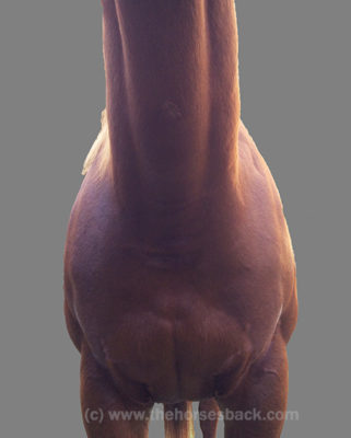
Horses extend their backs and stretch forward when eating from height. Indoor and outdoor spaces need to be large enough for them to do this.  (c) thehorsesback.com

Naysayers are often heard grumbling that horses ‘shouldn’t hollow their backs’ and that ‘high feeding is bad’ for this reason.

However, if you observe other animal species such as dogs and cats, they can all be seen stretching their spines by rounding and then hollowing.

And where would our own Yoga classes be without the classic ‘cat / cow’ pose?  Range of motion is healthy, natural and necessary.

Our domestic horses are often denied any type of variable feeding as we feed hay and hard feeds on the ground, and many horses are kept in paddocks that have limited access to appealing trees, shrubs, and long grass to facilitate variable feeding.

So how can we help?

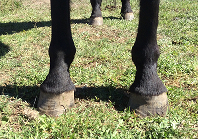
Horses with good spinal mobility appear happy to eat from any angle on a hill. (c) Tanja Kraus


Offer Your Horses Variable Feeding Positions

Provide opportunity for variable feeding, with the following examples:

  • High hay fixed
  • High hay swinging
  • High feeder on rail
  • High feeder with ramp
One flake of hay per day in a swinging high net keeps this herd, which lives out, actively mobilizing their necks. (c) Sally Taylor

If hay has to go on the ground, you can make use of the natural lie of the land.

  • Feed on slope uphill / downhill
  • Hay scattered around to encourage movement
  • Multiple haynets and feeders to encourage movement
This mare stands square while eating on a gentle slope and raising her cervicothoracic spine. Note that she could stand at the top and eat virtually on the level if she preferred to. (c) thehorsesback.com

The Benefits of Variable Feeding Positions

Basically, we can try any variation from the usual static grazing position.

Feeding in variable positions can also be a useful ‘passive physio’ technique. They may find it harder at first, but it gets easier as their bodies become more supple and mobile.

It follows that horses being rehabbed from injury or restriction can be fed to encourage beneficial postures that activate or stretch particular muscles or body areas.

Gypsy needed encouragement to load an injured forelimb. Unlike most horses, she had been loading her hindquarters rather than her forehand. The gate encourages her to come forward and raise the lower neck. (c) thehorsesback.com

It’s important to note here that I have had some equine dentists express concern as they have seen terrible damage to horses’ teeth (namely racehorses) who are fed only in high feeders.

But, those horses are stabled and fed only in up positions, and therefore the grazing position is not a part of their daily routine. A recent study suggests there is no negative dental effect in horses using haynets for a period of one year [1].

Below, these horses are fully mobilising their necks while eating from a shared large small-hole haynet – and from the ground. Note the near squared position of the grey mare, who suffered a fractured pelvis as a yearling.

We can also reverse engineer this – if your horse has an asymmetry, or difficulty working in one direction, is the way you position their feed having a negative effect?

Horses, when given the freedom to choose, graze approximately 80% of the time, and browse 20% of the time, and this balance should be sought when providing variable feeding positions for their benefit.


Solid and safe ramps and pedestals can be created to replicate environmental factors. (c) Footloose Barefoot Hooves

I have adopted variable feeding positions for my own horses, student horses, and for horses when they come in for training.

I’ve taken photos and filmed their start of variable feeding, and as they progress, and I have noticed a significant improvement in their posture while eating, along with their resting and grazing stances.

Depending on what you are trying to achieve there are many options, and the best part is the horse is doing their own physio in a gentle way.
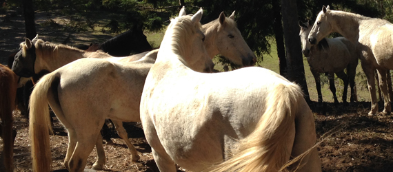
 

Read more about enrichment in this blog post:  How Well Are We Doing? Why Some Horses Thrive While Others Just Get By
Reference
[1] Johnson L, Martinson K, Keener L, DeBoer, M. A preliminary study: Effect of hay nets on horse hay usage, dental wear, and dental conditions in mature adult horses. Journal of Equine Veterinary Science; 2023;124;104366; doi.org/10.1016/j.jevs.2023.104366.

Filed Under: Bodywork, Guest Posts, Sharon May-Davis, Viewpoint Tagged With: Equine Physio, equine posture, GA, horse posture, natural feeding horses, passive physio horses, Sharon May-Davis, tanja kraus, Tanja Kraus Horsemanship, variable feeding positions

Why I Now Believe My Warmblood Gelding Had the C6-C7 Problem – Reader Story

November 16, 2017 by Jane @ THB 14 Comments

“I went through three years of horror…”

So speaks a Warmblood owner, reflecting upon a truly dreadful period in her life. Like many other owners, she has gained belated understanding of her horse’s problems after reading about research findings into the C6-C7 malformation (article) led by Australian gross anatomist, Sharon May-Davis.

Sadly, this is a bittersweet moment: there is a temporary relief as everything finally makes sense, yet with it comes the realisation that there is nothing that can be, or could have been, done for a treasured horse bearing the more extreme version of this malformation.

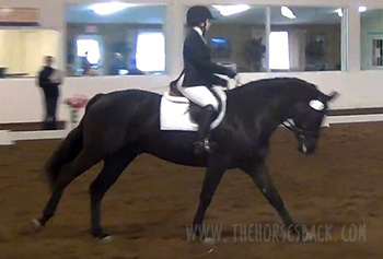
Here, Diana from Michigan, USA, gives her story of the desperation and – ultimately – heartache she experienced when trying to find out what was wrong with her horse of a lifetime, a young Oldenburg gelding, “the kindest horse I ever had”.
Note: We are not identifying the lines of the individual horse in this story. If you’re thinking we should, perhaps remember that it’s a litigious world out there.

But for now, over to Diana.

© All text copyright of Jane Clothier, www.thehorsesback.com. No reproduction of partial or entire text without permission. Sharing the link back to this page is fine. Please contact me for more information. Thank you!


Diana’s Story

I purchased a stunning 3-year-old Oldenburg to fulfill my dream of developing a young horse through the upper levels in dressage. I cashed in the 401K [retirement savings plan] for the horse and bought a Schleese saddle.
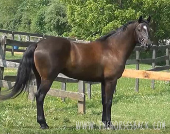
For the first two years (ages 3 and 4) we were champion every time out.

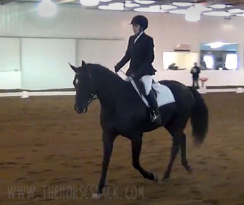
At age 5, however, my trainer noticed he began to drag his left hind toe. Then his impulsion began to deteriorate, and months later he became too unstable to safely handle. He was euthanized at 7 years old.

Looking back, I now believe that all his quirks were directly related to this problem. I am going to briefly list the oddities I experienced with this wonderful horse.

I am not a vet or tech, and am only sharing my experiences in the event that it may help someone else, because my journey was one of the most emotionally draining experiences in my life.


Possible Signs of this Warmblood’s C6-C7 Problem

I purchased my gelding on his 3rd birthday and he’d had 30 days under saddle. The oddities of my gelding: he was extremely sweet and willing – the kindest horse I ever had.

He required a tremendous amount of leg every step. Although there seemed to be no ‘self-carriage’, I attributed this to his being green and still learning.
He would buck EVERY TIME he was saddled (I was very conscientious about slowly tightening the girth, tried every type of girth, pads, had adjustable saddle, etc).

Again, I attributed the bucking to youthfulness, and would let him buck out for a few laps, and then wondered if I had created a habit, because this occurred every schooling.

  • If he wasn’t lunged first, he would explode into a non-rideable rodeo with absolutely no warning.
  • He had a very difficult time bending to the left – he would try, but would drift.
  • Two separate times after mounting at the block, he felt like he arched or sucked his back up about 3 inches before walking off.
  • After 25 mins of schooling he would start rooting at the reins, or if the lesson was a short one, as soon as I’d give him his head to cool out.
  • He was a stumbler – not often – again, I attributed it to youth.
  • He could not be shod or trimmed without being sedated.
  • Once, a friend said it looked like he couldn’t put his head down to reach hay. Of course, I thought she was nuts until I witnessed it first hand.

Questions, thoughts or comments? Join us at The Horse’s Back Facebook Group. 


Investigating the C6-C7 Problem

During my discovery phase, he was diagnosed with moderate PSSM. So, he and I spent 9 months testing sugar levels in hay, with turnout in a dry lot.

There were periods he would barely walk forward when leading, and at the time I thought he was tying up, but it didn’t seem like what I had read, and I have never witnessed a horse tying up.
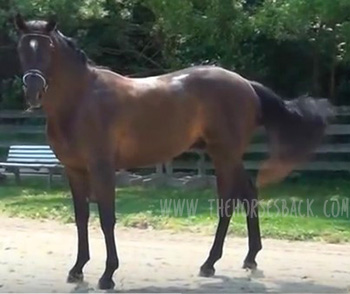
Then in his latter days, his neurological issues became more obvious with each passing day.

I did have a myelogram done on him, and it revealed compression of the spinal cord at C3 and C4.

Vets at Michigan State University did finally identify malformations of both C6 and C7 through radiographs. Their report stated:

The intervertebral foramen is severely reduced at C6-C7. A minimal amount of osseous proliferation is noted along the dorsal aspect of the articular processes at C5-C6.

Interpretation: Multifocal cervical articular degenerative joint disease (C4-T1), most severe at C6-7.”

I had a very difficult time with the decision to euthanize him, because his neurological issues would fluctuate. Some days, no evidence all. On other days, he would fall against the stall wall when turning him around in the stall.

In any event, I euthanized him, believing that he was experiencing severe pain from the narrowed foramen at C6 and C7.

I believe they were the reasons for the bucking, for needing to be sedated for the farrier, for not being able to lower his head, for rooting on the reins, for barely walking forward when I thought he was tying up, and the bizarre lifting of his back. He also had a very difficult time holding his right lead, and would swap unless you really held him.

I am so, so glad I ran across this website and article because I will go to sleep tonight knowing that euthanizing was for sure the right thing to do. Thank you!

Diana


If you’re about to purchase a horse that falls into the ‘at risk’ category for this malformation, do your due diligence as a buyer, and have the caudal cervical vertebrae radiographed as part of your pre-purchase examination. A protocol for radiographing for the malformation is on this site.

The following papers also cover radiographs and CT imaging of the C6 malformation:

Ex Vivo Computated Tomographic Evaluation of Morphology Variations in Equine Cervical Vertebrae, Veraa, S. et al,  Veterinary Radiology & Ultrasound, Vol. 57, Issue 5

Prevalence of Anatomical Variation of the Sixth Cervical Vertebra and Association with Vertebral Canal Stenosis and Articular Process Arthritis in the Horse,
Spriet, M. and M Aleman, Veterinary Radiology & Ultrasound, Vol. 57, Issue 3


If you wish to comment, please feel free to do so below, but please do so with respect for an owner who found herself in a nightmare situation. Alternatively, come over the blog’s discussion group on Facebook.

Filed Under: Bodywork, Sharon May-Davis Tagged With: Anatomy, c6 c7, cervical vertebrae, oldenburg, Sharon May-Davis, warmblood, wobbler syndrome, wobblers

By Popular Demand: Here’s How You X-Ray for the C6-C7 Malformation

August 16, 2017 by Jane @ THB 2 Comments

News about the malformation of the C6 and C7 vertebrae has understandably raised a lot of concern. There are major implications for both buyers and breeders of TB and TB-derived breed horses, as outlined in the original article, All You Need to Know About the Hidden C6-C7 Malformation That’s Bringing Horses Down. In response, Sharon May-Davis has very kindly provided us with the English version of a paper she published in the Journal of Japanese Local Government Racing, outlining a preliminary protocol for radiographing this area of the neck and identifying the malformation. (Yes, a single racing authority has taken this issue on board!)

So without further ado, here is the paper in its entirety, the only adaptations being the positioning of images to better fit this site’s blog format.

© All text copyright of the published authors. No reproduction of partial or entire text without permission. Sharing the link back to this page is fine. Please contact me for more information. Thank you!


Preliminary Radiographic Protocols for Identifying Congenital Malformations of the Caudal Cervical Vertebrae

Authors: May-Davis S.E.R., Minowa F., Monoe S.


Abstract

In 2014, a published study based on dissections and skeletal examinations noted that 19:50 Thoroughbred horses had a congenital malformation of the 6th cervical vertebra (C6). In addition, it was found that in those 19 Thoroughbred horses expressing a congenital malformation of C6, 9 displayed a concurrent congenital alformation of the 7th cervical vertebra (C7). In this study, 3 Thoroughbred horses and 1 Thoroughbred type were clinically examined; 3 were radiographed for limb abnormalities and 2:3 radiographed for the congenital malformation of C6 and C7 prior to euthanasia. Upon dissection, 3:4 expressed a congenital malformation of C6 with 2:3 displaying a concurrent CM of C7. These 2 horses were positively radiographed for the CM of C6 and C7 prior to dissection. The radiographs of C6 were taken in direct lateral orientation with 0 degree of elevation and revealed the absence of the caudal ventral tubercle (CVT) of C6. Re-positioning the horse’s forelimbs caudally with an outstretched neck, C7 was radiographed at a 30° oblique lateral angle in a cranial to caudal direction with 0 degree of elevation; the transverse process of C5 remained cranial to the beam. The caudal aspect of the plate was positioned medial the Cranial deep pectoral and rotated vertically to expand the field of view of the cervical vertebrae, whilst remaining perpendicular to the beam.


Introduction

In recent times, 3 Australian studies reported congenital malformations of C6, C7, the 1st sternal rib, along with their associative soft tissue structures. Furthermore, it was noted that these congenital malformations were predominantly breed related and more specifically, to Thoroughbred horses or derivatives thereof. In addition, reports of forelimb proprioceptive dysfunction, neurological impediment and gait deficits were concurrently noted (May-Davis [a,b], May-Davis and Walker). The congenital malformations of C6 appeared as either a left or right unilateral absence of the caudal ventral tubercle (CVT) or as a bilateral absence of the CVT (Figure 1).


Left: Normal. Centre: Absent right CVT. Right: Bilaterally absent CVT

Figure 1. View of the ventral aspect of C6, cranial aspect top. Photo credit: Sharon May-Davis


The congenital malformation of C7 appeared with either a fully transposed CVT from C6 onto the ventral surface of C7 or a partial transposition of the CVT from C6 onto the ventral surface of C7 (Figure 2) (May-Davis [a]).


Left: Normal. Centre: Transposed right CVT. Right: Bilaterally transposed CVT

Figure 2. View of the ventral aspect of C7, cranial aspect top. Photo credit: Sharon May-Davis


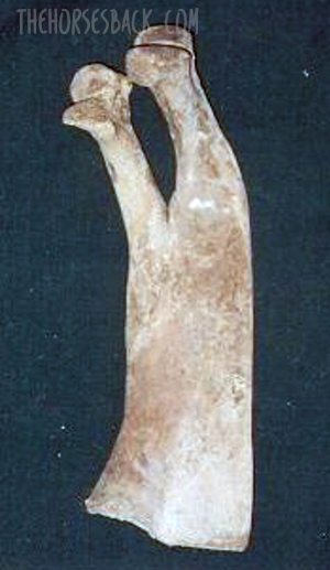
The congenital malformation of the 1st sternal rib appeared in multiple presentations with gross anatomic variations including; an absent 1st sternal rib; bifid Tuberculum costae (Figure 3); bifid Sternochondral articulation onto the sternum; flared shaft; normal 1st sternal rib inserting onto the cranial branch of a bifid Sternochondral articulating 2nd sternal rib; straight costal shaft and an articulating rudimentary Tuberculum costae with a ligamentous extension replacing the bony shaft and attaching to a rudimentary Sternochondral articulation onto the sternum (May-Davis [b]).

Figure 3. Left costal bifid Tuberculum costae. Photo credit: Sharon May-Davis

Associative soft tissue structures varied according to the presentation of the congenital malformation with the most noted being, the Longus colli muscle, Scalene muscles and neural vessels such as the Phrenic nerve and Brachial plexus (May-Davis [b], May-Davis and Walker).

The congenital malformations noted in these 3 studies were determined via dissection and skeletal examination. They were based on 2 specific skeletal variations in C6 and C7 plus any variation to normal of the 1st sternal rib.

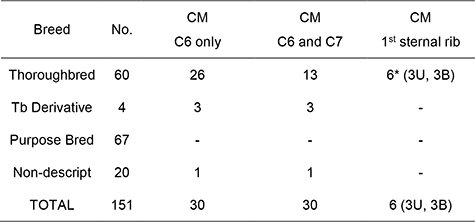
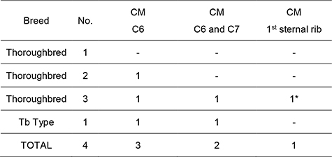
The congenital malformations could appear in C6 as a singular expression with no other anomalies, however, the congenital malformation of C7 only occurred when the CVT was absent on C6. The congenital malformations of the 1st sternal rib only occurred in the presence of the congenital malformation of C6 and C7 (Table 1) (May-Davis [b]).

With this in mind, the premise that if a congenital malformation exists in C6 then there is a 50% chance that transposition will occur on C7. In this format, it would stand to reason that there could also be an anomalous rib. Therefore, this study is designed to identify the absent CVT on C6 and investigate its transposition onto C7.


Table 1. Noted observations of 151 horses of mixed gender aged between 0 (stillborn) and 30, exhibiting a congenital malformation of C6, C7 and the 1st sternal rib.


Materials and Methods

After evaluation and clinical examination, the 2 horses (1 Thoroughbred and 1 Thoroughbred type) were restrained in cross ties. Detomidine Hydrochloride (Detomovet CEVA Animal Health Pty Ltd) was administered via left jugular vein venepuncture. The dosage was determined by the clinician’s experience and in consideration of the individual’s current health status.

The radiographs were obtained by utilising a Porta 100 HF High Frequency portable x-ray unit (7.8kgs). The kV range was 40-100kV with an mA range of 20-30 mA and a mAs range of 0.3 – 20 mAs. The images were captured on a wireless Rayence 1012WCA Medical Image processing unit (26cm x 32.5cm) and computerised with Vetview Digital Diagnostic Imaging software.

The radiographic angles were obtained with the horse’s neck in full extension and the forelimbs behind the vertical in standing position. With the clinician standing with the x-ray unit on the left side of the neck and the plate positioned on the right side of the neck, the distance between the x-ray unit and plate was 80cms. The x-ray unit and plate remained lateral to the cervical vertebrae determined by the transverse processes of C5.

The first view is a direct lateral at 90 degrees, with 0 degree of elevation whilst maintaining the transverse process of C5 to the left of the beam. 

Remaining lateral to the cervical vertebrae with a 0 degree of elevation, the second view is taken with the x-ray unit positioned at a 30° oblique lateral angle in a cranial to caudal direction with the transverse process of C5 cranial to the beam. The caudal aspect of the plate is positioned medial the Cranial deep pectoral and rotated vertically to expand the field of view of the cervical vertebrae, whilst remaining perpendicular to the beam.

Upon the radiographic evaluation of C6, radiographs of C7 are obtained. The 4 horses were then euthanized and dissected so to verify the radiographic findings. Only those pertaining to the congenital malformation of C6 and C7 are reported in this study.


Results

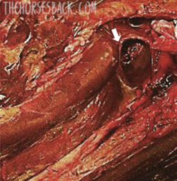
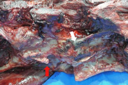
Figure 4. A compressed trachea (white arrow) in Thoroughbred No. 2 at C6 / C7. Photo credit: Sharon May-Davis

Upon euthanasia of the 4 horses, Thoroughbred No.1 was normal. Thoroughbred No.2 presented with an absent CVT in C6 and trachea compression (Figure 4).

Thoroughbred No. 3, presented with a C6 and C7 congenital malformation and a malformed 1st sternal rib. The Thoroughbred type also presented with a congenital malformation of C6 and C7 (Table 2).


Table 2. Noted observations via dissection of the congenital malformation of C6, C7 and the 1st sternal rib.


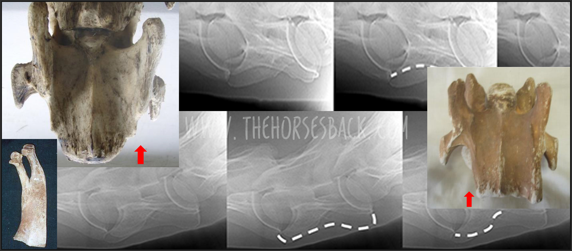
Left: Radiographic view of C6. Centre: The white line denotes a normal CVT. Right: The white denotes an absent CVT.

Figure 5. The radiographic lateral view of C6 depicting a unilaterally absent CVT in C6. Image credit: Sharon May-Davis


Thoroughbred No. 3 C7 radiograph (Figure 6) clearly presents the observer with a transposition of the CVT from C6 onto the ventral surface of C7, furthermore it denotes a reciprocal deviation in the trachea at the point of this transposition.


Left: Radiographic view of C7. Centre: The white line denotes the unilateral transposition of the CVT from C6 onto the ventral surface of C7. Right: The white line denotes the deviation in the trachea.

Figure 6. The radiographic view of C7 depicting a unilaterally transposition of the CVT from C6. Image credit: Sharon May-Davis


The Thoroughbred type (Figure 7) exhibits the absent CVT in C6. The radiograph clearly indicates an absent CVT that is unilateral in its presentation.


Left: Radiographic view of C6. Centre: The white line denotes a normal CVT. Right: The white denotes an absent CVT.

Figure 7. The radiographic lateral view of C6 depicting a unilaterally absent CVT in C6. Photo credit: Sharon May-Davis.


The Thoroughbred type C7 radiograph (Figure 8) clearly presents the observer with a transposition of the CVT from C6 onto the ventral surface of C7, furthermore it denotes a reciprocal deviation in the trachea at the point of this transposition.


Left: Radiographic view of C7. Centre: The white line denotes the unilateral transposition of the CVT from C6 onto the ventral surface of C7. Right: The white line denotes the deviation in the trachea.

Figure 8. The radiographic view of C7 depicting a unilaterally transposition of the CVT from C6. Photo credit: Sharon May-Davis.


Figure 9. Right: Absent CVT on C6 (white arrow) and transposed CVT from C6 onto the ventral surface of C7. Photo credit: Sharon May-Davis

Upon dissection, the Thoroughbred type displayed identical congenital malformations of C6 and C7 as depicted in the radiographs (Figure 9).


Discussion

This study was conducted with the view to obtain specific radiographic evidence of the congenital malformation of C6 and C7. Radiographic views of the 1st sternal rib were not possible due to thick and dense musculature in the region. The views obtained of C6 and C7 with exact radiographic angles proved that this region can be radiographed with precision so that the practitioner can make a clear diagnosis should a horse present with a neurological deficit, as previously published (May-Davis

[a, b], May-Davis and Walker]). Furthermore, the asymmetry of the cervical vertebra could exacerbate arthritis in the articular process joints as previously noted (May-Davis [a]). In addition, the incidental finding of the compression of the trachea is an important factor to consider when Thoroughbred racehorses present with compromised airways. An explanation for this occurring is that the Longus colli muscle hypertrophies in support of the weakened structures and due to its proximity to the Trachea, it impinges upon its dorsal surface. This impingement is further exacerbated by the ventral projection from C7 of the transposed CVT from C6.

Since the first publication in 2014, retrospective studies were conducted that added another dimension to the current research. Italian and American studies noted that Warmbloods, Quarter Horses and Arabs were also afflicted with this condition (Santinelli et. al 2016 and DeRouen et.al 2016). It is significant to note that these breeds present the majority of the ridden horse population in Japan extending from Racing, Showjumping, Dressage to Eventing and Endurance. However, a specific Dutch Warmblood study was conducted with fresh cadaver’s that were portioned and CT scanned (Veraa et.al 2016), this also included Oldenburg horses. The congenital malformation of C6 and C7 was present in both breeds with a noted malformed 1st sternal rib in a Dutch Warmblood. Combined percentages of these 3 studies in conjunction with the 3 Australian studies and the current study has the congenital malformation of C6 at; Warmbloods 30%, Quarter Horses 16% and Thoroughbred horses over 40%. 

Aside from the Arabian who is an ancestor to the Thoroughbred, the Warmblood breeds and Quarter Horses all have Thoroughbred lineage in the back line of their breeding. Thus, implying that this condition is heritable as noted by May-Davis [b] and more systemic than just one breed. With this in mind, all critical events should be assessed with the knowledge that a potential congenital malformation could exist in C6, C7 and the 1st sternal rib, as previously noted. With the largest population of horses in Japan being Thoroughbreds, it would be a recommendation to note the studies of several countries including Japan and the severity in percentage of this systemic congenital malformation. Especially in relation to Thoroughbred’ racehorses racing and cornering at speed.


Conclusion

This study showed that of the 4 horses investigated, 3:4 horses displayed a CM of C6, 2:3 displayed the congenital malformation in C6 and C7, and 1:2 a congenital malformation of the 1st sternal rib. Even with a small sample of Thoroughbreds, studies from other countries must be measured and it would therefore be a recommendation to radiograph for this condition in the caudal cervical vertebrae in a pre-purchase examination. The purpose of which would be to eliminate this condition so that riders and handlers are not put at risk.


Author contributions

Sharon May-Davis, Fumiko Minowa and Sadae Monoe wrote and reviewed this article.


Conflict of Interest

The authors have no conflict of interest in the preparation or presentation of this original research article.


Acknowledgement

The author wishes to thank Christine Gee for her professional advice on the manuscript. The Nippon Veterinary and Life Sciences University for the use of their facilities and to those authors / editors/ publishers of those articles, journals and books cited in this manuscript.


References

Derouen A, Spriet M, Aleman M. Prevalance of anatomical of the sixth cervical vertebra and association with vertebral canal stenosis and articular process osteoarthritis in the horse. Vet Radiol Ultrasound, Vol. 00, 2016, pp 1–5.

May-Davis SER. The Occurrence of a Congenital Malformation in the Sixth and Seventh Cervical Vertebrae Predominantly Observed in Thoroughbred Horses. J Equine Vet Sc 2014; 34:1313-17.

May-Davis SER. The Congenital Malformation of the 1st Sternal Rib. J Equine Vet Sc 2014; 34:1313-17.

May-Davis SER, Walker C. Variations and implications of the gross morphology in the Longus colli muscle in Thoroughbred and Thoroughbred derivative horses presenting with a congenital malformation of the sixth and seventh cervical vertebrae. J Equine Vet Sc 2015; 35:560-8.

Santinelli I, Beccati F, Pepe M. Anatomical variation of the spinous and transverse processes in the caudal cervical vertebrae and the first thoracic vertebra in horses. EVJ 48 (2016) 45–49.

Veraa S, Bergmann W, van den Belt A-J, Wijnberg I, Back W. Ex vivo computed tomographic evaluation of morphology variations in equine cervical vertebrae. Vet Radiol Ultrasound, Vol. 00, 1–7.


Filed Under: Bodywork, Sharon May-Davis Tagged With: C6, C7, cervical vertebrae, GA, homepage, TBs, Thoroughbred

All You Need to Know About the Hidden C6-C7 Malformation That’s Bringing Horses Down

July 16, 2017 by Jane @ THB 71 Comments


What if you were to learn that your horse is living with a hidden malformation? A skeletal abnormality that could be affecting it every day, changing the way it moves, creating a string of other physical problems, and possibly underlying the hard-to-pinpoint problems you’ve been noticing for months or even years ?

And that might even be causing a level of inherent instability that could be putting the rider in danger?

Sadly, this isn’t a hypothetical question. Instead it’s a reality that is only now being slowly uncovered.

And like the proverbial stone rolling down a mountain, the issue is gathering momentum as the equine industry, owners, breeders and researchers learn about it.

  • It’s a skeletal malformation and it can’t be corrected.
  • It’s congenital, ie inherited, so is present from birth.
  • It has been in some lines of TBs for hundreds of years.
  • It creates biomechanical issues due to asymmetry and lack of anchor points for key muscles.
  • At its worst, it can contribute to neurological issues such as Wobbler syndrome.
  • Some horses are so unstable, they are more prone to falling (not good news for jockeys).
  • It can cause constant pain and associated behavioural changes.
  • It’s primarily found in Thoroughbreds, Thoroughbred crosses and Warmbloods, but has also been identified in European breeds, Quarter Horses, Arabs and Australian Stock Horses.


Note: in the years since this post was written with direct support from Dr Sharon May-Davis, research has moved on. The body of literature now includes papers from Sharon and also from other researchers who do not reach similar conclusions about the issues caused by this malformation. I have included a list of journal papers and webinars on the topic at the end of the post, for reference if you wish to deepen your knowledge. You can also go view the full list here (opens in a fresh tab).


The problem behind this is ECVM, a congenital malformation of the C6 and C7 cervical vertebrae (ie, base of neck) – and it’s pretty nasty.

I’ve written about the work of Sharon May-Davis on this blog before and here I’m going to do so again. Through her many dissections per year, gross anatomist Sharon has become the first person to comprehensively document and quantify this problem.

In doing so, and publishing her findings in peer-reviewed journals, she has triggered a minor research avalanche as others take up the subject.

Update: since this article was published in July 2017, this malformation has been labelled Equine Complex Vertebral Malformation (ECVM). Some amendments have been made to include this term. To preserve existing links to the page, I’ve not updated the title and URL.

© All text copyright of the author, Jane Clothier, https://thehorsesback.com. No reproduction of partial or entire text without permission. Sharing the link back to this page is fine. Please contact me for more information. Thank you!


A hidden problem: this OTTB had the C6-C7 malformation but presented few outward signs.


Twenty years + of research into ECVM

Sharon May-Davis’s path with this research began some 20 years ago. In February 1996, a Thoroughbred called Presley came down unimpeded in a race in Grafton, NSW, fracturing his pelvis, a hock bone, and right front fetlock.

Three years later, Sharon examined his bones, and saw something strange in his last two cervical vertebrae and his first ribs.

Fast forward to 2014, when Sharon published the first of her four peer-reviewed papers in the Journal of Equine Veterinary Science, concerning a congenital malformation in the sixth and seventh cervical (neck) vertebrae.

Although the problem had been mentioned briefly in papers, this was the first time that a researcher had accurately described and quantified the problem in its various forms.

Sharon’s unique perspective, gained as an anatomist who dissects between 15 and 20 horses per year, had certainly placed her in a position to do so.


The horse’s seven cervical vertebrae – made simple

Horses have seven vertebrae in their necks, labelled C1 to C7. Of these, four have unique shapes. Most horse people are familiar with C1, the first vertebrae known as the atlas, as it can be both seen and felt by hand with its distinctive ‘wing’ at the top of the neck.

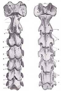
Cervical vertebrae from C1 (top) to C7 (bottom), view from above (L) and below (R).

Almost as well-known is C2, the second vertebrae, known as the axis.

Both atlas and axis have unique shapes for a special reason: they support the heavy skull and anchor the muscles that control the head’s movement.

Heading down the neck, C3, C4 and C5 are broadly similar in shape, with each being a bit shorter and blockier than the one above.

However, C6 and C7 are both slightly different on the ventral (lower) side, for here they provide insertion points for muscles arising from the chest.

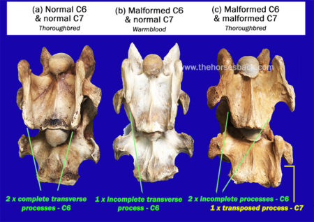
  • C6 has transverse processes (the protrusions extending outwards) that are different to those of neighbouring bones, with two distinctive ridges running the vertebrae’s length. C6 also has two large transverse foramen, the openings that the arteries pass through.
  • C7 is the shortest and squattest cervical vertebrae of all. Its transverse processes are shorter, while there are also two facets that articulate with the first ribs. C7 has no transverse foramen.

At least, that’s how the vertebrae should be in a normal horse.


So, what is wrong with the malformed C6 and C7 vertebrae in ECVM?

In certain horses, these last two vertebrae are rather different, being malformed.

Sharon has identified the manifestations of this problem as a congenital (inherited) malformation affecting some Thoroughbred horses, and horses with Thoroughbred blood in their ancestry.

In C6, there is a problem with the two ridges of the transverse processes, as one or both can be partially absent.

When both are partially missing, it is common for one or two ridges (ie, parts of the transverse processes) to appear on C7 instead.

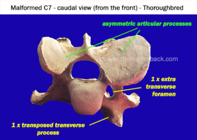
Also, the articular processes (the oval surfaces on the upper side, where each vertebrae links to its neighbours) can be radically different sizes. There can also be an additional arterial foramen or two.

The level of asymmetry can be radical.

The secondary problems the ECVM malformation causes

Being at the base of the neck, the asymmetry of C6 and C7 can cause alignment problems all the way up the vertebral column, leading to osteoarthritis of the articular facets.

It can also contribute to Wobbler Syndrome (Cervical Vertebral Stenotic Myelopathy), due to narrowing and/or malalignment of the vertebral foramen/canal, the opening through which the spinal cord passes. Not all Wobbler cases have this particular malformation, though.

A further problem is that the lower part of the longus colli muscle, which is involved in flexing the neck, would normally insert on the transverse processes of C6 and C7. When these processes are malformed, the normal insertions are not possible.

This means there is a serious symmetry problem in the junction of the thorax and neck, which can have a deeper effect on the horse’s neurology and proprioception, as well as respiration.

Asymmetry and narrowed vertebral foramen (canal) contribute to DJD and Wobbler Syndrome.


In a few cases, horses with both the C6 and C7 problem also have malformations of the first sternal rib, on one or both sides. This can cause problems beneath the scapular and further issues with muscular attachments.

Associated stability problems can have far-reaching consequences for the horse, not to mention some serious safety issues for the rider. The safety issue can’t be stated often enough.


Why isn’t ECVM – the C6-C7 problem – more widely known ?

Why hasn’t this problem been noticed in regular veterinary interventions?

The answer is quite simple. While neurological issues may have been diagnosed, the exact cause has often remained hidden.

Both Thoroughbred horses and Warmbloods are known to have higher incidences of Wobbler Syndrome than other breeds, and while this is certainly not always due to C6-C7 malformation, the malformation has been found in some when dissected.

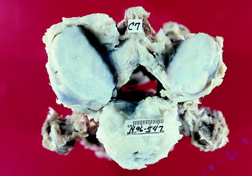
For example, the following dissection image appears in a veterinary account of large animal spinal cord diseases. It clearly shows a malformed C7 vertebrae, very similar to the one in the above image, but without giving any further categorisation.

The difficulty lies in the deep location of the lower cervical vertebrae. While normal radiographs can show all or some of C6, they are unable to penetrate the deeper tissues beneath the shoulder to image C7.

Photo from another online article: the familiar asymmetry of a malformed C7 is clearly visible in a dissected set of vertebrae.  (c) veteriankey.com (click image to access full article.)


Nevertheless, the malformation can be identified in radiographs of C6, once you know where to look.

Since Sharon’s first paper appeared, the School of Veterinary Medicine, University of California, Davis, has reviewed its history of radiographs from horses with Wobbler Syndrome.

Researchers found that 24 cases out of 100 (close to 25%) showed malformation of one or both C6 transverse processes. This study also clarified how to identify the problem on standard radiographs of C6.

In another study, the Faculty of Veterinary Medicine, Utrecht University, completed CT scans on horses’ necks and found the various forms of the malformation in 26 horses out of 78 (33%). Unlike radiographs, the CT scans enabled identification of the C7 and first rib issues, although of course this imaging was conducted post mortem.

Is this rare, or are many horses affected?

While the problem has been identified primarily in TBs, it affects most breeds with TB blood in the ancestry to some degree.

Sharon May-Davis reports that to date, published, peer-reviewed journal papers have tallied 136 out of 471 horses as exhibiting congenital malformation of C6.

These have been in a range of breeds including Thoroughbreds (39%), Thoroughbred crosses (27%), Warmbloods and European breeds (30%), Quarter Horses (11%), and Arabs (11%). Standardbreds have also shown the problem, although the numbers included in studies are very small.

A common question is whether it’s known which TB lines predominantly carry this problem. The answer is: Yes. However, it is now so disseminated amongst the modern equine population beyond TBs, that it is of little help to identify them.

It must be remembered that these horses are those already brought to veterinary attention and/or euthanized for a related or unrelated reason, so the percentages may be higher than those for the general horse population. At the same time, the malformation might have played a major part in the horses’ decline, due to the many locomotory and postural problems it can lead to.


How do we identify these horses in life?

It’s all very well looking at these bones post mortem, you might say. Yet how can I tell if my horse has this problem? Or a horse that I might want to buy?

Some answers are forthcoming. As Sharon has frequently assessed horses before dissecting them – usually from video – she has been able to observe that many of these horses lack stability. (Indeed, in many cases, it is this very instability has directly led to the horse being euthanized, and ending up on the dissection table.)

As her research has progressed, she has also been able to identify many biomechanical and locomotion traits that make these horses ‘suspicious’ or at least ‘of interest’. Unsurprisingly, these problems have been particularly noticeable in horses with both a malformed C6 and C7.

For owners and equine professionals, here are some signs that can raise initial suspicions. All can also be caused by other problems, so a group of signs is more common than an individual indication.

  • Some of these horses have a problem with standing square in front, and will always keep one foot further forward. This can persist despite all attempts to improve the horse’s body and to train the horse to halt squarely.
  • Horses with the more serious malformations will often stand base-wide. Such horses can become very unbalanced on uneven ground, and sometimes in work. They easily become unbalanced when a hoof practitioner works on a forefoot. 


A bilateral C6 – unilateral C7 horse showing a toe-out stance and hoof distortion. The ventral part of the transverse process was transposed onto the left side of C7.


  • With such asymmetry in the skeletal structure, these horses have serious lateral flexion issues that can’t be overcome. When required to elevate the forehand, many will experience difficulties, due to the absence of correctly inserted musculature and incorrect articulation through the joints of the lower neck. 
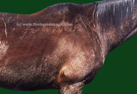
The horse may have one very prominent, widely positioned scapula.
  • A high level of asymmetry may be seen in the shoulders, with one scapula sometimes positioned very wide, with no improvement after chiro, osteopathy or bodywork. This is particularly so with the C6-C7 problem and associated first sternal rib abnormalities.
The ventral aspect of the neck may show some scoliosis.


  • There may be scoliosis along the entire spine.
  • There may be an obvious scoliosis to the underside of the neck.
  • The problem may lead to heavily asymmetric loading of the forefeet, so may be accompanied by a severe high foot/low foot issue (this is not in itself a sign of the C6-C7 problem).

If you suspect your horse has the C6-C7 issue

First, note that many horses do just fine with a C6 problem. It is those with the bilateral C6 and unilateral/bilateral C7 issue that tend to show the more worrying problems.

If your horse is showing ongoing signs of instability, it’s important to seek veterinary advice, so that neurological issues can be ruled out. (As this a recently recognised problem, it may be worth printing out the abstracts from the journal articles listed at the end of this page and handing them over.)

If the more severe malformations are identified by radiograph, it is important to remember that in some cases this can cause discomfort and pain to the horse, and it is not going to improve over time.

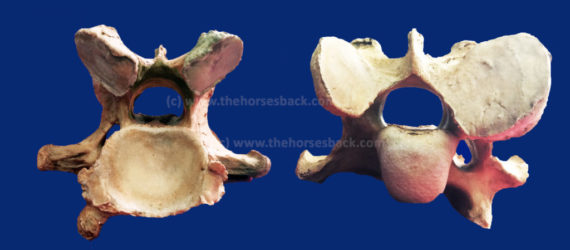
On the contrary, the cervical vertebrae of some older horses with the C6 and C7 malformations often display advanced osteoarthritis of the articular processes, as shown in the header image of a 19-year-old Thoroughbred’s malformed C7.


Where does this knowledge take us?

At the moment, that question is wide open. The findings published by Sharon May-Davis have triggered ongoing research on an international level. There are certainly ramifications for breeders in more than one equine sporting industry.

See Sharon May-Davis’s December 2020 presentation on ECVM here (article continues below):

Connections have been made with a number of falls on the racetrack that have caused injury, and worse, to both horse and jockey, as well as other runners. Similar things can be said for the sport of eventing, where unforced errors can have equally catastrophic effects.

It is entirely possible that at higher levels, pre-purchase examination radiographs will come to include a check on C6. While it’s not possible to radiograph the deeply positioned C7, we do at least know that this will only be present if the C6 anomaly exists.

Vets in some countries are proving faster at picking this up than others. While papers are being published, it clearly takes some time for information to filter down.

And until more is known, this problem is being unknowingly propagated every breeding season.

Of course, many horses harbouring the milder manifestations of this problem at C6 level are functioning very well. All horse owners can do is be aware that this issue exists, make use of this information if a problem arises, and await further research findings.

Literature on the malformation

In no particular order: the following peer reviewed journal articles address ECVM, as it is now known. Please remember when reading papers that no single paper proves one thing – a review of many papers and critical thinking is always essential. Apologies for not having standardized the formatting, but the links should be present.

  • The Occurrence of a Congenital Malformation in the Sixth and Seventh Cervical Vertebrae Predominantly Observed in Thoroughbred Horses, May-Davis, Sharon, Journal of Equine Veterinary Science , Volume 34 , Issue 11 , 1313 – 1317
  • Variations and implications of the gross morphology in the longus colli muscle in thoroughbred and thoroughbred derivative horses presenting with a congenital malformation of  the Sixth and Seventh Cervical Vertebrae May-Davis, S, Walker, C. Journal of Equine Veterinary Science 35 (7), 560-568
  • Characterization of the Caudal Ventral Tubercle in the Sixth Cervical Vertebra in Modern Equus ferus caballus. May-Davis, S., Dzingle, D, Saber, E., Blades Eckelbarger, P. Animals 13 (14), 2384
  • Morphology of the Ventral Process of the Sixth Cervical Vertebra in Extinct and Extant Equus: Functional Implications, May-Davis, S., Hunter, R, White, R. Animals 13 (10), 1672
  • Variations and Implications of the Gross Morphology in the Longus colli Muscle in Thoroughbred and Thoroughbred Derivative Horses Presenting With a Congenital Malformation of the Sixth and Seventh Cervical Vertebrae, May-Davis, Sharon et al., Journal of Equine Veterinary Science , Volume 35 , Issue 7 , 560 – 568
  • Congenital Malformations of the First Sternal Rib, May-Davis, Sharon, Journal of Equine Veterinary Science , Volume 49 , 92 – 100
  • Ex Vivo Computated Tomographic Evaluation of Morphology Variations in Equine Cervical Vertebrae,  Veraa, S. et al,  Veterinary Radiology & Ultrasound, Vol. 57, Issue 5
  • Prevalence of Anatomical Variation of the Sixth Cervical Vertebra and Association with Vertebral Canal Stenosis and Articular Process Arthritis in the Horse, Spriet, M. and M Aleman, Veterinary Radiology & Ultrasound, Vol. 57, Issue 3
  • Anatomical Variation of the Spinous and Transverse Processes in the Caudal Cervical Vertebrae and the First Thoracic Vertebra in Horses, Santinelli, I. et al, Equine Veterinary Journal, Vol. 48, 45–49
  • Congenital variants of the ventral laminae of the sixth and seventh cervical vertebrae are not associated with clinical signs or other radiological abnormalities of the cervicothoracic region in Warmblood horses. Dyson S, Phillips K, Zheng S, Aleman M. Equine Veterinary Journal, 2025 Vol.2 :419-430
  • Neck pain but not neurologic disease occurs more frequently in horses with transposition of the ventral lamina from C6 to C7. Henderson CS, Story MR, Nout-Lomas YS. J Am Vet Med Assoc. 2024 May 29;262(9):1215-1221


Filed Under: Bodywork, Sharon May-Davis Tagged With: C6, C7, ECVM, equine dissection, GA, homepage, Sharon May-Davis, skeletal malformation, slider, Thoroughbred anatomy

How The Anatomy Books (Unintentionally) Fail Us Over The Nuchal Ligament

July 13, 2014 by Jane @ THB 5 Comments

nuchal-ligament-header-1

The nuchal ligament is a soft tissue structure that is widely discussed in dressage circles. Unsurprisingly, given its deep location, relatively few of us get to cast eyes on it or feel it directly under our hands.

It’s equally unsurprising, then, that most of us don’t realize that the image we hold in our heads is somewhat different to the reality of the ligament inside our horse.

 © All text copyright of the author, Jane Clothier, www.thehorsesback.com. No reproduction of images, partial or entire text without permission. Sharing the link back to this page is fine. Please contact me for more information. Thank you!

I have recently been fortunate enough to attend another dissection with renowned Australian gross anatomist (and she will point out repeatedly that despite this title, she is not gross – or, at least, not that often), Sharon May-Davis.

In this dissection workshop, Sharon had yet another opportunity to show us that an aspect of textbook anatomy is incorrect.

Yes, apparently there are many points where this is the case.

Where the nuchal ligament is and what it connects

The structure in question is the nuchal ligament, or the nuchal ligament lamellae to be exact.

George Stubbs illustration
George Stubbs, 1777, showed the NLL attaching from C2 to C7.

To quickly explain, the funicular part of the nuchal ligament is the cord-like part that runs from the withers to the occiput (back of skull). The lamellae is the fibrous sheet-like part that extends from the funicular part to the cervical (neck) vertebrae.

According to the majority of anatomy diagrams and textbooks, it extends down to attach to the cervical vertebrae, from C2 to C7.

According to Sharon, it doesn’t. And here’s why.

Findings on the nuchal ligament’s true location

In this study of 35 horses on the dissection table, Sharon found:

  • No cases where the attachments were from C2 to C7.
  • No horses where the attachments were from C2 to C6.
  • In all 35 horses, the attachments were from C2 to C5.
  • And in 9 of the 35, the attachments to C5 consisted of thin and feeble fibers.
  • The horses were of a mixture of identifiable breeds, aged 2 to 28 years old.

So, why do the majority of anatomical drawings of the deeper structures of the horse show something different?

When received knowledge can be a problem

Nuchal ligament, 5yo TB [click to enlarge]
Many of today’s illustrators are referring to illustrations that have themselves been amended from earlier illustrations.

(The header image for this site’s most viewed post, The Disturbing Truth About  Neck Threadworms and Your Itchy Horse, shows an inaccurate rendering of this ligament, as do most of the other illustrations I used. Dang!)

Inaccuracy is a recognized problem when it comes to received knowledge – was this anomaly due to an earlier artist’s error, or was it a characteristic of some 17th century horses that has been progressively bred out over subsequent centuries?

  • And this raises the question of which structure, exactly, is supporting the base of the neck of the horse in motion? Read more about m. Spinalis cervicis in this post, Meet Spinalis, the Forgotten Muscle in Saddle Fitting.
  • And how does this awareness inform current training approaches that require horses to raise themselves into self-carriage?

The findings from this study are in a peer-reviewed paper by Sharon May-Davis and Janeen Kleine currently in press with the Journal of Equine Veterinary Science. The paper includes a detailed review of illustrations in equine anatomy literature, an explanation of the study, and a thought-provoking discussion on the implications for our understanding of equine biomechanics.

Variations and implications of the gross anatomy in the equine nuchal ligament lamellae, Sharon May-Davis, Janeen Kleine, Journal of Equine Veterinary Science 30 June 2014 (Article in Press DOI: 10.1016/j.jevs.2014.06.018)

Have you read about Sharon’s findings on arthritis of the humeroradial (elbow) joint in all ridden or driven horses?


Questions, thoughts or comments? Join us at The Horse’s Back Facebook Group. 


Filed Under: Bodywork, Sharon May-Davis Tagged With: equine anatomy, equine bodywork, GA, horse anatomy, Nuchal ligament, Sharon May-Davis

Footer

Contact Me

Send your questions this way…

Get in Touch

  • Store Support
  • Shipping & Returns
  • Contact

Copyright © 2025 · Parallax Pro on Genesis Framework · WordPress · Log in

MAY Red Light Products Now In Stock! Dismiss