• Skip to main content
  • Skip to footer

The Horses Back

Body Talk for Thinking Owners

  • Home
  • Articles
  • BEMER
  • Store
    • Store
    • Shipping
    • Returns
    • Support
  • Equine Healthworks
    • Bodywork for Horses
    • Reviews
    • Leave a Review
  • About & Contact
    • About
    • Contact

Bodywork

An Unwelcome Side Effect: Transitional Vertebrae in Horses

May 1, 2018 by Jane @ THB 24 Comments


They can lead to scoliosis, spinal arthritis, flexion and straightness problems, saddle fit issues, secondary lameness, hoof problems and soft tissue trauma. So, what on earth are transitional vertebrae, and why haven’t we heard more about them?

To answer the first part of that question, transitional vertebrae are hybrids that appear where one group of vertebrae changes to another. They show mixed features of each group.

They can be found along the spine, where:

  • the cervical (neck) meet the thoracic vertebrae,
  • the thoracic meet the lumbar vertebrae,
  • the lumbar meet the sacral vertebrae (sacrum),
  • where the sacrum meets the caudal vertebrae (tail bones).

As for why we’ve not heard much about them, the answer is probably that they’re rarely identified while a horse is alive.

However, they can lead to some very real problems in the living horse due to the asymmetry they cause along the spine – and they’re far more common than you might think.

A transitional vertebra at L1. (c) J. Clothier

 The affected process or rib can hurt when the horse bends into it, as the abnormal rib/process is literally ‘stabbing’ into soft tissue.”

© All text copyright of the author, Jane Clothier, https://thehorsesback.com.


Thoracic and lumbar transitional vertebrae

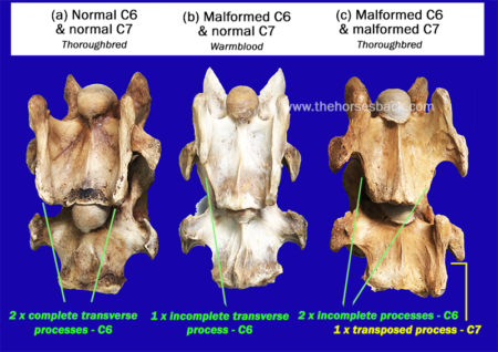
Here are the three main types of variation, as shown in this diagram from one of the few research papers to mention this issue.

Here, we’re going to look at the first two – labeled A and B – which are the most common manifestations.

The three kinds of thoracolumbar transitional vertebrae. (c) American Journal of Veterinary Research. (Annotated in green by J. Clothier) Haussler, K.K., Stover, S.M., Willits, N.H. Developmental variation in lumbosacropelvic anatomy of Thoroughbred racehorses (1997); American Journal of Veterinary Research, 58 (10), pp. 1083-1091

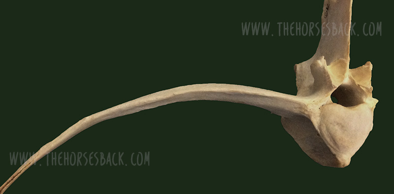
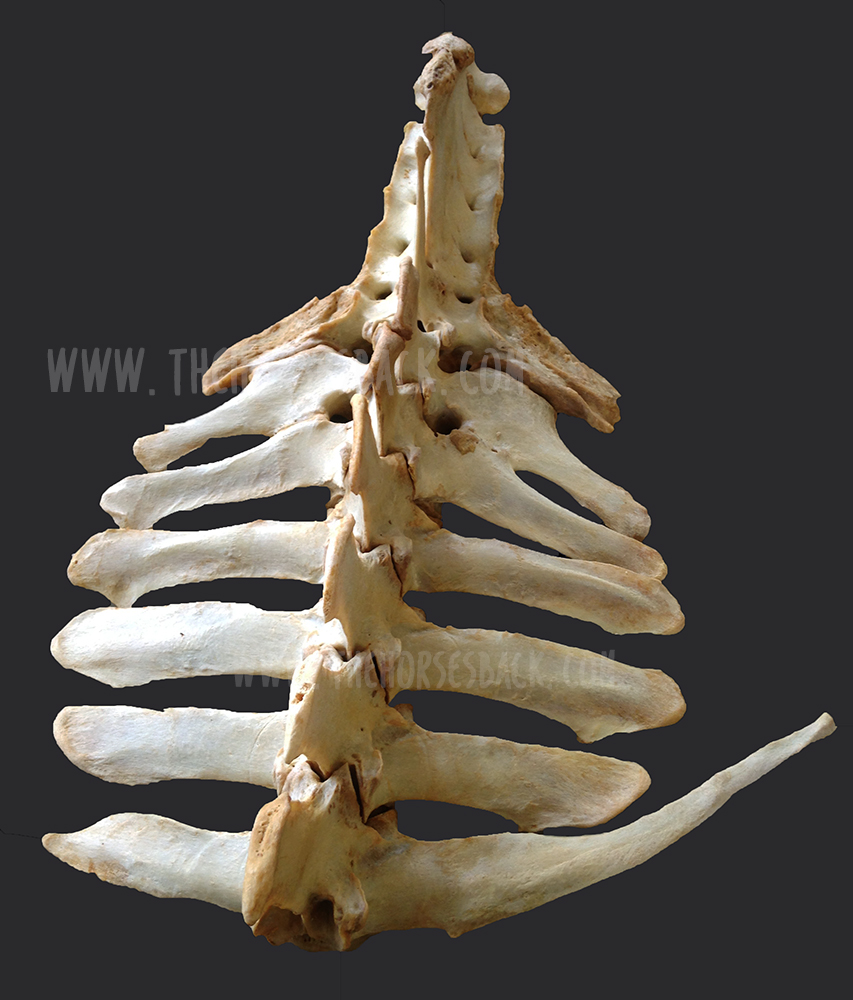
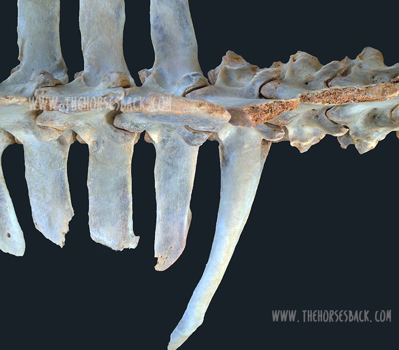
A ‘process-like rib’ at T18

Labeled ‘A’ in the above diagram, this is a transitional vertebra at T18 (the last thoracic vertebrae) – a rib that thinks it might be a transverse process, lacking an articulation or joint with the vertebral body.

A normal facet on one side, a non-articulated process-like rib on the other (c) J. Clothier

Instead, the process-like rib is solidly attached, meaning there is no independent movement whatsoever. At its end point, it’s joined by costal cartilage to the preceding rib, partially restricting that rib’s movement, too.

This is a problem, as the caudal ribs are not directly attached to the sternum because they need to move more.

The abnormality can be on one or both sides of the vertebra, although single side is most common.

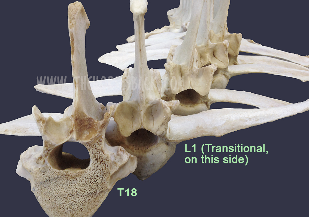
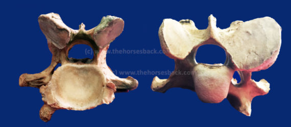
A ‘rib-like process’ at L1

Labeled ‘B’ in the above diagram, this is a transitional vertebra at L1 (the first lumbar vertebrae). Again, it’s usually one-sided, although two sides also occur.

Here, we’re looking at a transverse process that rather than being fairly short, wide and flat, instead extends outwards like a misshapen rib. There’s no articulation with the vertebral body.

The first lumbar vertebrae (L1) of this Quarter Horse mare is a transitional vertebra. (c) Melissa Longhurst, www.equinebodybalance.com.au 

The above image shows an abnormal L1 found in a Quarter Horse mare. This mare was asymmetric throughout her body, and had a history of unsoundness both fore and rear throughout her lifetime.

Effect on the horse

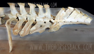
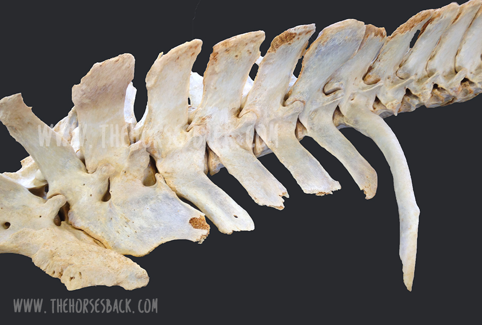
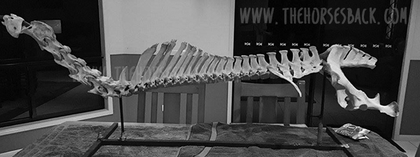
Scoliosis is the major effect of transitional vertebrae. It’s an asymmetry that in these cases can be lifelong and permanent.

I’ve seen it a few times now in skeletons and on horses that have subsequently been euthanized for unrelated reasons – the spine curves in the affected direction, ie. the horse’s ‘short side’ is the same as the abnormal rib/process that is causing restriction.

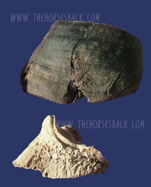
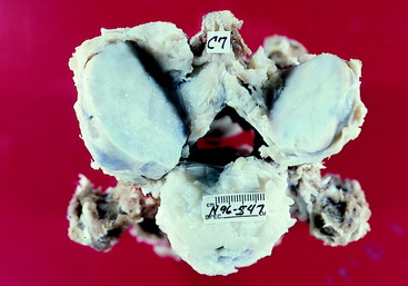
The above bones were from a TB gelding who was in his late teens. Over his lifetime, the additional pressure on the side of the abnormal L1 had caused greater bone development in the vertebra further forward. In this photo, T18, the last thoracic vertebra, has been cut to show this impact.

Cases are highly individual and the degree of impact depends on how abnormal the vertebra is, plus other factors affecting the horse’s musculoskeletal balance – including tack and riders. However, we can consider the following points.

There can be an obvious localized effect:

  • The affected process or rib can hurt when the horse bends into it, as it is literally ‘stabbing’ into soft tissue.
  • The attachments of the deep, short muscles involved in segmental stabilization at L1 and T18 are affected, also affecting proprioception and posture.
  • The abdominal muscles involved in breathing and flexion during locomotion are restricted over an affected T18.
  • The diaphragm inserts onto T18, meaning its function is also affected.
L1 transitional vertebra on the left side causing scoliosis along the spine, including the sacrum. (c) Melissa Longhurst, www.equinebodybalance.com.au


This can affect overall spinal health and biomechanics:

  • Scoliosis means that bending to the affected side can be uncomfortable, while bending to the opposite side can be highly limited.
  • Achieving straightness may be impossible. Scoliosis can extend through the withers and into the neck.
  • Impinging transverse processes and vertebral arthrosis at other vertebral joints further limit movement.
  • These restrictions make lifting the back problematic. 

And then there can be a host of secondary effects:

  • In the heavily pregnant mare, existing discomfort due to a T18 may worsen.
  • Achieving saddle fit is difficult on an asymmetric horse with scoliosis.
  • Abnormal loading can lead to recurrent lameness and persistent hoof issues.
  • Unrelated pathologies can scale up uncontrollably, as the horse cannot compensate effectively.


More on this Topic

Take a closer look at the vertebrae featured in this article (Equine Healthworks is my practice page in NSW, Australia – also on Facebook.)


Questions, thoughts or comments? Join us at The Horse’s Back Facebook Group. 


Can we spot transitional vertebrae in the living horse?

Yes, sometimes.

Unilateral transitional vertebra at T18. (c) J. Clothier

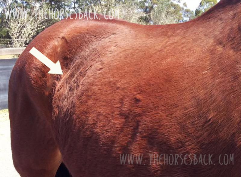
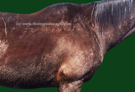
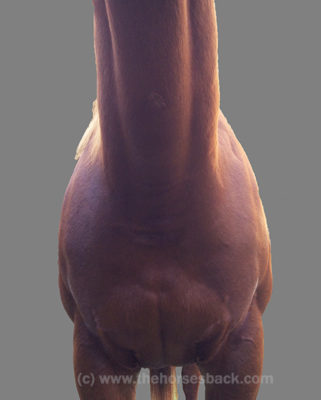
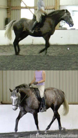
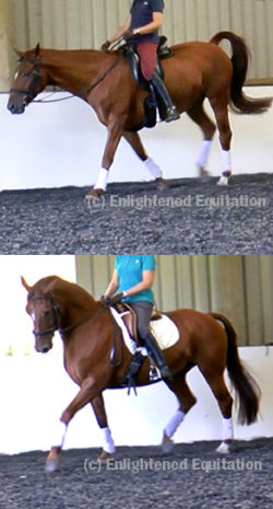
As this TB mare (above and below) was unable to maintain weight due to the physical stresses she was experiencing, her rib outline was fairly clear.

In her case, the last rib felt wider and flatter than the other ribs. The space between the rib and the point of hip was also noticeably narrower on the affected side (although this would be true of any horse with scoliosis, it’s a matter of putting the picture together, sign by sign).

The problem is visible here. This mare’s body condition and tension reflects the stresses caused by the T18 transitional vertebra, which was later confirmed at necropsy. (c) J. Clothier

There were other reasons for suspicion. Even when all the surrounding tissue was relaxed, there was no ‘spring’ when the rib was palpated with a flat hand. That’s not definitive, but it’s a cause for concern.

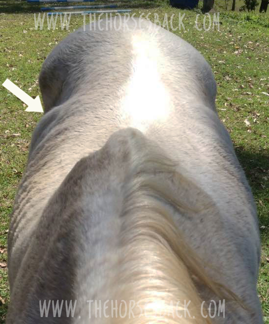
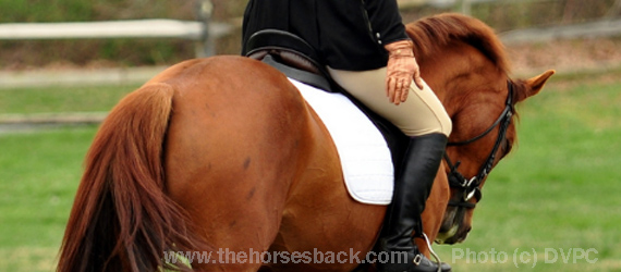
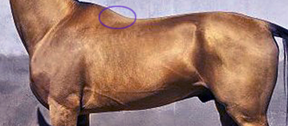
Do something that most people never do – stand on a fence or mounting block and take a photo down the horse’s spine, when it’s standing square…”

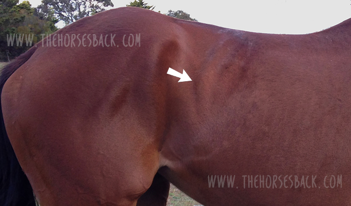
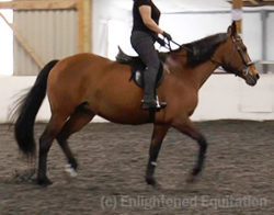
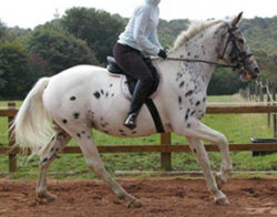
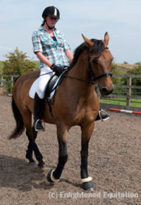
This veteran grey Arabian, below, is one I’d also consider a suspect. Again, we can see a very obvious protruding last rib on the offside and a lack of straightness. Even with musculoskeletal bodywork and spinal mobilization, the rib remained just as pronounced.

Arabian mare with a suspect rib. Photo: J. Clothier

Incidentally, I’ve also worked on this horse’s offspring, and the younger horse has the same profile to the ribs, on the same side, accompanied by a history of inexplicable back pain – and lack of straightness. 

Note: It’s important to eliminate other causes first, as horses will often have this appearance at the last rib, without it being caused by a transitional vertebra. What’s happening is that the rib is protruding because the vertebra is immobilised in a rotated position. When chiropractic, osteopathy or bodywork restores mobility to the spine, the rib returns to its normal position. 
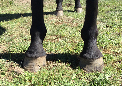
 

Ongoing hoof issues

In the bay TB mare, spinal asymmetry (scoliosis, with bend to the right) had led to excessive loading of the near fore. This was no doubt compounded by constantly training and racing in a clockwise direction, plus the classic long toe/low heel frequently found in ex-racehorses.

As a result, her near fore had constant hoof wall separation, bacterial infection (seedy toe / white line disease) and a deep P3 problem that would never come right.

Here’s the hoof capsule and P3. Yes, the poor girl suffered, despite extensive efforts to reconstruct that hoof.

P3 and hoof capsule, near fore, TB mare. Photo: J. Clothier


Patreon members can view videos of this mare and further photos. Go to: www.patreon.com/thehorsesback for more details.


The TB gelding mentioned earlier also had chronic issues in the opposing fore hoof, with wall separation, damage to P3 and evidence of earlier laminitis.


How many horses are affected?

Who knows? The study mentioned earlier (Haussler et al, 1997) found that 22% of Thoroughbreds examined at necropsy, having died or been euthanized at the racetrack, had thoracolumbar transitional vertebrae.

Transitional vertebra at T18 (above ground skeleton, damaged by scavengers)    (c) J. Clothier


To date, I’ve come across 3 in above-ground skeletons (2 x T18, 1 x L1), plus one in a horse later euthanized (1 x T18). These were TBs and Australian Stock Horses.

And as mentioned, I’ve suspected the T18 issue here and there amongst clients’ horses.

Although found mostly in Thoroughbreds, transitional vertebrae are seen across a range of breeds. And certainly, with equine dissection having taken off in quite a big way in the equine care industry, more and more of these anomalies are being observed.


Questions, thoughts or comments? Join us at The Horse’s Back Facebook Group. 


Should we be concerned?

The answer is, inevitably, both yes and no.

On the positive side, if the numbers harbouring this problem are as high as it seems, we have to assume that many horses are coping just fine. 

For as with any musculoskeletal anomaly, horses can compensate very well.

However, when another problem is added to the mix, things can head south very quickly indeed.

And it can all happen without us ever knowing that a skeletal anomaly is an underlying factor. When this happens, owners often have a lot of unanswered questions about their horses – and often large vets bills.

Transitional vertebrae at T18. (c) J. Clothier

It’s the TB or TB-derived breed horse that is most likely to present this (although not exclusively). If you’re buying one and you view a horse with an obvious T18 that really stands out, you might want to get that checked.

At the very least, do something that most people never do – stand on a fence or mounting block and take a photo down the horse’s spine, when it’s standing square or close to square.

If there’s a clear scoliosis along the spine, be cautious (this is a good rule of thumb anyway, no matter what the cause is). If you see an overly pronounced rib on the concave side, be doubly cautious.

And if you believe your horse may have one, the answer is the same as always: be aware, take a 360 degree approach in ensuring that hooves, tack, training and riding are as good as they can be, and your horse will have the best possible chance of functioning well without cause for concern.

(c) Melissa Longhurst, www.equinebodybalance.com.au


Filed Under: Bodywork Tagged With: equine anatomy, equine bodywork, equine vertebrae, GA, Thoroughbred, transitional vertebrae

All You Need to Know About the Hidden C6-C7 Malformation That’s Bringing Horses Down

July 16, 2017 by Jane @ THB 72 Comments


What if you were to learn that your horse is living with a hidden malformation? A skeletal abnormality that could be affecting it every day, changing the way it moves, creating a string of other physical problems, and possibly underlying the hard-to-pinpoint problems you’ve been noticing for months or even years ?

And that might even be causing a level of inherent instability that could be putting the rider in danger?

Sadly, this isn’t a hypothetical question. Instead it’s a reality that is only now being slowly uncovered.

And like the proverbial stone rolling down a mountain, the issue is gathering momentum as the equine industry, owners, breeders and researchers learn about it.

  • It’s a skeletal malformation and it can’t be corrected.
  • It’s congenital, ie inherited, so is present from birth.
  • It has been in some lines of TBs for hundreds of years.
  • It creates biomechanical issues due to asymmetry and lack of anchor points for key muscles.
  • At its worst, it can contribute to neurological issues such as Wobbler syndrome.
  • Some horses are so unstable, they are more prone to falling (not good news for jockeys).
  • It can cause constant pain and associated behavioural changes.
  • It’s primarily found in Thoroughbreds, Thoroughbred crosses and Warmbloods, but has also been identified in European breeds, Quarter Horses, Arabs and Australian Stock Horses.


Note: in the years since this post was written with direct support from Dr Sharon May-Davis, research has moved on. The body of literature now includes papers from Sharon and also from other researchers who do not reach similar conclusions about the issues caused by this malformation. I have included a list of journal papers and webinars on the topic at the end of the post, for reference if you wish to deepen your knowledge. You can also go view the full list here (opens in a fresh tab).


The problem behind this is ECVM, a congenital malformation of the C6 and C7 cervical vertebrae (ie, base of neck) – and it’s pretty nasty.

I’ve written about the work of Sharon May-Davis on this blog before and here I’m going to do so again. Through her many dissections per year, gross anatomist Sharon has become the first person to comprehensively document and quantify this problem.

In doing so, and publishing her findings in peer-reviewed journals, she has triggered a minor research avalanche as others take up the subject.

Update: since this article was published in July 2017, this malformation has been labelled Equine Complex Vertebral Malformation (ECVM). Some amendments have been made to include this term. To preserve existing links to the page, I’ve not updated the title and URL.

© All text copyright of the author, Jane Clothier, https://thehorsesback.com. No reproduction of partial or entire text without permission. Sharing the link back to this page is fine. Please contact me for more information. Thank you!


A hidden problem: this OTTB had the C6-C7 malformation but presented few outward signs.


Twenty years + of research into ECVM

Sharon May-Davis’s path with this research began some 20 years ago. In February 1996, a Thoroughbred called Presley came down unimpeded in a race in Grafton, NSW, fracturing his pelvis, a hock bone, and right front fetlock.

Three years later, Sharon examined his bones, and saw something strange in his last two cervical vertebrae and his first ribs.

Fast forward to 2014, when Sharon published the first of her four peer-reviewed papers in the Journal of Equine Veterinary Science, concerning a congenital malformation in the sixth and seventh cervical (neck) vertebrae.

Although the problem had been mentioned briefly in papers, this was the first time that a researcher had accurately described and quantified the problem in its various forms.

Sharon’s unique perspective, gained as an anatomist who dissects between 15 and 20 horses per year, had certainly placed her in a position to do so.


The horse’s seven cervical vertebrae – made simple

Horses have seven vertebrae in their necks, labelled C1 to C7. Of these, four have unique shapes. Most horse people are familiar with C1, the first vertebrae known as the atlas, as it can be both seen and felt by hand with its distinctive ‘wing’ at the top of the neck.

Cervical vertebrae from C1 (top) to C7 (bottom), view from above (L) and below (R).

Almost as well-known is C2, the second vertebrae, known as the axis.

Both atlas and axis have unique shapes for a special reason: they support the heavy skull and anchor the muscles that control the head’s movement.

Heading down the neck, C3, C4 and C5 are broadly similar in shape, with each being a bit shorter and blockier than the one above.

However, C6 and C7 are both slightly different on the ventral (lower) side, for here they provide insertion points for muscles arising from the chest.

  • C6 has transverse processes (the protrusions extending outwards) that are different to those of neighbouring bones, with two distinctive ridges running the vertebrae’s length. C6 also has two large transverse foramen, the openings that the arteries pass through.
  • C7 is the shortest and squattest cervical vertebrae of all. Its transverse processes are shorter, while there are also two facets that articulate with the first ribs. C7 has no transverse foramen.

At least, that’s how the vertebrae should be in a normal horse.


So, what is wrong with the malformed C6 and C7 vertebrae in ECVM?

In certain horses, these last two vertebrae are rather different, being malformed.

Sharon has identified the manifestations of this problem as a congenital (inherited) malformation affecting some Thoroughbred horses, and horses with Thoroughbred blood in their ancestry.

In C6, there is a problem with the two ridges of the transverse processes, as one or both can be partially absent.

When both are partially missing, it is common for one or two ridges (ie, parts of the transverse processes) to appear on C7 instead.

Also, the articular processes (the oval surfaces on the upper side, where each vertebrae links to its neighbours) can be radically different sizes. There can also be an additional arterial foramen or two.

The level of asymmetry can be radical.

The secondary problems the ECVM malformation causes

Being at the base of the neck, the asymmetry of C6 and C7 can cause alignment problems all the way up the vertebral column, leading to osteoarthritis of the articular facets.

It can also contribute to Wobbler Syndrome (Cervical Vertebral Stenotic Myelopathy), due to narrowing and/or malalignment of the vertebral foramen/canal, the opening through which the spinal cord passes. Not all Wobbler cases have this particular malformation, though.

A further problem is that the lower part of the longus colli muscle, which is involved in flexing the neck, would normally insert on the transverse processes of C6 and C7. When these processes are malformed, the normal insertions are not possible.

This means there is a serious symmetry problem in the junction of the thorax and neck, which can have a deeper effect on the horse’s neurology and proprioception, as well as respiration.

Asymmetry and narrowed vertebral foramen (canal) contribute to DJD and Wobbler Syndrome.


In a few cases, horses with both the C6 and C7 problem also have malformations of the first sternal rib, on one or both sides. This can cause problems beneath the scapular and further issues with muscular attachments.

Associated stability problems can have far-reaching consequences for the horse, not to mention some serious safety issues for the rider. The safety issue can’t be stated often enough.


Why isn’t ECVM – the C6-C7 problem – more widely known ?

Why hasn’t this problem been noticed in regular veterinary interventions?

The answer is quite simple. While neurological issues may have been diagnosed, the exact cause has often remained hidden.

Both Thoroughbred horses and Warmbloods are known to have higher incidences of Wobbler Syndrome than other breeds, and while this is certainly not always due to C6-C7 malformation, the malformation has been found in some when dissected.

For example, the following dissection image appears in a veterinary account of large animal spinal cord diseases. It clearly shows a malformed C7 vertebrae, very similar to the one in the above image, but without giving any further categorisation.

The difficulty lies in the deep location of the lower cervical vertebrae. While normal radiographs can show all or some of C6, they are unable to penetrate the deeper tissues beneath the shoulder to image C7.

Photo from another online article: the familiar asymmetry of a malformed C7 is clearly visible in a dissected set of vertebrae.  (c) veteriankey.com (click image to access full article.)


Nevertheless, the malformation can be identified in radiographs of C6, once you know where to look.

Since Sharon’s first paper appeared, the School of Veterinary Medicine, University of California, Davis, has reviewed its history of radiographs from horses with Wobbler Syndrome.

Researchers found that 24 cases out of 100 (close to 25%) showed malformation of one or both C6 transverse processes. This study also clarified how to identify the problem on standard radiographs of C6.

In another study, the Faculty of Veterinary Medicine, Utrecht University, completed CT scans on horses’ necks and found the various forms of the malformation in 26 horses out of 78 (33%). Unlike radiographs, the CT scans enabled identification of the C7 and first rib issues, although of course this imaging was conducted post mortem.

Is this rare, or are many horses affected?

While the problem has been identified primarily in TBs, it affects most breeds with TB blood in the ancestry to some degree.

Sharon May-Davis reports that to date, published, peer-reviewed journal papers have tallied 136 out of 471 horses as exhibiting congenital malformation of C6.

These have been in a range of breeds including Thoroughbreds (39%), Thoroughbred crosses (27%), Warmbloods and European breeds (30%), Quarter Horses (11%), and Arabs (11%). Standardbreds have also shown the problem, although the numbers included in studies are very small.

A common question is whether it’s known which TB lines predominantly carry this problem. The answer is: Yes. However, it is now so disseminated amongst the modern equine population beyond TBs, that it is of little help to identify them.

It must be remembered that these horses are those already brought to veterinary attention and/or euthanized for a related or unrelated reason, so the percentages may be higher than those for the general horse population. At the same time, the malformation might have played a major part in the horses’ decline, due to the many locomotory and postural problems it can lead to.


How do we identify these horses in life?

It’s all very well looking at these bones post mortem, you might say. Yet how can I tell if my horse has this problem? Or a horse that I might want to buy?

Some answers are forthcoming. As Sharon has frequently assessed horses before dissecting them – usually from video – she has been able to observe that many of these horses lack stability. (Indeed, in many cases, it is this very instability has directly led to the horse being euthanized, and ending up on the dissection table.)

As her research has progressed, she has also been able to identify many biomechanical and locomotion traits that make these horses ‘suspicious’ or at least ‘of interest’. Unsurprisingly, these problems have been particularly noticeable in horses with both a malformed C6 and C7.

For owners and equine professionals, here are some signs that can raise initial suspicions. All can also be caused by other problems, so a group of signs is more common than an individual indication.

  • Some of these horses have a problem with standing square in front, and will always keep one foot further forward. This can persist despite all attempts to improve the horse’s body and to train the horse to halt squarely.
  • Horses with the more serious malformations will often stand base-wide. Such horses can become very unbalanced on uneven ground, and sometimes in work. They easily become unbalanced when a hoof practitioner works on a forefoot. 


A bilateral C6 – unilateral C7 horse showing a toe-out stance and hoof distortion. The ventral part of the transverse process was transposed onto the left side of C7.


  • With such asymmetry in the skeletal structure, these horses have serious lateral flexion issues that can’t be overcome. When required to elevate the forehand, many will experience difficulties, due to the absence of correctly inserted musculature and incorrect articulation through the joints of the lower neck. 
The horse may have one very prominent, widely positioned scapula.
  • A high level of asymmetry may be seen in the shoulders, with one scapula sometimes positioned very wide, with no improvement after chiro, osteopathy or bodywork. This is particularly so with the C6-C7 problem and associated first sternal rib abnormalities.
The ventral aspect of the neck may show some scoliosis.


  • There may be scoliosis along the entire spine.
  • There may be an obvious scoliosis to the underside of the neck.
  • The problem may lead to heavily asymmetric loading of the forefeet, so may be accompanied by a severe high foot/low foot issue (this is not in itself a sign of the C6-C7 problem).

If you suspect your horse has the C6-C7 issue

First, note that many horses do just fine with a C6 problem. It is those with the bilateral C6 and unilateral/bilateral C7 issue that tend to show the more worrying problems.

If your horse is showing ongoing signs of instability, it’s important to seek veterinary advice, so that neurological issues can be ruled out. (As this a recently recognised problem, it may be worth printing out the abstracts from the journal articles listed at the end of this page and handing them over.)

If the more severe malformations are identified by radiograph, it is important to remember that in some cases this can cause discomfort and pain to the horse, and it is not going to improve over time.

On the contrary, the cervical vertebrae of some older horses with the C6 and C7 malformations often display advanced osteoarthritis of the articular processes, as shown in the header image of a 19-year-old Thoroughbred’s malformed C7.


Where does this knowledge take us?

At the moment, that question is wide open. The findings published by Sharon May-Davis have triggered ongoing research on an international level. There are certainly ramifications for breeders in more than one equine sporting industry.

See Sharon May-Davis’s December 2020 presentation on ECVM here (article continues below):

Connections have been made with a number of falls on the racetrack that have caused injury, and worse, to both horse and jockey, as well as other runners. Similar things can be said for the sport of eventing, where unforced errors can have equally catastrophic effects.

It is entirely possible that at higher levels, pre-purchase examination radiographs will come to include a check on C6. While it’s not possible to radiograph the deeply positioned C7, we do at least know that this will only be present if the C6 anomaly exists.

Vets in some countries are proving faster at picking this up than others. While papers are being published, it clearly takes some time for information to filter down.

And until more is known, this problem is being unknowingly propagated every breeding season.

Of course, many horses harbouring the milder manifestations of this problem at C6 level are functioning very well. All horse owners can do is be aware that this issue exists, make use of this information if a problem arises, and await further research findings.

Literature on the malformation

In no particular order: the following peer reviewed journal articles address ECVM, as it is now known. Please remember when reading papers that no single paper proves one thing – a review of many papers and critical thinking is always essential. Apologies for not having standardized the formatting, but the links should be present.

  • The Occurrence of a Congenital Malformation in the Sixth and Seventh Cervical Vertebrae Predominantly Observed in Thoroughbred Horses, May-Davis, Sharon, Journal of Equine Veterinary Science , Volume 34 , Issue 11 , 1313 – 1317
  • Variations and implications of the gross morphology in the longus colli muscle in thoroughbred and thoroughbred derivative horses presenting with a congenital malformation of  the Sixth and Seventh Cervical Vertebrae May-Davis, S, Walker, C. Journal of Equine Veterinary Science 35 (7), 560-568
  • Characterization of the Caudal Ventral Tubercle in the Sixth Cervical Vertebra in Modern Equus ferus caballus. May-Davis, S., Dzingle, D, Saber, E., Blades Eckelbarger, P. Animals 13 (14), 2384
  • Morphology of the Ventral Process of the Sixth Cervical Vertebra in Extinct and Extant Equus: Functional Implications, May-Davis, S., Hunter, R, White, R. Animals 13 (10), 1672
  • Variations and Implications of the Gross Morphology in the Longus colli Muscle in Thoroughbred and Thoroughbred Derivative Horses Presenting With a Congenital Malformation of the Sixth and Seventh Cervical Vertebrae, May-Davis, Sharon et al., Journal of Equine Veterinary Science , Volume 35 , Issue 7 , 560 – 568
  • Congenital Malformations of the First Sternal Rib, May-Davis, Sharon, Journal of Equine Veterinary Science , Volume 49 , 92 – 100
  • Ex Vivo Computated Tomographic Evaluation of Morphology Variations in Equine Cervical Vertebrae,  Veraa, S. et al,  Veterinary Radiology & Ultrasound, Vol. 57, Issue 5
  • Prevalence of Anatomical Variation of the Sixth Cervical Vertebra and Association with Vertebral Canal Stenosis and Articular Process Arthritis in the Horse, Spriet, M. and M Aleman, Veterinary Radiology & Ultrasound, Vol. 57, Issue 3
  • Anatomical Variation of the Spinous and Transverse Processes in the Caudal Cervical Vertebrae and the First Thoracic Vertebra in Horses, Santinelli, I. et al, Equine Veterinary Journal, Vol. 48, 45–49
  • Congenital variants of the ventral laminae of the sixth and seventh cervical vertebrae are not associated with clinical signs or other radiological abnormalities of the cervicothoracic region in Warmblood horses. Dyson S, Phillips K, Zheng S, Aleman M. Equine Veterinary Journal, 2025 Vol.2 :419-430
  • Neck pain but not neurologic disease occurs more frequently in horses with transposition of the ventral lamina from C6 to C7. Henderson CS, Story MR, Nout-Lomas YS. J Am Vet Med Assoc. 2024 May 29;262(9):1215-1221


Filed Under: Bodywork, Sharon May-Davis Tagged With: C6, C7, ECVM, equine dissection, GA, homepage, Sharon May-Davis, skeletal malformation, slider, Thoroughbred anatomy

5 Ways Your Seat Can Screw Up Your Horse Without You Even Noticing

January 26, 2017 by Guest Author 2 Comments


In this Guest Post, the UK’s ‘Classical Seat’ trainer Heather Moffett looks at how the rider’s seat can make movement harder for the horse. It’s something that saddle fitters know about, but it’s often the last thing riders think of: the riding position as much as the saddle fit affects their horse’s back and movement. 

Heather Moffett has over 40 years’ instruction experience. Chiefly following the French school of classical equitation, which focuses on dressage as an art form rather than competition, she is best known as an authority on the Classical Seat. 


We all keep hearing about ‘connection’.  It’s a current buzzword and usually means connecting with your horse on the ground, either through loose or in-hand work.

Many riders assume this will enable them to achieve the same connection once mounted.

There is nothing wrong with that. However, they often then wonder why they lose that connection – sometimes literally, complete with the saddle(!) – once they start riding. The fact is that they are, in ignorant bliss, impeding the horse!


The Disconnected Seat

If the rider is out of sync with the horse’s movement, the flow and harmony will always be disrupted as the horse struggles to balance the rider as well as himself.

Or, he endures the discomfort of a rider sitting like a lump of lead, driving with the seat against his sensitive back, or bouncing stiffly in the saddle.

The horse has only one way to show his discomfort or pain, as he is mute, and that is by behaviour that is then construed as ‘misbehaviour’.

I often ask riders who kick and hit their horses if they would do the same to their dog. The dog is able to cry out in pain. The horse cannot, and it is his muteness, throughout history, that has led to his downfall and still does to this day.


What Gets My Goat is This

I’ve been a specialist trainer/teacher of the Classical seat for 46 years. In that time, there has been little interest in increasing knowledge of the seat and refinement of the aids.

So many teachers and trainers say that it is necessary to ignore imperfections in the seat until “the horse is going well”. Then they wonder why the horse never progresses, or why force must be resorted to, in order to make the horse submit!

Yes, there is that word submit (or submission) that’s a requirement in a dressage test.

My own teachers soon found that it worked far better to encourage my cooperation in school rather than forcing me into submission! And a horse is no different.

I would like to see the word submission in tests replaced with willing cooperation.

How different would the expression be on many a horse’s face, if he were trained as a partner, and not as an adversary?

So, are you screwing up your horse with your seat, without even noticing? Here are the 5 top points I’d like you to think about.


1. Saddle Fit Woes (Yes, Again)

Poor saddle fit can cause the seat to tip back.

No surprises for this one. Saddle fit is probably the most obvious thing that needs to be right, but many saddles are a long way from perfect. That’s true even in my home of the UK, where saddles are most often professionally fitted.

But, what amazes me is the number of saddles I see with clients coming to me for lessons that have faults that make it damn near impossible for the rider to sit either straight or in balance!

Here are the worst offenders.

The saddle is too narrow. This is still one of the most common faults – it pinches the horse and tips the rider’s pelvis backward, aiding a chair seat rather than the ear/shoulder/hip/heel line which is the only position of balance.

Try walking with your butt stuck out behind you and knees up, as though sitting on a chair! It’s hard enough to even remain upright! Your weight will be over the cantle region, making it difficult for the horse to lift and round his back, and causing him to go hollow.

Stirrup bars are, almost without exception, even on many dressage saddles, too far forwards. This is why the rider is constantly being nagged by instructors to “get that lower leg back”!

I do wonder why instructors and even top dressage trainers, never seem to notice that all of their students are not anatomically challenged and there might just be something wrong with the saddle design and balance!! (Don’t get me started!!)


2. An Insecure Seat
Sucks (for Both Horse and Rider)

7yo Oldenburg x Irish Sport Horse. 1st day to 2nd day of HM course.

Here’s what I mean by insecure. Riders are not taught to absorb and sync with the horse’s movement.

We hear:

“Sit deeper!”

“Relax your back!”

“Go with the movement!”

“Follow the horse’s movement!”

Well, usually if you are following something you are behind it!

Is it any wonder beginners are confused and often never learn to move in sync with the horse?

If this describes you, fear not. I have had riders here on my horse movement simulator workshops who’ve been riding 10, 20 or more years, and still they have never learned to move in sync with the horse.


3. Saddle Seat Glue Hasn’t Been Invented Yet

Gripping the saddle blocks movement.

If you’re bouncing around, or driving with both seat bones to achieve some adhesion to the saddle, or sitting on your back pockets and collapsing the rib cage, you will be making your horse’s life more difficult.

It is so NOT rocket science to learn this!

But until teachers are trained to teach it, the situation will not improve!!

And it is not just novice riders who block their horse through incorrect adhesion to the saddle.

Look at the nodding head, flailing legs (usually with spurs attached) riders to be seen even in the Grand Prix dressage arena…


4. Horses Have the Low Down on Our Weight Issues…

How many times do you hear it said that a horse can feel a fly land on his back? So how much more can he feel his rider, whether good or bad?

For me, my aim – both as a rider myself and also as a trainer – is to be as little burden on the back of my horse as possible.

The horse copes with the crooked rider. Day 1 of HM course.

I aim to do this by sitting lightly, but deeply, in sync with his movement.

If the rider is crooked, possibly due to a problem with the saddle, or is asymmetric due to their own physical problem, the horse suffers.

He has to cope with this and compensate, usually by going crookedly himself.

And there’s more. The use of the rider’s body as a primary aid, is so rarely taught. Yet when utilised, it is the most invisible aid of all. Combined with the seat bones moving in sync with the horse’s back, it is the secret to an elegant harmonious seat, that appears to be doing nothing.

That’s when the horse and rider glide through all the movements as though they are one being – like a Centaur.


5. ‘Feel’ Begins in your Backside (I Mean It!)

Hanoverian X. 1st and 2nd day of HM course.

‘Connection’ means being able to feel, and not just when working from the ground. We all have nerve endings in our backsides – if you are taught what to feel and how to feel, it is within the grasp of any rider, even beginners.

And ‘aid’ really means ‘help’. If you learn to use aids that make biomechanical sense to the horse, they do become truly invisible as the horse becomes more and more sensitive with correct training.

BUT, if the horse hasn’t been schooled to respond to specific aids, then is it any wonder he is confused and ‘misbehaves’? It’s a bit like us lazy Brits here, shouting at foreigners in the hope they will understand English then getting annoyed when they don’t!

Your seat can genuinely aid (help) your horse. This happens when you’re taught not only the hand and leg aids, but also:

  • the weight aids for turning,
  • the seat aid for collecting and for downward transitions,
  • the precise positioning of the torso in lateral work and circles/ bends, etc.

At this point, riding becomes a whole language, which almost all horses quickly understand. Why? Because it is working with, not against, their own body.

Moving in sync with the horse allows the rider to learn ‘feel’, that term that often seems to imply that only a favoured few have the ability to learn it.

Rubbish!


So, in closing, if you wish to have true connection with your horse, you need to:

a) Absolutely not screw up either his back or his brain,

b) Learn to ride to the best of your ability, and

c) Treat your horse as a partner and friend, and not as a tool merely to win the next rosette.

If winning happens as a by-product of good riding, even better, but if your horse is not progressing, look to your own riding and equipment before you blame your horse. Get these 5 points sorted and you’ll be well on your way to true connection with your horse!


Filed Under: Bodywork, Guest Posts, Saddle Fit Tagged With: classical dressage, classical seat, GA, saddle fit

Meet Spinalis, the Forgotten Muscle in Saddle Fitting

December 20, 2016 by Jane @ THB 43 Comments

Spinalis Header

It’s barely mentioned in saddle fit or anatomy books, yet the muscle Spinalis cervicis can hugely impact on the spinal health and movement of the horse, particularly with poor tack fit.

Meet muscle Spinalis cervicis et thoracis, a far more important muscle than is generally realized. As a deep muscle, it’s influential in mobilizing and stabilizing that hidden area of the spine at the base of the neck, the cervico-thoracic junction, deep between the scapulae.


Where to Find this Muscle

As part of the deeper musculature, Spinalis is as hidden in books as it is in life. Usually, it’s a single entry in the index.

Spinalis Standard
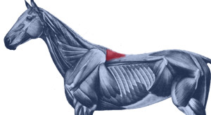
At best, it has no more than a bit part in anatomical illustrations,  usually as a small triangular area at the base of the withers. This is also where we can palpate it.

The reality is quite a bit more interesting. It’s actually a muscle of three parts – dorsalis, thoracis and cervicis. These names denote its many insertions, for it links the spinous processes of the lumbar, thoracic and cervical vertebrae.

  • Bradley_2.1
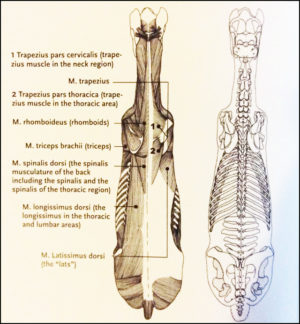
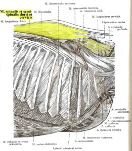
Further back along the spine, it lies medially to Longissimus dorsi, and in fact integrates with this larger, better known muscle, attaching to the processes of the lumbar and thoracic vertebrae.
  • When it reaches the withers, it becomes more independent, attaching to the processes of the first half dozen thoracic vertebrae (T1-T6). Here, the cervical and thoracic portions overlap and integrate to share a common attachment. (The part we palpate, at the base of the withers, is the thoracic section.)
  • Heading into the neck, as Spinalis cervicis, it attches to the last 4 or 5 cervical vertebrae (C3/C4-C7). Only the lamellar portion of the nuchal ligament runs deeper than this muscle.

Dissection 2
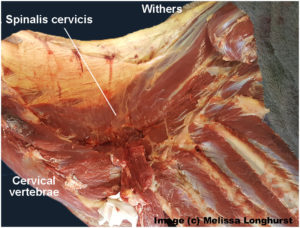
Its integration with other muscles is complex, and its close relationship with Longissimus dorsi partially explains why it doesn’t get much consideration as a muscle in its own right.

It is the more independent section, Spinalis cervicis, between withers and neck, that we are interested in, although its influence is present along the entire spine.

© All text copyright of the author, Jane Clothier, https://thehorsesback.com. 

What Does Spinalis Do? 

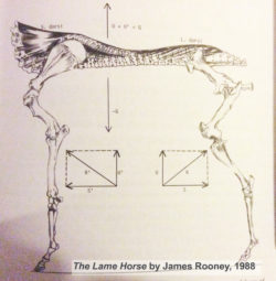
In his 1980s’ Guide to Lameness videos, Dr. James Rooney, first director of the Gluck Equine Research Center, University of Kentucky, referred to Spinalis as part of the suspension bridge of muscles supporting the spine (Longissimus dorsi achoring from the lumbosacral vertebrae, Spinalis thoracis et dorsalis from the upper thoracics). He also refers to this extensively in The Lame Horse (1988).

In fact, the suspension bridge analogy only really makes sense if Spinalis dorsi is considered.

Spinalis cervicis is usually credited with a role in turning the head to left to right, and raising the head.

Bradley Spinalis-1Older texts, such as Bradley’s 1922 veterinary dissection guide, Topographical Anatomy of the Horse, mention its role in stabilizing the spine.

This creates a point of interest. Given that the nuchal ligament (lamellar portion) doesn’t attach to C6 and frequently only weakly with C5 (see the findings of anatomist Sharon May-Davis, in this earlier article ), Spinalis cervicis suddenly appears pretty important in stabilizing and lifting the base of the neck, particularly as it does so at the point of greatest lateral bending.


Spinalis and Poor Saddle Fit

Anyone who has been involved in close examination of the horse’s back will recognize Spinalis thoracis where it surfaces close to the skin, on either side of the withers.

When a horse has been ridden in an overly tight saddle, this small area of muscle can become pretty hypertrophic – raised and hardened. Typically, the neighbouring muscles are atrophied. When Spinalis is palpated, the horse often gives an intense pain response, flinching down and raising the head.

GerdHeuschman
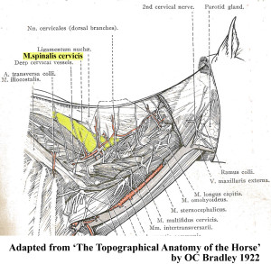
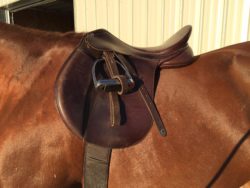
What often happens is this. An overtight saddle fits over the base of the withers like a clothes peg, pinching Trapezius thoracis and  Longissimus dorsi. However, it frequently misses Spinalis thoracis where it surfaces, wholly or partially within the gullet space. Often, the muscle is partially affected.

It’s as if the neighbouring muscles are under lockdown. Free movement of the shoulder is restricted and the horse’s ability to bear weight efficiently while moving is impeded. In response to the surrounding restriction and its own limitation, this muscle starts to overwork.

Result? The horse, which was probably already moving with an incorrect posture, hollows its back even further, shortening the neck and raising its head.  As this becomes even more of a biomechanical necessity, all the muscles work even harder to maintain this ability to move, despite the compromised biomechanics.

Working harder and compensating for its neighbours, Spinalis becomes hypertrophic. It is doing what it was designed to do, but it’s now overdoing it and failing to release. We now have a rather nasty vicious circle.


Spinalis photo

Here, Spinalis thoracis stands out due to atrophy of the surrounding musculature. In this TB, a clearly audible adjustment occurred in the C4-C5 area after the muscle was addressed. 


Vicious CircleThe Inverted Posture and Asymmetry

Of course, saddle fit is not the only cause of an inverted posture. However, any horse that holds its head and neck high for natural or unnatural reasons is more vulnerable to saddle fit issues, thus starting a cascade effect of problems.

Are there further effects of this hypertrophy? Consider the connections.

  • When saddles are too tight, they’re often tighter on one side than the other. This can be due to existing asymmetry in the horse, such as uneven shoulders, uneven hindquarters, scoliosis, etc.
  • On the side with greater restriction, the muscle becomes more more hypertrophic.
  • With its attachment to the spinous processes of the lower cervical vertebrae, there is an unequal muscular tension affecting the spine.
  • Without inherent stability, the neck and head are constantly being pulled more to one side than the other, with the lower curve of the spine also affected.
  • Base of neck asymmetry affects the rest of the spine in both directions and compromises the horses ability to work with straightness or elevation.
  • There is also asymmetric loading into the forefeet.
  • We haven’t even started looking at neurological effects…

This isn’t speculation. I have seen this pattern in horses I’ve worked on, many times over.


So, How Do We Help?

In working with saddle fit problems, the saddle refit may be enough to help the horse, if the riding is appropriate to restoring correct carriage and movement. Obviously, the horse’s musculoskeletal system is complex and no muscle can be considered in isolation. As other muscles are addressed through therapeutic training approaches, with correct lateral and vertical flexion achieved, M. spinalis will be lengthened along with the surrounding musculature.

I hold with a restorative approach:

  1. Refit the saddle, preferably with the help of a trained professional,
  2. Remedial bodywork, to support recovery from the physical damage,
  3. Rest the horse, to enable healing of damaged tissue and lowering of inflammation, and
  4. Rehabilitate the horse, through the appropriate correct training that elevates the upper thoracics while improving lateral mobility.

This is particularly important where saddle fit has been a major contributor to the problem. I have frequently found that in these cases,correction will take longer to achieve, as the debilitating effects of poor saddle fit (especially long-standing issues) can long outlast the change to a new, better-fitting saddle. In bodywork terms, the hypertrophic M. spinalis cervicis is often the last affected muscle to let go.

It’s as if Spinalis cervicis is the emergency worker who will not leave until everyone else is safe.


Bodywork Notes

I am fortunate, in that my modalities enable the gentle release of joints through a non-invasive, neuromuscular approach.  The responses I’ve had from horses when M. spinalis cervicis et thoracis has been addressed in isolation have been hugely informative.


** Questions, thoughts or comments? Join us at The Horse’s Back Facebook Group. 


Appendix: Spinalis in the Textbooks

I’m going to add Spinalis references to this post on a regular basis, as I come across them. It’s interesting to see how much, or how little, the muscle is referenced in various textbooks.


Equine Back Pathology

This image, from Equine Back Pathology, ed. F Henson 2009, shows acute atrophy of Longissimus dorsi due to neurological damage. It’s still possible to see the raised attachment/origin of Spinalis cervicis et thoracis – the highlighting is mine. Spinalis does not appear in the book’s index. (added 23 Dec 2016)


nuchal and spinousI have also altered this image, in order to show M. spinalis cervicis more clearly. This is Fig 2.16 from Colour Atlas of Veterinary Anatomy Vol 2, The Horse, R Ashdown and S Done. Spinalis cervicis is within the bounded area and it’s possible to see how it overlies the lamellar part of the nuchal ligament, lamellar portion. (added 23 Dec 2016)


S&G
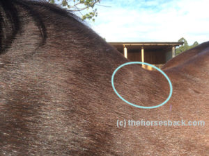
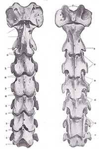
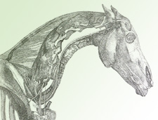
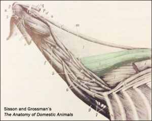
The muscle is tinted green in this image from Sisson and Grossman’s The Anatomy of Domestic Animals, Volume 1, fifth edition 1975.  Here, it is labelled Spinalis et semi-spinalis cervicis. This anatomical figure is credited to an earlier text, Ellenberger and Baum, 1908. (added 23 Dec 2016)


James Roony dedicates two pages to the ‘suspension bridge’ theory of the vertebral column in The Lame Horse (1988). His interest is in Spinalis dorsii section of the muscle and its effect behind the withers, in conjunction with  Longissimus dorsii. (added 4 Jan 2017)


Schleese diagramMaster Saddler Jochen Schleese refers to Spinalis dorsi and its function in stabilizing the withers in Suffering in Silence, his passionate book about saddle fitting from 2014. “This muscle area is especially prone to significant development – especially with jumpers – because it is continually contracted to accommodate the shock of landing”. The surface area of the muscle is indicated in the anatomical figure, reproduced here. (added 23 Dec 2016)


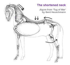
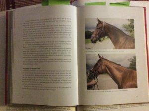
In his seminal text addressing issues of modern dressage training, Tug of War, 2007, Gerd Heuschmann includes Spinalis cervicis in the triangle formed by the rear of the rear of the cervical spine, the withers, and the shoulder blades, “… an extensive connection between the head-neck axis and the truck… it explains how the position and length of the horse’s neck directly affects the biomechanics of the back.” (added 31 Dec 2016)
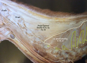
 

Filed Under: Bodywork, Saddle Fit Tagged With: Anatomy, equine bodywork, forgotton muscle, GA, homepage, saddle fit, saddle fitting, slider, spinalis, spinalis cervicis, spinalis dorsalis, spinalis thoracis

How The Anatomy Books (Unintentionally) Fail Us Over The Nuchal Ligament

July 13, 2014 by Jane @ THB 5 Comments

nuchal-ligament-header-1

The nuchal ligament is a soft tissue structure that is widely discussed in dressage circles. Unsurprisingly, given its deep location, relatively few of us get to cast eyes on it or feel it directly under our hands.

It’s equally unsurprising, then, that most of us don’t realize that the image we hold in our heads is somewhat different to the reality of the ligament inside our horse.

 © All text copyright of the author, Jane Clothier, www.thehorsesback.com. No reproduction of images, partial or entire text without permission. Sharing the link back to this page is fine. Please contact me for more information. Thank you!

I have recently been fortunate enough to attend another dissection with renowned Australian gross anatomist (and she will point out repeatedly that despite this title, she is not gross – or, at least, not that often), Sharon May-Davis.

In this dissection workshop, Sharon had yet another opportunity to show us that an aspect of textbook anatomy is incorrect.

Yes, apparently there are many points where this is the case.

Where the nuchal ligament is and what it connects

The structure in question is the nuchal ligament, or the nuchal ligament lamellae to be exact.

George Stubbs illustration
George Stubbs, 1777, showed the NLL attaching from C2 to C7.

To quickly explain, the funicular part of the nuchal ligament is the cord-like part that runs from the withers to the occiput (back of skull). The lamellae is the fibrous sheet-like part that extends from the funicular part to the cervical (neck) vertebrae.

According to the majority of anatomy diagrams and textbooks, it extends down to attach to the cervical vertebrae, from C2 to C7.

According to Sharon, it doesn’t. And here’s why.

Findings on the nuchal ligament’s true location

In this study of 35 horses on the dissection table, Sharon found:

  • No cases where the attachments were from C2 to C7.
  • No horses where the attachments were from C2 to C6.
  • In all 35 horses, the attachments were from C2 to C5.
  • And in 9 of the 35, the attachments to C5 consisted of thin and feeble fibers.
  • The horses were of a mixture of identifiable breeds, aged 2 to 28 years old.

So, why do the majority of anatomical drawings of the deeper structures of the horse show something different?

When received knowledge can be a problem

Nuchal ligament, 5yo TB [click to enlarge]
Many of today’s illustrators are referring to illustrations that have themselves been amended from earlier illustrations.

(The header image for this site’s most viewed post, The Disturbing Truth About  Neck Threadworms and Your Itchy Horse, shows an inaccurate rendering of this ligament, as do most of the other illustrations I used. Dang!)

Inaccuracy is a recognized problem when it comes to received knowledge – was this anomaly due to an earlier artist’s error, or was it a characteristic of some 17th century horses that has been progressively bred out over subsequent centuries?

  • And this raises the question of which structure, exactly, is supporting the base of the neck of the horse in motion? Read more about m. Spinalis cervicis in this post, Meet Spinalis, the Forgotten Muscle in Saddle Fitting.
  • And how does this awareness inform current training approaches that require horses to raise themselves into self-carriage?

The findings from this study are in a peer-reviewed paper by Sharon May-Davis and Janeen Kleine currently in press with the Journal of Equine Veterinary Science. The paper includes a detailed review of illustrations in equine anatomy literature, an explanation of the study, and a thought-provoking discussion on the implications for our understanding of equine biomechanics.

Variations and implications of the gross anatomy in the equine nuchal ligament lamellae, Sharon May-Davis, Janeen Kleine, Journal of Equine Veterinary Science 30 June 2014 (Article in Press DOI: 10.1016/j.jevs.2014.06.018)

Have you read about Sharon’s findings on arthritis of the humeroradial (elbow) joint in all ridden or driven horses?


Questions, thoughts or comments? Join us at The Horse’s Back Facebook Group. 


Filed Under: Bodywork, Sharon May-Davis Tagged With: equine anatomy, equine bodywork, GA, horse anatomy, Nuchal ligament, Sharon May-Davis

  • « Go to Previous Page
  • Page 1
  • Interim pages omitted …
  • Page 3
  • Page 4
  • Page 5
  • Page 6
  • Go to Next Page »

Footer

Contact Me

Send your questions this way…

Get in Touch

  • Store Support
  • Shipping
  • Returns
  • Contact

Copyright © 2026 · Parallax Pro on Genesis Framework · WordPress · Log in