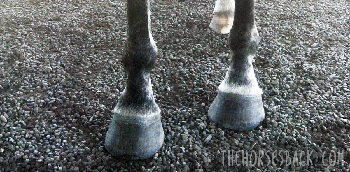

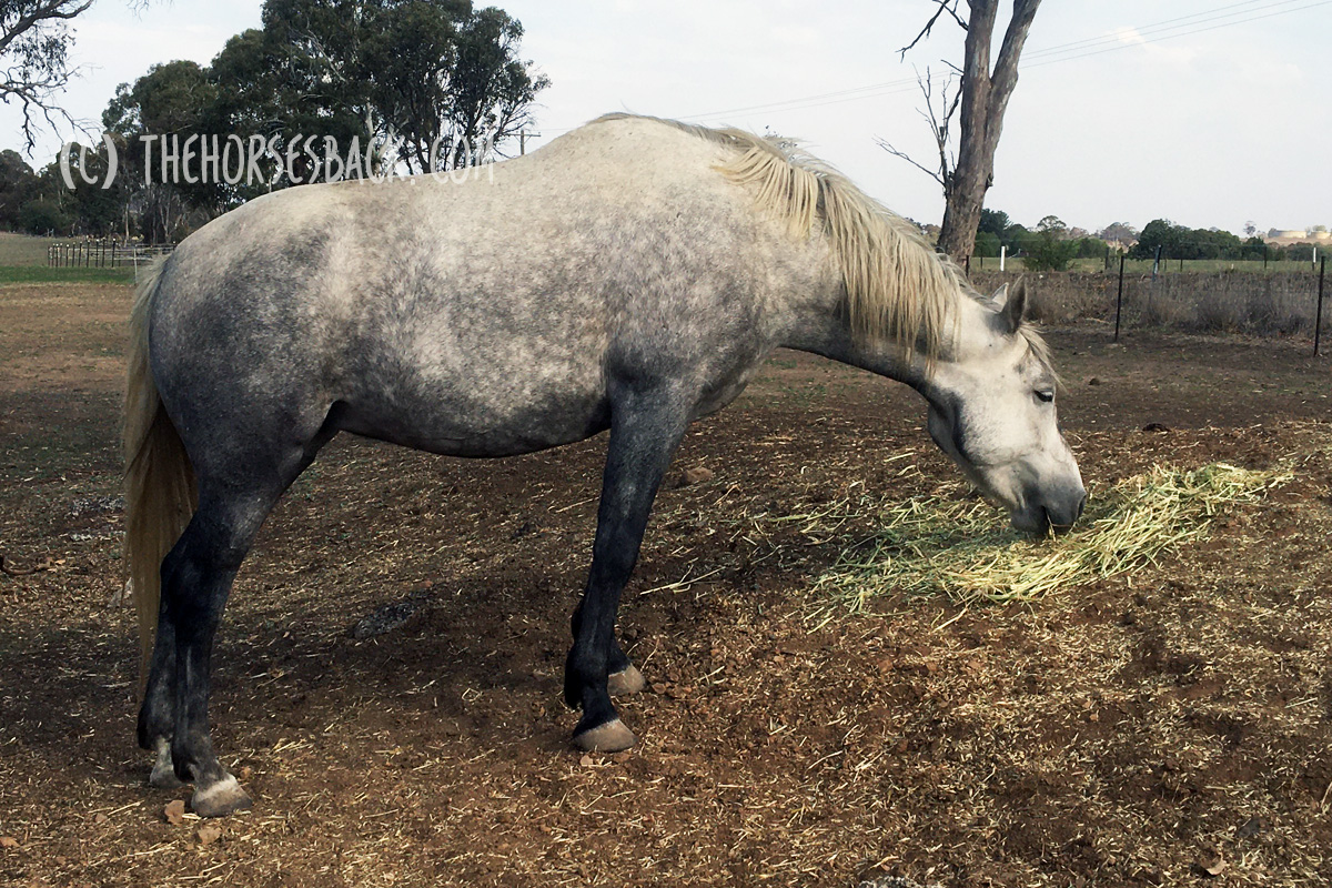
Sorting out that high-low hooves situation. It’s the farrier’s job, isn’t it?
Well yes, they’re clearly the primary professional. But there are plenty of things you can also do to help.
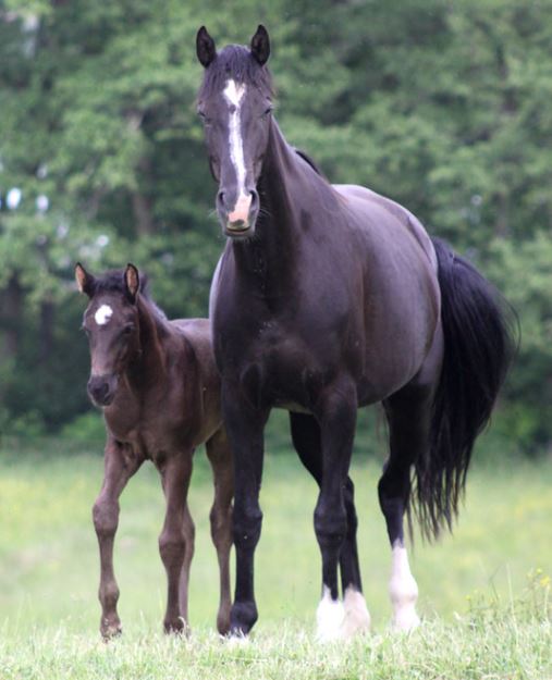
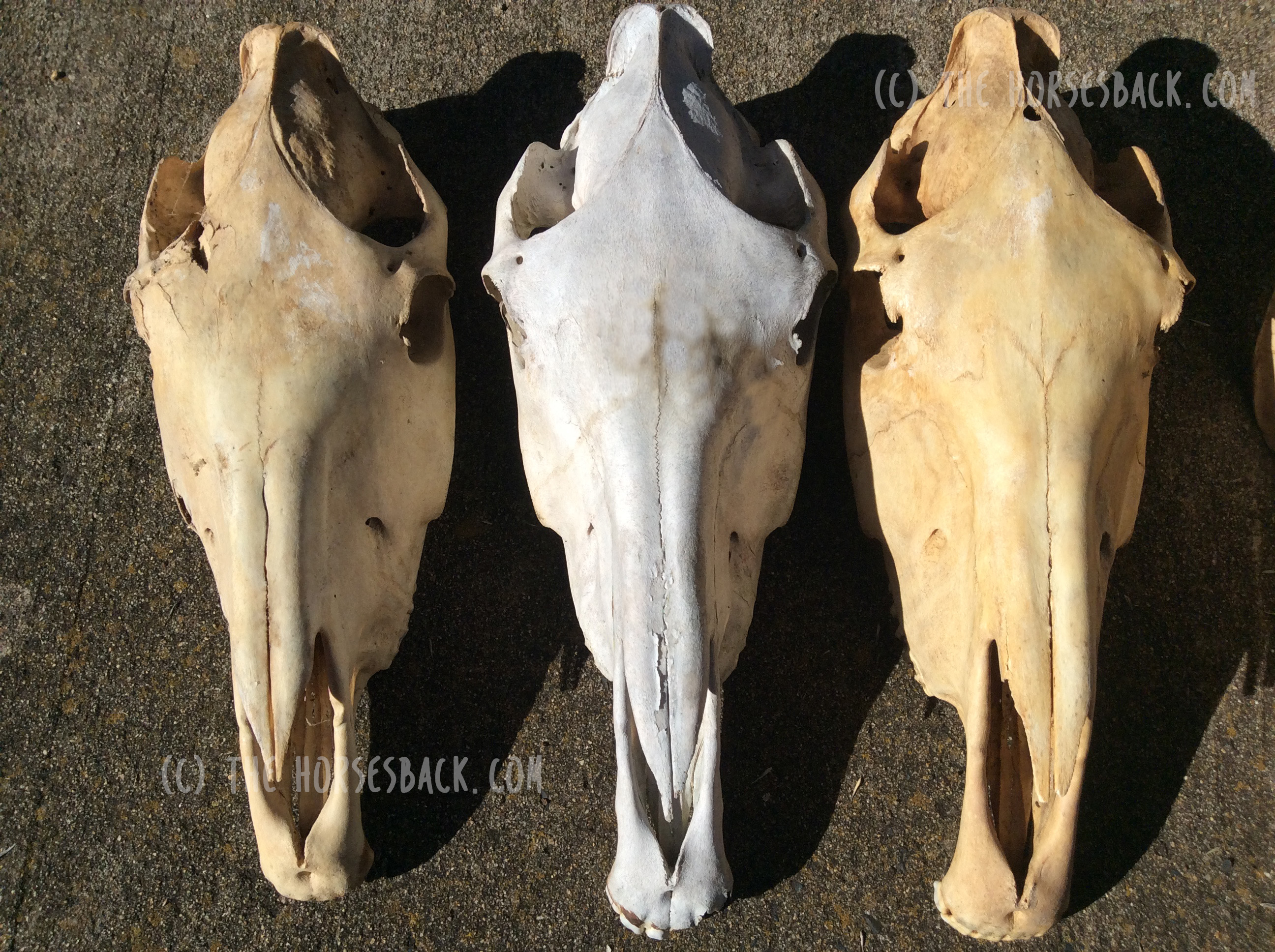
As their horse’s bodyworker, I’m often the first to tell an owner that unbalanced forefeet are causing problems right through the body.
Related issues can include ringbone, carpal arthritis, shoulder asymmetry, base of neck arthritis, atlas rotation, TMJ issues, spinal rotation, scoliosis and even sacroiliac dysfunction – all depending on the severity and duration of the hoof issue.
That’s not me being dramatic. In an older horse, that’s absolutely the type of problems I can find when there’s a long term high hoof.
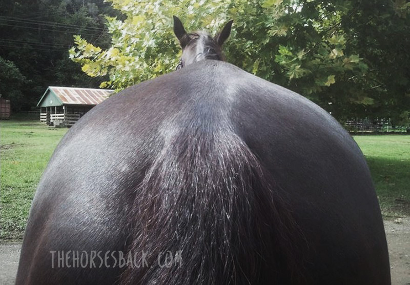
And don’t forget, there are the saddle fit issues that go along with all of that.
Here, in plain speak, is the list of rehab tips that I offer my clients, so that they can help their farrier to help their horse.
© All text copyright of the author, Jane Clothier, https://thehorsesback.com. No reproduction of partial or entire text without permission. Sharing the link back to this page is fine. Please contact me for more information. Thank you!

Before We Start: Choose Your Hoofcare Professional Well
It’s obvious, but this is essential.
Engage a hoof professional who walks the walk as well as talking the talk.
This is someone who can tell you about hoof function and how the ‘normal’ hoof works during loading and movement.
This is someone who frequently updates their professional training.
Professionals who take continuing education are informed about current research into hoof function, as well as methods to address problems.
They know a lot more about current best practice and are more likely to demonstrate it, too.
This most definitely isn’t someone who relies solely on the apprenticeship they completed many years ago.
How it helps:
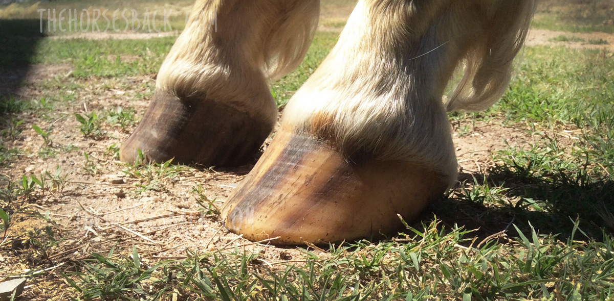
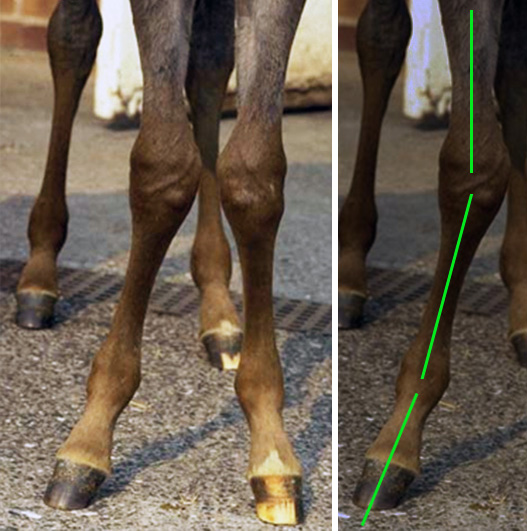
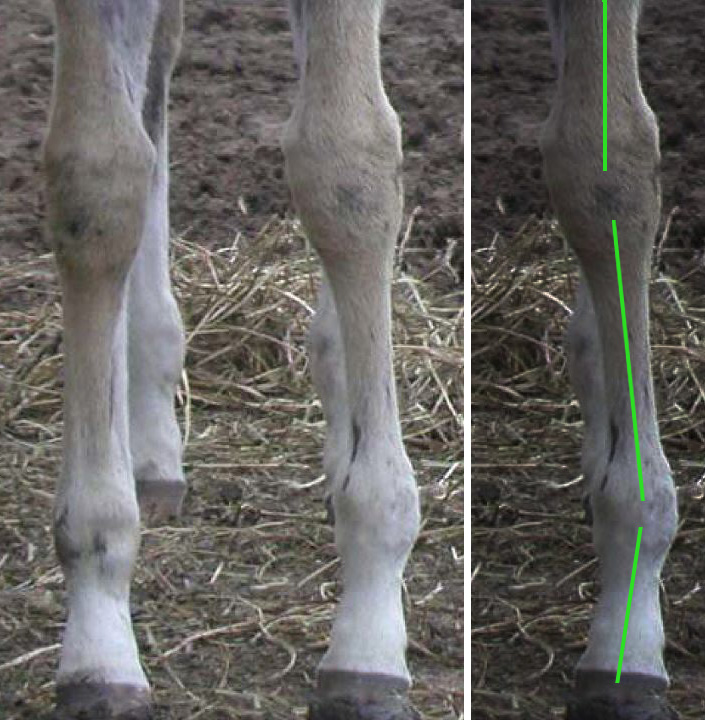
Skilled hoofcare professionals will aim to achieve a similar height in the two front hooves, even though the hoof angles may be different when viewed from the side.
They will trim each hoof according to its underlying structure, making corrections where needed.
They will NOT simply try to create similar angles and toe length, which creates stresses in a high hoof, and sets your horse up for numerous hoof and body problems.
With a skilled hoofcare practitioner on board, there is plenty more you can do to help your horse through this process of change. The more effort you put in, the more your horse will improve.
1. Use Variable Feeding Positions
Variable feeding positions were first described by Sharon May-Davis, who recognised their value as a form of passive physio.
This one is easy: hang a haynet at poll height to replicate eating from trees. This encourages the horse to stand square while eating.
You’ll gain the best results from small hole nets, as your horse will twist her head from left to right while eating. She’ll also drop down to eat hay from the ground.
Doing so activates the deep muscles beneath the neck vertebrae. It also activates the muscles beneath both shoulder blades as she shifts her weight one foot to the other.
If you feed more than one net a day, place the other at chest height.
How it helps:
Think thoracic sling and freeing up the restrictions that have arisen from stabilising the weight over different height limbs, as well as adopting a ‘scissor’ position to eat (which will have contributed to the problem in the first place).
2. Introduce Different Surfaces
The hoof balance is changing, but there may still be asymmetries in your horse’s body that are slow to shift.
Standing your horse on different and unfamiliar surfaces can make the body’s self-adjustments happen quicker.
If you’re able to put some down, gravel provides wonderful under-hoof stimulation. Sand too. Your horse can tilt his hooves toe-down, heel-down or side-down, as he wishes.
Interlocking mats are also effective and can be used anywhere, as can commercially made physio mats and pads.
Watch your horse become curious, start relaxing and yawning, and you know that body adjustments are happening from within.
How it helps:
It can take the nervous system a while to wake up and catch up with what’s new.
The horse has sensory nerves in the feet, and new surfaces provide different proprioceptive.
It’s not just the hoof – changes in balance are registered from the many neuroreceptors of the lower leg.
As the parasympathetic nervous system (‘rest and repair’) is activated, musles are allowed to relax and reset.
3. Feed on a Slope
Again, think thoracic sling. The horse must open up the spine at the cervico-thoracic junction (base of neck).
It’s also next to impossible to adopt a scissor position (the foreleg grazing stance, see no. 7 below) when eating on a slope.
How it helps:
Your horse is encouraged to load into the front hooves equally, left and right.
Finally, the soft tissue structures at the back of the leg will be gently stretched on the limb with the upright hoof.
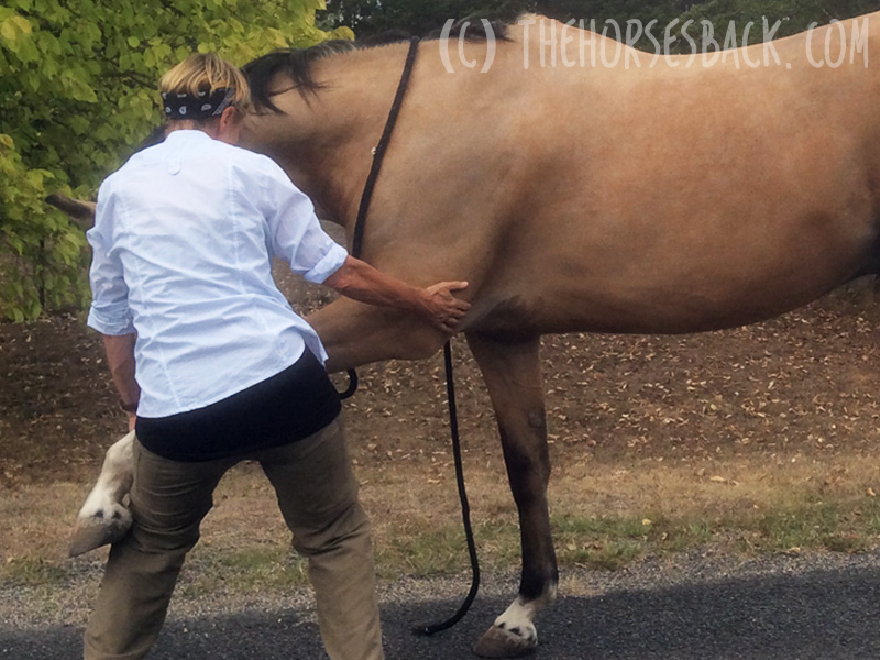
4. Rock the Withers
Standing alongside your horse when she is standing square, gently rock the withers from side to side.
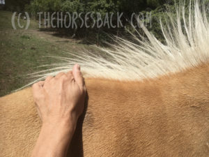
Hold one or two wither processes at a time (feel for the ‘buttons’) and swing gently from one side to another.
This makes the horse load into one forefoot, then back into the other.
Do this from both sides to ensure equal work, as most of us are either stronger pullers or stronger pushers.
How it helps:
The horse is being asked to mobilise between the shoulder blades. When there’s restriction, she’ll raise a hoof on the swing away from that leg.
Easing any restriction will help with relaxing the shoulder on the high side and allowing the joint angles to open out.
She may dislike the exercise at first if it’s uncomfortable, but it will improve over time. You are also gently mobilising the spine deep to the shoulders.
5. Massage the Neck Muscles
When there’s a high foot, the restriction and reduced movement on that side (the stride and therefore leg swing is often shorter) will transfer up through the shoulders to the neck.
 Tension also builds because the horse is trying – and failing – to balance their weight centrally.
This is a secondary effect, caused by the horse’s changed posture.
Pay particular attention to the fine muscles behind the poll, which will be imbalanced.
How it helps:
Releasing muscular tension will help the horse to move to a more symmetrical posture through training and postural improvement.
6. Good Old Carrot Stretches
Use pieces of carrot to encourage stretches to either side.
This will help your horse to become more familiar with loading into both forehooves to a more equal degree.

There are many variations on these stretches and everyone has an opinion!
If you try to ensure that your horse is standing square in front, these stretches will help, whatever the exact approach.
How it helps:
The horse is stretching out the shoulder and neck muscles, including those beneath the shoulder blade, which are going to be more restricted on one side than the other.
This action will also stimulate and activate the deep spinal muscles, bring fresh ‘body awareness’ to the region (ie, proprioception).
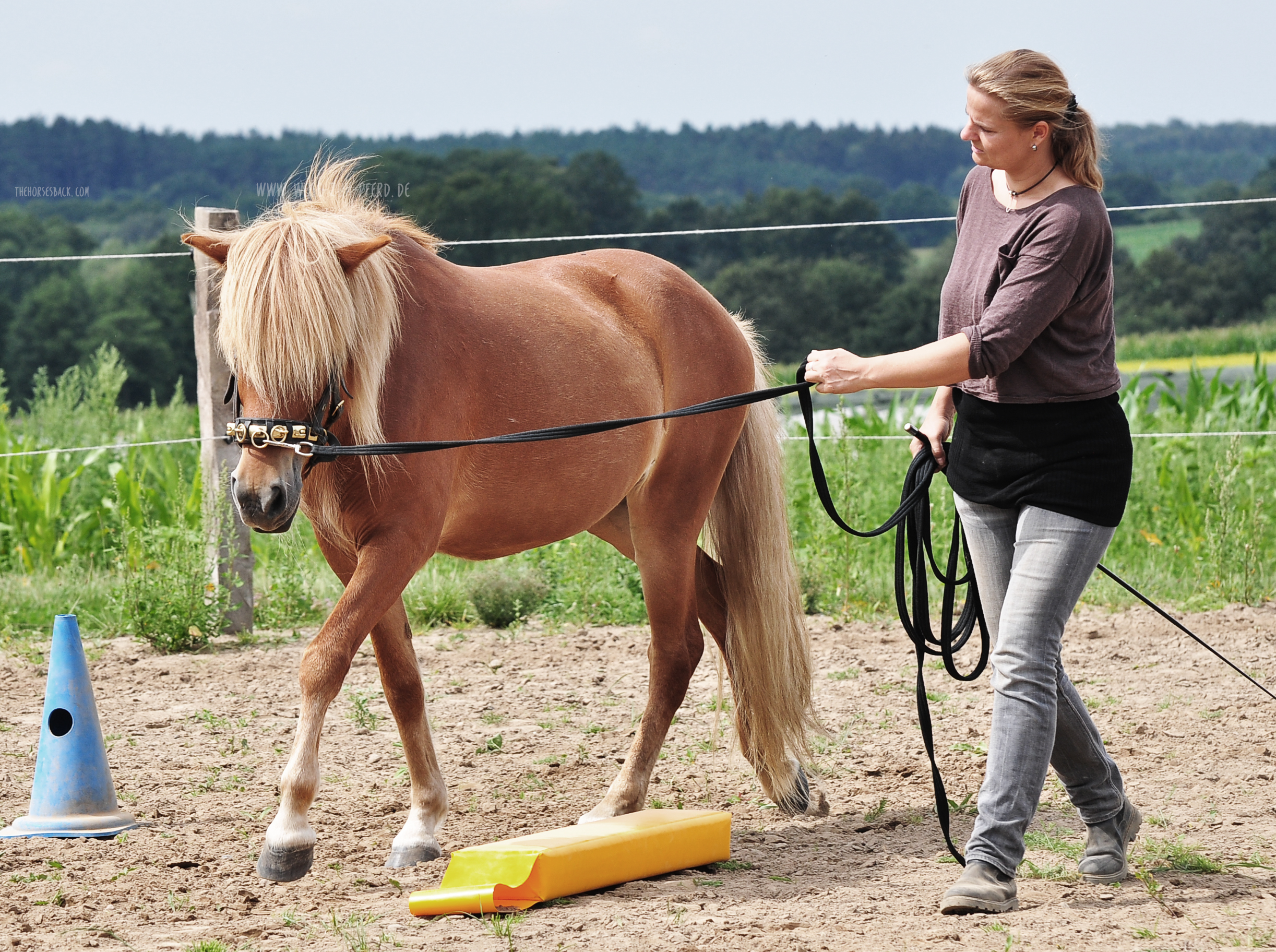
7. In-Hand Grazing Tricks
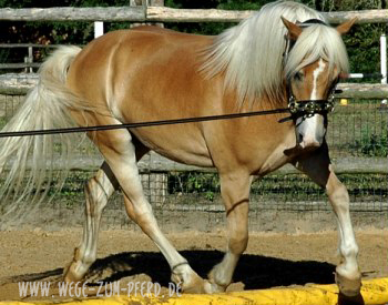
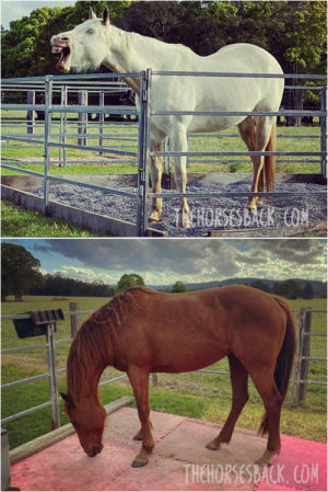
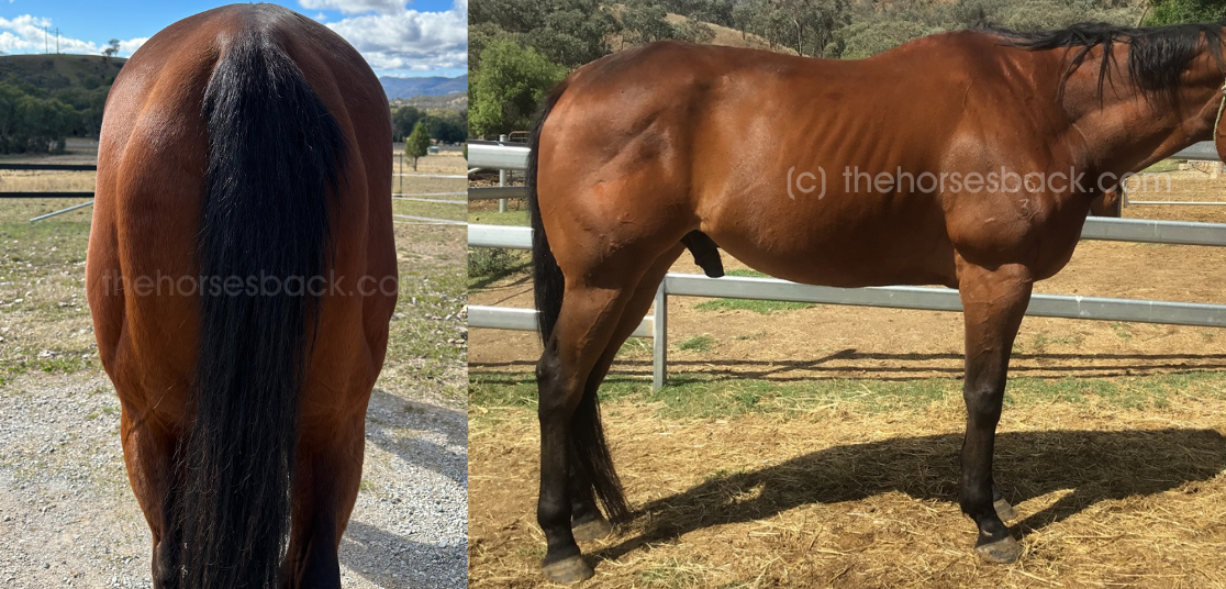
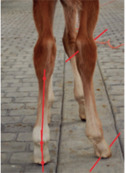
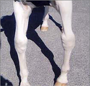
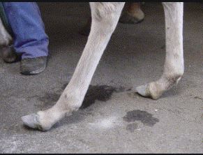
Does your horse have an obvious ‘scissor’ grazing stance, as in the first photo (right)?
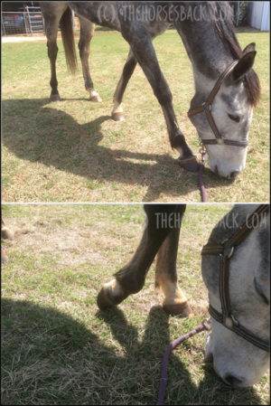
If you horse’s high hoof is always the one at the back, then there is more you can do.
Try to spend up to 20 minutes a day feeding your horse in-hand.
Walk with her and let her graze, but ONLY allow her to do so when the upright foot is placed either level with the lower foot, or further ahead.
This means lots of stepping forward, stopping, stepping again, until she’s stood as you’d like.
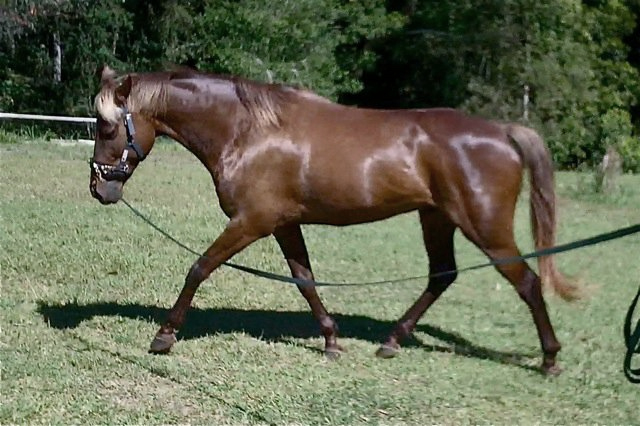
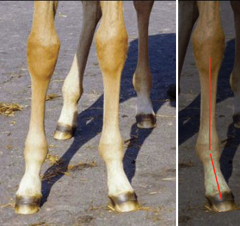
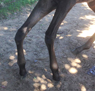
This will be hard for your horse at first, as you can see here, but should become easier over time.
How it helps:
As grazing positions became long term, they also familiar and are adopted habitually. It’s like folding our arms or crossing our legs in one direction.
As the hooves become balanced, the horse will continue adopting the ‘old’ position.
This is a way of reprogramming your horse as the hooves start to become more balanced.
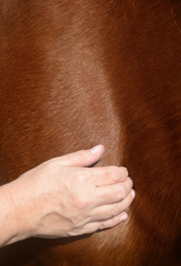
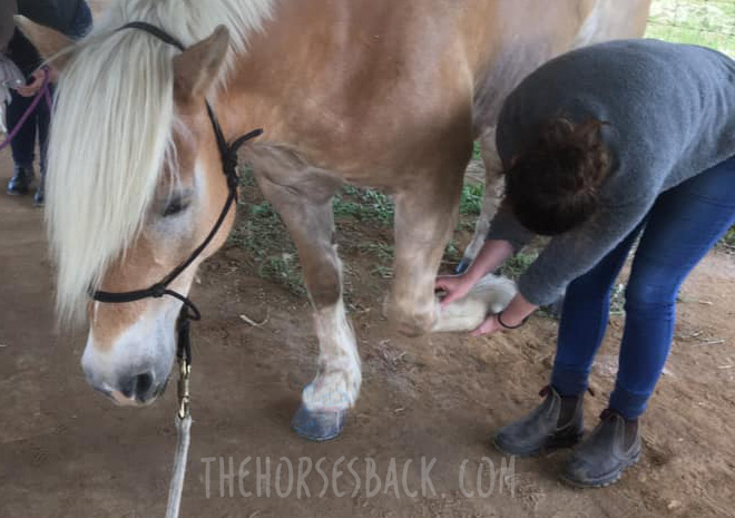
8. Daily Leg Stretches
This is where little and often really helps. Perform gentle leg stretches forward.
Don’t haul the leg, but allow your horse to take up the last inch or so herself.
If there’s a lot of tension, go very gently and don’t stretch to the limit.
Let the cannon hang vertically from the knee, so you’re only extending the upper leg.
A hand behind the elbow will help and your encourage your horse to ‘pop’ the last part of the stretch.
Don’t force it. It’s not about physically lengthening muscles: you’re allowing signals to reach the brachial plexus (nerve centre behind the shoulder blade).
Watch out for your horse looking down and touching his own knee as you start doing this, almost as if he’s surprised to see it there.
How it helps:
This is far more than a muscle stretch! You are influencing the horse’s nervous system’s awareness of the forelimb’s joints and muscles, and the leg’s position in relation to the body and then the ground.
This is proprioception and it will benefit from some help in resetting itself at shoulder joint level.
This ‘resets’ the leg and improves awereness around various joints’ range of motion.
9. Joint Mobilization
The hoof height is changing and becoming more equalized, meaning there’ll be changes higher up the legs as well.
The high hoof side will have to open out a bit, while the low hoof side will be slightly less extended than before.
This involves the soft tissue structures around joints, particularly those of the fetlock and pasterns.
Do you ever see horses shaking their hooves or rotating their lower limb? No.
Your horse is likely to love you for gently rotating and flexing the lower joints, allowing structures to move in different ways, where previously they have been limited.
Pasterns above an upright side are likely to feel stuck. Above an underrun hoof, they will feel loose.
How it helps:
Mobilizing the joints through gentle rotation helps to restore range of motion where it has been restricted.
Spaces will be opened up that allow fluid to circulate, lubricating the joint and bringing more freedom to tendon movement.
This all helps the horse to stand above the changed hoof, positioning the leg closer to where it needs to be.
10. Work Over Poles
Many horses with long toes have a tendency to trip. Once again, as the hoof balance changes, they need to adjust their sense of ‘where their feet are’.
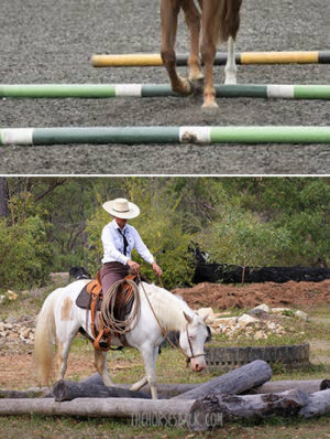
Working over poles helps your horse to focus on individual hoof placement, in terms of both stride length and height of motion.
This can be done over ground and raised poles, or over logs on the trail.
For stronger input, back your horse over poles.
Lead him over with one fore foot, then back with that foot. Lead him over with both forefeet, then back with both. And so on, until the horse knows how to navigate during back-up with all four feet.
How it helps:
Once again, it’s about the proprioceptive input.
As well as ‘equalizing’ your horse’s awareness of its feet, you’re correcting for any lasting effects of pain.
Pain in one hoof and the joints above it has a negative effect on proprioception. Same if two hooves are affected by pain.
By doing this kind of work, you’re helping your horse’s nervous system to ‘square up’, along with the feet.
There’s more – there’s always more. For example, make sure that nutrition is adequately providing the building blocks for hoof growth, development and strength.
As you get going, you’ll find that these are not so much short term changes for rehabilitation, but part of a move towards a more proactiveway of managing your horse.
(c) Jane Clothier, thehorsesback.com

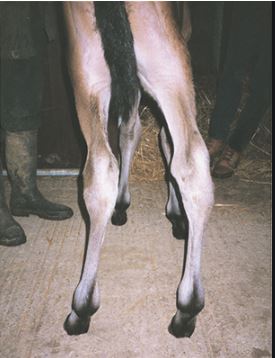
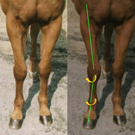
 What we’re looking at is a combination of ALDs: carpal valgus and varus in front, and/or a tarsal valgus and varus behind.
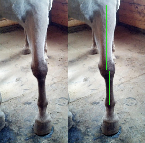
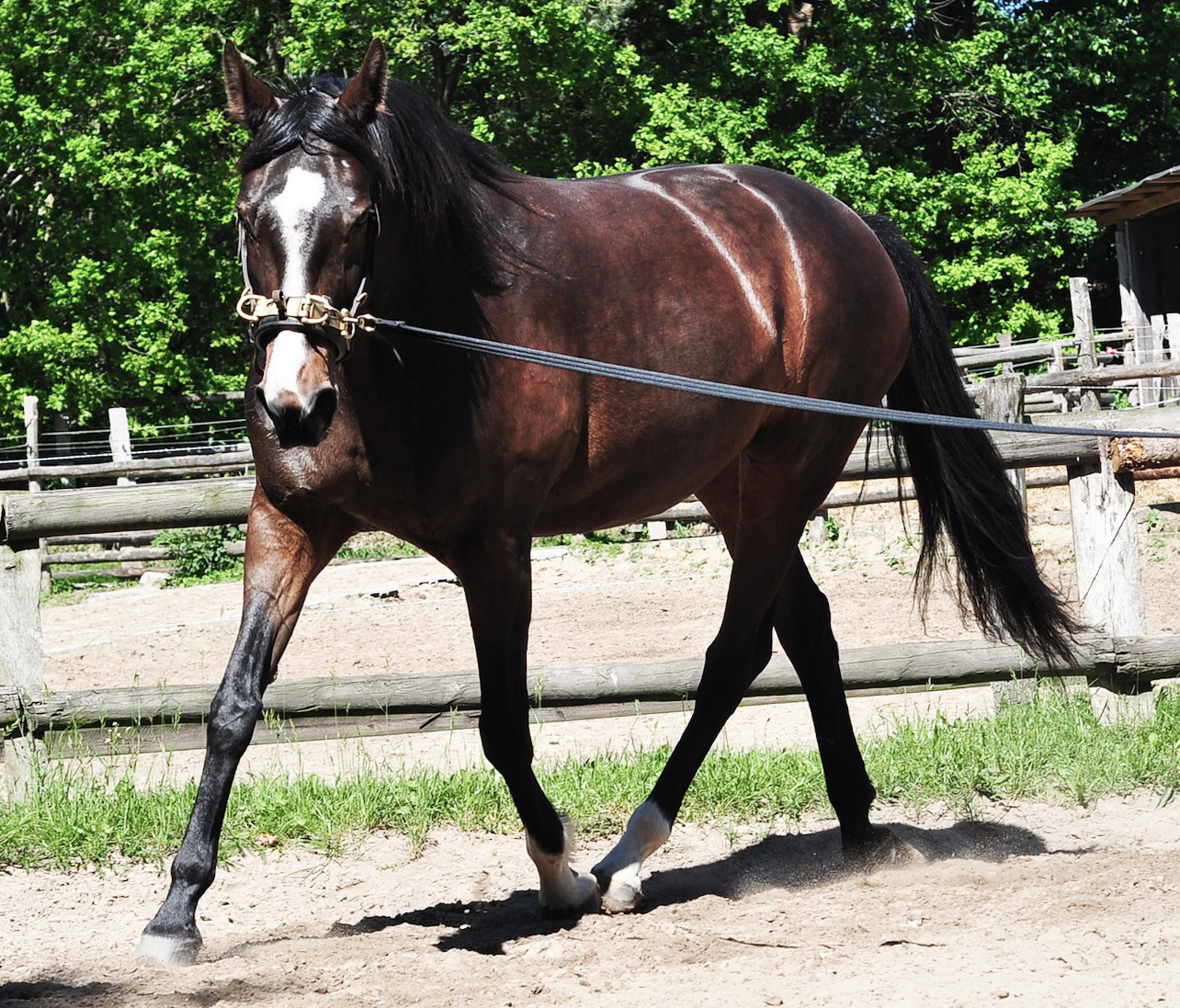
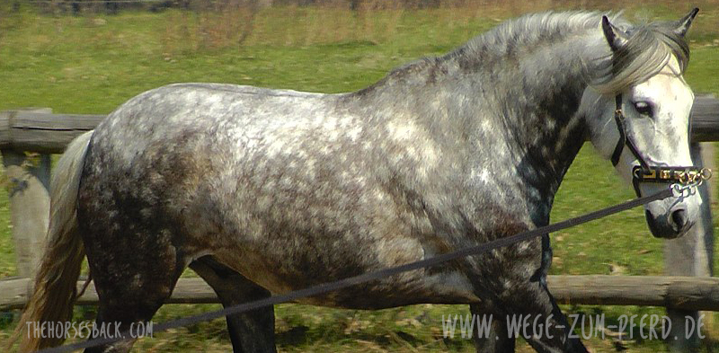 Thinking as a bodyworker, if there were one thing I could change in the training of ridden horses, it would be the way that many people lunge their animals.
 Here is a simple and affordable resource that will help you to do it better:
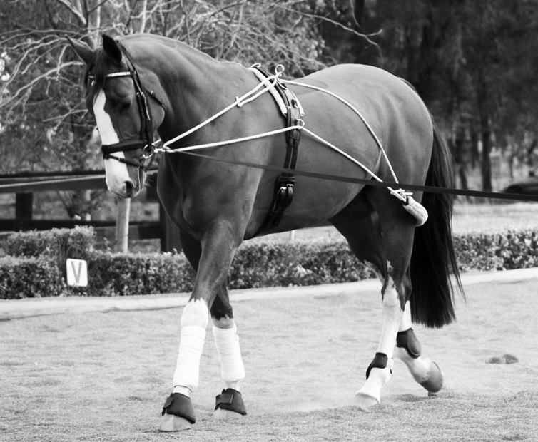
 In the last few years, a renewed interest in a more classical, biomechanically correct approach to training has brought simple, in-hand training techniques to wider audiences. Older methods that aimed to prepare horses for ridden careers, which have been overlooked in the great rush to do everything faster, have come back into focus.
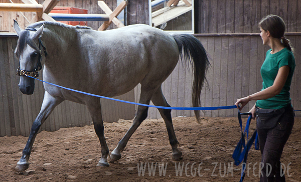
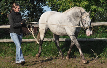 Biomechanically correct lungeing helps to develop the back and teach self-carriage, which makes it humane as well as effective.